Managing the Peritoneal Surface Component of Gastrointestinal Cancer: Part 2
Until recently, peritoneal carcinomatosis was a universally fatalmanifestation of gastrointestinal cancer. However, two innovations intreatment have improved outcome for these patients. The new surgicalinterventions are collectively referred to as peritonectomy procedures.During the peritonectomy, all visible cancer is removed in an attemptto leave the patient with only microscopic residual disease. Perioperativeintraperitoneal chemotherapy, the second innovation, is employed toeradicate small-volume residual disease. The intraperitoneal chemotherapyis administered intraoperatively with moderate hyperthermia.Part 1 of this two-part article, which appeared in the January issue,described the natural history of gastrointestinal cancer with carcinomatosis,the patterns of dissemination within the peritoneal cavity, andthe benefits and limitations of peritoneal chemotherapy. Peritonectomyprocedures were also defined and described. Part 2 discusses the mechanicsof delivering perioperative intraperitoneal chemotherapy andthe clinical assessments used to select patients who will benefit fromcombined treatment. The results of combined treatment as they vary inmucinous and nonmucinous tumors are also discussed.
ABSTRACT: Until recently, peritoneal carcinomatosis was a universally fatal manifestation of gastrointestinal cancer. However, two innovations in treatment have improved outcome for these patients. The new surgical interventions are collectively referred to as peritonectomy procedures. During the peritonectomy, all visible cancer is removed in an attempt to leave the patient with only microscopic residual disease. Perioperative intraperitoneal chemotherapy, the second innovation, is employed to eradicate small-volume residual disease. The intraperitoneal chemotherapy is administered intraoperatively with moderate hyperthermia. Part 1 of this two-part article, which appeared in the January issue, described the natural history of gastrointestinal cancer with carcinomatosis, the patterns of dissemination within the peritoneal cavity, and the benefits and limitations of peritoneal chemotherapy. Peritonectomy procedures were also defined and described. Part 2 discusses the mechanics of delivering perioperative intraperitoneal chemotherapy and the clinical assessments used to select patients who will benefit from combined treatment. The results of combined treatment as they vary in mucinous and nonmucinous tumors are also discussed.
Part 1 of this two-part article, which appeared in the January issue of ONCOLOGY, described the natural history of peritoneal carcinomatosis including the dissemination associated with its evolution. Treatment issues related to the use of intraoperative intraperitoneal chemotherapy and peritonectomy procedures were also described. In part 2, the mechanics of delivering perioperative intraperitoneal chemotherapy are reviewed, and factors affecting the success of the combined treatment approaches are presented. Four clinical assessments that assist patient selection for combined treatment protocols are also discussed.
Techniques for Perioperative Intraperitoneal Chemotherapy
Hyperthermic Intraoperative Intraperitoneal Chemotherapy
FIGURE 1
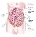
Tubes and Drains Used for Heated Intraoperative Intraperitoneal Chemotherapy
In order to initiate hyperthermic intraoperative intraperitoneal chemotherapy, a series of tubes and drains (four drainage tubes and a single inflow catheter) must be placed within the peritoneal cavity through the abdominal wall (Figure 1). To prevent leakage of these tubes as they exit the abdominal skin, a purse string suture is used. Generally, the inflow catheter is placed at a site thought to be at highest risk for recurrent disease, because the greatest heat will be generated at this site.
FIGURE 2

Coliseum Technique for Heated Intraoperative Intraperitoneal Chemotherapy FIGURE 3
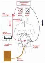
Perfusion Circuit for Heated Intraoperative Intraperitoneal Chemotherapy
Following tube placement, the selfretaining retractor is partially dismantled. It is reassembled to construct a frame approximately 4 in. above the anterior abdominal wall. A heavygauge monofilament suture is used to elevate the skin edges on the selfretaining retractor, and thereby, create a reservoir for chemotherapy solution within the abdomen (Figure 2).
A perfusion circuit is necessary to maintain a temperature of approximately 41.5oC within the peritoneal cavity (Figure 3). Inflow and outflow tubes are connected to roller pumps and a heat exchanger. A heater/cooler, which maintains a water temperature of 48oC flowing through the heat exchanger, is part of the apparatus. For most drugs, a 90-minute perfusion is indicated to achieve a maximal cytotoxic effect.
The drugs used intraoperatively for hyperthermic chemotherapy are mitomycin (Mutamycin), cisplatin, and doxorubicin or oxaliplatin (Eloxatin). The standardized orders utilized at the Washington Cancer Institute for this therapy are shown in Table 1. Following the completion of the intraoperative chemotherapy, the self-retaining retractor is again positioned. At this time, all intestinal anastomoses are completed, seromuscular repairs of the bowel are performed, and the abdomen is closed. Usually, a fifth closedsuction drain is placed within the subcutaneous space. The skin is closed with a watertight, running monofilament suture so that no leakage of fluid occurs from the abdomen postoperatively.
Most patients who receive heated intraoperative intraperitoneal chemotherapy are also given early postoperative intraperitoneal fluorouracil (5-FU) or paclitaxel. The catheters for drug instillation and abdominal drainage must be kept clear of blood clot, fibrin clot, and tissue debris. To accomplish this, an abdominal lavage utilizing the same tubes and drains as used for the heated intraoperative chemotherapy is initiated in the operating room. Large volumes of fluid are rapidly infused and then drained from the abdomen after a short dwell time. The standardized orders for immediate postoperative lavage are shown in Table 2.
Early Postoperative Intraperitoneal Chemotherapy
TABLE 1
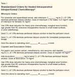
Standardized Orders for Heated Intraoperative Intraperitoneal Chemotherapy TABLE 2

Standardized Orders for Immediate Postoperative Peritoneal Lavage TABLE 3
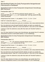
Standardized Orders for Early Postoperative Intraperitoneal Fluorouracil or Paclitaxel
If the patient recovers well from the cytoreductive surgery and intraperitoneal heated chemotherapy, then early postoperative intraperitoneal chemotherapy is initiated. At the Washington Hospital Center, early postoperative chemotherapy is initiated in all patients except those with very early disease and a low likelihood of tumor recurrence. The drugs employed in this chemotherapy are cell-cycle-dependent drugs. The standardized orders for early postoperative intraperitoneal 5-FU and early postoperative intraperitoneal paclitaxel are presented in Table 3. All the intra-abdominal catheters are withdrawn after fluid drainage is substantially reduced prior to the patient's discharge from the hospital.
Selection of Patients for Treatment Using Quantitative Prognostic Indicators
The greatest impediment to achieving long-term benefits from combined treatment with cytoreductive surgery and perioperative intraperitoneal chemotherapy is improper selection of patients. Patients with advanced disease experience minimal benefit and significant morbidity and mortality. Given the risks and benefits for patients with a large volume of invasive cancer, elective cytoreductive surgery should be withheld in this subgroup unless performed by the most experienced surgical teams.
Excluding pseudomyxoma peritonei and cystic mesothelioma, extensive cytoreductive surgery and aggressive intraperitoneal chemotherapy are not likely to produce a lasting benefit in patients with advanced peritoneal surface cancer from a gastrointestinal primary. Rapid recurrence of the peritoneal surface disease combined with progression of lymph nodal, liver, or systemic disease are likely to interrupt any long-term benefit. Asymptomatic patients with small-volume peritoneal carcinomatosis should be referred for treatment.
In the past, peritoneal carcinomatosis was a fatal disease process. The only assessment required was the presence or absence of carcinomatosis. Currently, four important clinical assessments of peritoneal surface malignancy are used to select patients who will benefit from the combined treatment: (1) histopathologic typing to assess the invasive character of the malignancy, (2) preoperative computed tomography (CT) of the chest, abdomen, and pelvis with maximal oral and intravenous contrast, (3) peritoneal cancer index determination, and (4) completeness of cytoreduction (CC) score.
Histopathologic Type and Biologic Aggressiveness of a Peritoneal Surface Malignancy
Noninvasive tumors arising in the appendix (pseudomyxoma peritonei) or from the peritoneal surface itself (cystic mesothelioma) may have largevolume accumulations within the abdomen and pelvis, and yet, may be made visibly disease-free with the use of peritonectomy procedures. Also, these noninvasive malignancies are extremely unlikely to metastasize via lymphatic channels to lymph nodes or via the bloodstream to liver or other systemic sites. Therefore, combined treatment with curative intent in patients with a large mass of widely disseminated pseudomyxoma peritonei or minimally aggressive peritoneal mesothelioma is appropriate. Pathology review and an assessment of the invasive vs nonaggressive nature of a malignant process are essential to treatment planning.[1]
Preoperative CT Scan
A preoperative CT scan of the chest, abdomen, and pelvis is required in planning treatment of a peritoneal surface malignancy. This radiologic examination is essential to exclude liver or systemic metastases and pleural surface spread. Unfortunately, the CT scan is an inaccurate test by which to quantitate nonmucinous carcinomatosis distribution and volume. The malignant tissue progresses as a layer on the peritoneal surfaces and conforms to the normal contours of the abdominopelvic structures-quite different from the metastatic process in the liver or lung, which show up as three-dimensional spherical tumor nodules and can be accurately assessed by CT.[2]
FIGURE 4
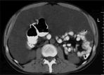
Computed Tomography Scan of a Patient With Adenomucinosis FIGURE 5
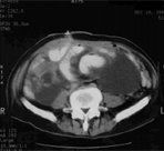
Computed Tomography Scan of a Patient With Intermediate-Grade Mucinous Adenocarcinoma
Fortunately, the CT scan is helpful in imaging mucinous adenocarcinoma on peritoneal surfaces.[3] These tumors produce large volumes of mucoid material, readily distinguishable from normal structures by anatomic location, shape, and density. A knowledgeable radiologic interpretation can also distinguish patients with a high likelihood of complete cytoreduction from those who would be designated for incomplete resections. The CT scan excludes patients who are unlikely to receive the benefit of a potentially curative approach from an elective operative intervention. Interventions in patients with advanced disease would be performed for symptom management, and the surgery would be of a palliative, minimally aggressive nature.
The two radiologic criteria found to be most useful in excluding carcinomatosis patients from elective intervention are segmental obstruction of the small bowel and the presence of tumor nodules greater than 5 cm in diameter on small bowel surfaces or directly adjacent to small bowel mesentery in the jejunum or upper ileum. Tumor involvement of the small bowel at the terminal ileum is not thought to be a contraindication to elective surgery, because disease at this site can be resected as part of the cytoreductive surgery.
These criteria reflect radiologically the pathobiology of carcinomatosis. Obstructed segments of bowel indicate an invasive malignancy where portions of small bowel lack peristalsis or become narrowed. Large, greater than 5 cm, mucinous tumor nodules on the small bowel or its mesentery indicate that the cancer is not redistributed away from the intestinal surface by peristaltic motion. Difficulties with dissection of the mucinous tumor from the small bowel signify the need for a palliative effort (Figures 4 and 5).
Peritoneal Cancer Index
FIGURE 6

Peritoneal Cancer Index
The third prognostic assessment of peritoneal surface malignancy is the peritoneal cancer index, which is a quantitative indicator of prognosis derived from the integration of the size of the peritoneal implant and the distribution of nodules on the peritoneal surface (Figure 6). This index should be used in the treatment decision-making process while the abdomen is being completely explored. The choice between a definitive cytoreduction and a palliative debulking is greatly influenced by the peritoneal cancer index.
To arrive at a score, the size of the intraperitoneal nodules must be assessed in all abdominopelvic regions; the number of nodules is not scored, only the size of the largest nodule. A lesion score of zero (LS-0) means that no malignant deposits are visualized; an LS-1 signifies the presence of tumor nodules less than 0.5 cm in size; an LS-2 indicates that tumor nodules between 0.5 and 5.0 cm are present; and an LS-3 indicates the presence of tumor nodules greater than 5.0 cm in any dimension. If there is a confluence (layering) of tumor, the lesion size is scored as 3.
To assess the distribution of peritoneal surface disease, an LS is determined for each of the 13 abdominopelvic regions. The summation of the LS score is the peritoneal cancer index for that patient. The maximal score is 39 (13 * 3).
To date, the peritoneal cancer index has been validated in four separate situations: Ottow and colleagues used it successfully to quantitate intraperitoneal tumor in a murine peritoneal carcinomatosis model.[4] Portilla and coworkers showed that it could be used to predict long-term survival in patients with peritoneal carcinomatosis from colon cancer who are undergoing a second cytoreduction.[ 5] Berthet and coworkers showed that it predicted benefits for the treatment of peritoneal sarcomatosis from recurrent visceral and retroperitoneal sarcoma.[6] Sugarbaker showed that it could be used to predict the likelihood of long-term survival in colon cancer patients undergoing combined treatment.[7] In these studies, patients with a favorable prognosis had a peritoneal cancer index of less than 13.
• Exceptions to the Rule-Exceptions to the rules for using the peritoneal cancer index have been established. First, noninvasive malignancy on peritoneal surfaces may be completely cytoreduced even though the peritoneal cancer index is as high as 39. Diseases such as pseudomyxoma peritonei, cystic peritoneal mesothelioma, and grade 1 sarcoma fall into this category. With these minimally invasive tumors, the status of the abdomen and pelvis at completion of cytoreduction may have no relationship to the volume recorded at the time of abdominal exploration; ie, even though the surgeon explores an abdomen with a maximal peritoneal cancer index, it can be brought to an index of 0 by cytoreduction. In these diseases, the prognosis is only related to the condition of the abdomen after the cytoreduction (CC score).
A second caveat for using the peritoneal cancer index is related to cancer at crucial anatomic sites. For example, a small volume of invasive cancer incompletely resected from the common bile duct will result in a poor prognosis despite a low peritoneal cancer index. Invasion of the base of the bladder or unresectable disease on a pelvic sidewall may, by itself, result in residual invasive disease after maximal cytoreduction and eventuate in a poor prognosis. In other words, invasive cancer at crucial anatomic sites may function as systemic disease in the assessment of the prognosis with invasive cancer. Because only patients who undergo a complete cytoreduction can achieve long-term survival, residual disease at anatomically crucial sites will supersede a favorable peritoneal cancer index score.
Completeness of Cytoreduction Score
The most definitive assessment of prognosis to be used with peritoneal surface malignancy is the CC score. This information, however, is of less value to the surgeon in planning treatment than the peritoneal cancer index, because it is not available until after the cytoreduction is complete (the peritoneal cancer index is available at the time of abdominal exploration). If, during exploration, it becomes obvious that the cytoreduction will not be complete, the surgeon may decide that a palliative debulking, which will provide temporary symptomatic relief, is appropriate and may discontinue plans for an aggressive cytoreduction with intraperitoneal chemotherapy.
In both noninvasive and invasive peritoneal surface malignancy, the CC score is the major prognostic indicator. It has been shown to function with accuracy in mucinous appendiceal malignancy, peritoneal carcinomatosis from colon cancer, sarcomatosis, and peritoneal mesothelioma.[5-8]
FIGURE 7

Completeness of Cytoreduction Score
For gastrointestinal cancer, the CC score has been defined as follows: A CC score of zero (CC-0) indicates that no peritoneal seeding occurred during the complete exploration. A CC-1 score indicates that tumor nodules persisting after cytoreduction are smaller than 2.5 mm. (This is a nodule size thought to be penetrable by intracavitary chemotherapy.) Both CC-0 and CC-1 scores would, therefore, be designated for complete cytoreduction. A CC-2 score indicates residual tumor nodules between 2.5 mm and 2.5 cm. A CC-3 score indicates residual tumor nodules greater than 2.5 cm or a confluence of unresectable tumor nodules at any site within the abdomen or pelvis. CC-2 and CC-3 cytoreductions are considered incomplete (Figure 7).
Results of Combined Treatment
Appendiceal Mucinous Malignancy as a Paradigm
FIGURE 8
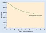
Survival of Patients With Established Peritoneal Surface Malignancy From a Perforated Appendiceal Malignancy
The best results of the treatment of peritoneal carcinomatosis from gastrointestinal malignancy are achieved in patients with mucinous epithelial malignancy of the appendix. Sugarbaker and Chang published their experience with 385 such patients treated over a 15-year period.[8] The survival rate was approximately 50% (Figure 8).
Several unique clinical features of the epithelial appendiceal malignancies have facilitated the favorable treatment results documented for this tumor: First, spread from appendiceal tumors usually occurs in the absence of lymph node and liver metastases. The primary tumor develops within a tiny lumen. Even small tumors early in the natural history of the disease will cause appendiceal obstruction and perforation, resulting in the release of tumor cells into the free peritoneal cavity. Symptoms or signs of the appendiceal perforation manifest in almost every patient before lymph node or liver metastases have occurred.
Second, these tumors exhibit a wide spectrum of invasion, with the majority demonstrating a noninvasive histology. Mucinous tumors that are minimally invasive (as in the pseudomyxoma peritonei syndrome) can be totally resected using peritonectomy procedures to achieve a CC-1 cytoreduction.
Third, the majority of these tumors are mucinous. The texture of the implants allows greater penetration by chemotherapy than occurs with solid tumors. Finally, the malignancy disseminates so that all of the disease is contained within the regional chemotherapy field. If the intraperitoneal chemotherapy is successful in eradicating microscopic residual disease on peritoneal surfaces, the patient will survive long term.
FIGURE 9
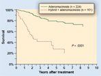
Survival of Patients With Appendiceal Malignancy and Peritoneal Surface Disease, by Histology FIGURE 10
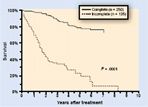
Survival of Patients With Appendiceal Malignancy and Peritoneal Surface Disease, by Completeness of Cytoreduction
In these patients, if a CC-1 cytoreduction is possible, eradication of microscopic residual disease by intraperitoneal chemotherapy determines long-term outcome. Survival is significantly correlated with the mucinous tumor morphology (adenomucinosis vs hybrid plus adenocarcinoma, Figure 9) and the CC score (Figure 10). In contrast to most studies in gastrointestinal cancer patients, the peritoneal cancer index and lymph node involvement are not determinate prognostic factors in patients with peritoneal dissemination of appendiceal mucinous tumors.
High Prior Surgical Score
As the combined treatment of carcinomatosis becomes more widely used, major changes in the management of cancer patients with peritoneal seeding must be considered. With this new approach of using peritonectomy for cytoreduction, one gains a concept for an intact peritoneum as the first line of defense against carcinomatosis.
Opening large tissue planes in the presence of free intraperitoneal cancer cells will jeopardize subsequent attempts at curative treatment. Cancer cells will implant within the cancer resection site and beneath the peritoneal surfaces. This implantation and cancer progression will occur beneath the peritoneum and be inaccessible to peritonectomy, which means that "iatrogenic invasion" may occur in the pelvic sidewall, along the course of the ureter, in and around the structures of the porta hepatis, and at other surgically traumatized sites. A patient with cancer progressing deep within the recesses of the abdomen and pelvis is no longer a candidate for peritonectomy and is unlikely to have successful combined treatment.
FIGURE 11
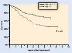
Survival of Patients With Appendiceal Malignancy and Peritoneal Surface Disease, by Extent of Prior Surgical Interventions
The impact of prior surgery on the survival of patients with epithelial appendiceal dissemination of peritoneal disease can be quantitatively assessed by the prior surgical score (PSS). This assessment estimates the extent of prior dissections before the definitive cytoreduction. Abdominopelvic regions 0 to 9 are scored for prior surgery. A PSS of zero (PSS-0) indicates that no major dissections occurred, and the diagnosis was achieved by biopsy only. A PSS-1 indicates dissection of one or two abdominopelvic regions, and a PSS-2 indicates dissection of three to five regions. A PSS-3 indicates an attempt at prior cytoreduction or extensive debulking in the absence of intraperitoneal chemotherapy. Figure 11 shows the detrimental effect of a PSS-3 on the outcome of combined treatment of appendiceal mucinous tumors.
• Surgical Modifications-One may conclude that the initial surgery in patients with peritoneal seeding should be modified. For example, in this new approach to gastrointestinal carcinomatosis, a patient with a perforated mucinous appendiceal malignancy who is found to have peritoneal seeding at the time the primary cancer is diagnosed should undergo a minimal surgical procedure. An appendectomy should be performed, the omental implants should be generously biopsied, and the abdomen should be closed for definitive combined treatment at a later time. A right colectomy should not be performed.
As a second example, if the patient has an obstructing colonic malignancy, an ostomy above the primary cancer would be appropriate. In a patient without obstructive symptoms and a diagnosis of colon cancer with carcinomatosis, definitive biopsy of peritoneal implants may be the only recommended procedure. Only the most debilitated patient, who is not a candidate for cytoreduction with intraperitoneal chemotherapy, should undergo definitive resection.
The optimal treatment of colon cancer with carcinomatosis requires resection of the primary cancer, peritonectomy of implants on visceral and parietal peritoneum in order to remove all visible evidence of diesase, and perioperative intraperitoneal chemotherapy. In the absense of an adequate management plan, minimal surgical intervention to avoid iatrogenic invasive disease is indicated. In an institution not adequately prepared to manage carcinomatosis, referral to a peritoneal surface treatment center would be appropriate.
Carcinomatosis From Colon Cancer
TABLE 4

Results of Combined Treatment of Peritoneal Carcinomatosis From Colon Cancer at Five Treatment Centers
In contrast to appendiceal malignancy, colorectal cancer most commonly shows an invasive histology, frequently disseminates to lymph nodes, liver, and systemic sites, and progresses on peritoneal surfaces as hard nodules that are less likely to be penetrated by heated chemotherapy solutions. Nevertheless, the high incidence of peritoneal seeding in this disease process and the excessive morbidity and mortality associated with this clinical situation have stimulated continued clinical efforts.
Reports from five institutions with at least 25 patients treated, for a total of 333 patients, are shown in Table 4.[9- 14] The combined mean follow-up was 33 months (range: 6-99 months); the mean survival at 3 years was 31% (range: 23%-47%). In all these reports, patients in whom complete cytoreductive surgery was possible had a median survival that far exceeded the survival in patients who had incomplete cytoreductive surgery.[15]
A phase III, prospective randomized study by Verwaal and colleagues in 105 patients deserves special attention.[ 11] After cytoreductive surgery and peritonectomy, 54 patients were treated with heated intraoperative intraperitoneal chemotherapy with mitomycin. In 51 patients, the administered treatment was the standard of care in Holland, a 5-FU/leucovorin regimen. Analyzing the data by an intent- to-treat principle at a median follow- up of 21.6 months (range: 3-44 months), 30 patients were alive in the experimental arm and 20 in the control arm. The Kaplan-Meier survival analysis showed a mean survival of 22.4 months for patients receiving the combined treatment. The 2-year survival was 43% in the experimental group and 16% in the systemic chemotherapy group (P = .032).
Carcinomatosis From Gastric Cancer
Gastric cancer is a rapidly progressing disease. Unfortunately, 30% of patients present with peritoneal seeding at the time their disease is diagnosed. Several groups, mostly in Japan and Korea, have attempted to formulate a management plan for gastric cancer patients with peritoneal seeding. In a majority of these patients, gastrectomy, peritonectomy, and perioperative intraperitoneal chemotherapy constitute a palliative strategy to prevent the adverse events caused by a retained primary gastric cancer and the formation of debilitating ascites.
With a peritoneal cancer index score of less than 10, treatment with gastrectomy and intraperitoneal chemotherapy will result in the patient's death after a median survival of 12 to 18 months. If the primary malignancy is retained, the complications of obstruction and starvation, bleeding, and perforation will cause the patient's death within 3 to 6 months, and debilitating ascites will markedly diminish quality of life. The conclusion of clinical research in this field is that gastrectomy, peritonectomy of obvious implants, and perioperative intraperitoneal chemotherapy prolong life but rarely result in long-term survival.[16]
TABLE 5

Adjuvant Treatment With Postoperative Intraperitoneal Chemotherapy in Gastric Cancer Patients With a Potentially Curative Resection
Perhaps the most promising use of intraperitoneal chemotherapy in gastric cancer is as an adjuvant measure with a potentially curative gastric resection. The results of such therapy have recently been summarized by Sugarbaker and coworkers (Table 5).[17-23] Improved survival has been demonstrated in prospective randomized trials and in trials with historical controls. The favorable impact on survival has been most evident in patients with stage III gastric cancer. In the study by Yu and colleagues, patients with stage III disease had an 18% 5-year survival with surgery alone vs 48% with gastrectomy plus early postoperative intraperitoneal chemotherapy.[ 23]
Morbidity and Mortality
The early results of treatment in these carcinomatosis patients were associated with a reasonable longterm survival when patients with peritoneal seeding were compared to other poor prognosis patients with pancreatic cancer, liver metastases from colorectal cancer, or abdominopelvic sarcoma. However, the initial morbidity and mortality rates were high. Sugarbaker and Jablonski reported that 26% of patients (19 out of 72) developed a bowel perforation postoperatively if they presented for treatment with obstruction, prior radiotherapy, or prior intraperitoneal chemotherapy.[24]
As a result of continued efforts to reduce complications, Stephens and colleagues reported a prospective study of morbidity and mortality in 200 consecutive patients who had undergone combined treatment.[25] In these more recent studies, three treatment- related deaths occurred (1.5%), and grade 3/4 complications developed in 27.0% of patients. Peripancreatitis was seen in 6% of patients, and the incidence of fistula decreased to 4.5%.
Ethical Considerations
The requirements for initiating a new program in peritoneal surface malignancy have been examined in a recent review.[26] Guidelines for the implementation of these complex new treatment strategies vary from institution to institution and country to country. However, without exception, studies of adjuvant intraperitoneal chemotherapy in patients with primary gastrointestinal cancer must be randomized and reviewed by a research board. Also, when a group first attempts to initiate treatment plans for carcinomatosis, a steep learning curve is associated with the new surgical procedures and the new technology.
FIGURE 12
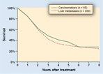
Comparison of the Survival of a Group of Patients With Colorectal Metastases to the Liver and a Second Group With Carcinomatosis
A "start-up protocol" approved by an institutional review board may prompt the members of the group to standardize the methods and familiarize themselves with the experience of others. Probably most importantly, selection criteria for patient treatment would be standardized. An omnibus protocol that allows aggressive peritonectomy and perioperative intraperitoneal chemotherapy in patients without hepatic or systemic dissemination and with small-volume peritoneal seeding seems reasonable. Such a protocol should be utilized for a limited time to treat between 10 and 20 patients.
Formal institutional review board protocols should not be required for the treatment of debilitating ascites, in light of the marked quality-of-life benefits demonstrated by McQuellon and colleagues.[ 27] Also, long-term survival of patients with peritoneal surface malignancy and a low peritoneal cancer index has been established. The survival of patients with resected liver metastases has been compared to that of patients with complete cytoreduction from carcinomatosis.[ 28] Indeed, a nearly identical survival has been shown for these two groups (Figure 12). If liver resection for metastases has been accepted as standard of practice in the absence of phase III studies, perhaps this favorable comparison of treatment outcome suggests that further phase III studies may not be necessary for colorectal carcinomatosis. The ethical implications of these comparisons require careful thought.
Disclosures:
The author(s) have no significant financial interest or other relationship with the manufacturers of any products or providers of any service mentioned in this article.
References:
1.
Ronnett BM, Yan H, Kurman RJ, et al:Pseudomyxoma peritonei associated with disseminatedperitoneal adenomucinosis have a significantlymore favorable prognosis than peritonealmucinous carcinomatosis. Cancer 92:85-91, 2001.
2.
Archer AG, Sugarbaker PH, Jelinek JS:Radiology of peritoneal carcinomatosis, inSugarbaker PH (ed): Peritoneal Carcinomatosis:Principles of Management, pp 263-288.Boston, Kluwer, 1996.
3.
Jacquet P, Jelinek JS, Chang D, et al: Abdominalcomputed tomographic scan in theselection of patients with mucinous peritonealcarcinomatosis for cytoreductive surgery. J AmColl Surg 181:530-538, 1995.
4.
Ottow RT, Steller EP, Sugarbaker PH, etal: Immunotherapy of intraperitoneal cancerwith interleukin 2 and lymphokine
•activatedkiller cells reduces tumor load and prolongssurvival in murine models. Cell Immunol104:366-376, 1987.
5.
Portilla AG, Sugarbaker PH, Chang D:Second-look surgery after cytoreduction andintraperitoneal chemotherapy for peritonealcarcinomatosis from colorectal cancer: Analysisof prognostic factors. World J Surg 23:23-29, 1999.
6.
Berthet B, Sugarbaker TA, Chang D, etal: Quantitative methodologies for selection ofpatients with recurrent abdominopelvic sarcomafor treatment. Eur J Cancer 35:413-419,1999.
7.
Sugarbaker PH: Successful managementof microscopic residual disease in large bowelcancer. Cancer Chemother Pharmacol43(suppl):S15-S25, 1999.
8.
Sugarbaker PH, Chang D: Results of treatmentof 385 patients with peritoneal surfacespread of appendiceal malignancy. Ann SurgOncol 6:727-731, 1999.
9.
Pestieau SR, Sugarbaker PH: Treatmentof primary colon cancer with peritoneal carcinomatosis:Comparison of concomitant vs delayedmanagement. Dis Colon Rectum43:1341-1346, 2000.
10.
Elias D, Blot F, El Otmany A, et al: Curativetreatment of peritoneal carcinomatosis arisingfrom colorectal cancer by complete resectionand intraperitoneal chemotherapy. Cancer92:71-76, 2001.
11.
Verwaal VJ, van Ruth S, de Bree E, etal: Randomized trial of cytoreduction and hyperthermicintraperitoneal chemotherapy versussystemic chemotherapy and palliative surgery in patients with peritoneal carcinomatosisof colorectal origin. J Clin Oncol 21:3737-3743, 2003.
12.
Witkamp AJ, de Bree E, Kaag MM, etal: Extensive cytoreductive surgery followedby intra-operative hyperthermia intraperitonealchemotherapy with mitomycin C in patientswith carcinomatosis of colorectal cancer. EurJ Cancer 37:979-984, 2001.
13.
Shen P, Levine EA, Hall J, et al: Factorspredicting survival after intraperitoneal hyperthermicchemotherapy with mitomycin C aftercytoreductive surgery for patients with peritonealcarcinomatosis. Arch Surg 138:26-33, 2003.
14.
Glehen O, Mithieux F, Oshinsky D, etal: Surgery combined with peritonectomy proceduresand intraperitoneal chemohyperthermiain abdominal cancers with peritonealcarcinomatosis: A phase II study. J Clin Oncol21:799-806, 2003.
15.
Glehen O, Gilly FN, Sugarbaker PH:New perspectives in the management ofcolorectal cancer: What about peritoneal carcinomatosis.Scand J Surg 92:178-179, 2003.
16.
Sugarbaker PH, Yu U, Yonemura Y: Gastrectomy,peritonectomy and perioperative intraperitonealchemotherapy: The evolution oftreatment strategies for abnormal gastric cancer.Semin Surg Oncol 21:233-248, 2003.
17.
Koga S, Hamazoe R, Maeta M, et al:Prophylactic therapy for peritoneal recurrenceof gastric cancer by continuous hyperthermicperitoneal perfusion with mitomycin C. Cancer61:232-237, 1988.
18.
Hamazoe R, Maeta M, Kaibara M: Intraperitonealthermochemotherapy for preventionof peritoneal recurrence of gastric cancer:Final results of a randomized controlled study.Cancer 73:2048-2053, 1994.
19.
Fujimura T, Yonemura Y, Muraoka K, etal: Continuous hyperthermic peritoneal perfusionfor the prevention of peritoneal recurrenceof gastric cancer: Randomized controlled study.World J Surg 18:150-155, 1994.
20.
Yonemura Y, Nonomiya I, Kaji M, et al:Prophylaxis with intraoperative chemohyperthemicagainst peritoneal recurrence of aserosal invasion-positive gastric cancer. WorldJ Surg 19:450-454, 1995.
21.
Ikeguchi M, Kondou A, Oka A, et al:Effects of continuous hyperthermic peritonealperfusion on prognosis of gastric cancer withserosal invasion. Eur J Surg 161:581-586,1995.
22.
Fujimoto S, Takahashi M, Mutou T, etal: Successful intraperitoneal hyperthermiachemoperfusion for the prevention of postoperativeperitoneal recurrence in patients withadvanced gastric carcinoma. Cancer 85:529-534, 1999.
23.
Yu W, Whang I, Chung HY, et al: Indicationsfor early postoperative intraperitonealchemotherapy of advanced gastric cancer: Resultsof a prospective randomized trial. WorldJ Surg 25:985-990, 2001.
24.
Sugarbaker PH, Jablonski KA: Prognosticfactors of 51 colorectal and 130 appendicealcancer patients with peritoneal carcinomatosistreated by cytoreductive surgery and intraperitonealchemotherapy. Ann Surg 221:124-132,1995.
25.
Stephens AD, Alderman R, Chang D, etal: Morbidity and mortality of 200 treatmentswith cytoreductive surgery and hyperthermicintraoperative intraperitoneal chemotherapyusing the Coliseum technique. Ann Surg Oncol6:790-796, 1999.
26.
Gonzalez-Bayon L, Sugarbaker PH,Gonzalez-Moreno S, et al: Initiation of a programin peritoneal surface malignancy. SurgOncol Clin N Am 12:741-753, 2003.
27.
McQuellon RP, Loggie BW, FlemingRA, et al: Quality of life after intraperitonealhyperthermic chemotherapy (IPHC) for peritonealcarcinomatosis. Eur J Surg Oncol 27:65-73, 2001.
28.
Sugarbaker PH: Carcinomatosis-is curean option? J Clin Oncol 21:762-764, 2003.