A Look Behind Development of Pafolacianine for Tumor Detection During Ovarian Cancer Surgery
Philip S. Low, PhD, discusses the obstacles he overcame while creating pafolacianine and what other cancers he hopes will be improved with the use of this agent.
Philip S. Low, PhD, the Presidential Scholar for Drug Discovery and Ralph C. Corley Distinguished Professor of Chemistry at Purdue University

Pafolacianine (Cytalux) was recently approved to help identify ovarian cancer lesions during surgical procedures. This new drug is said to work because it “lights up” cancer cells and allows clinicians to better identify these cells during surgery. When this treatment was used, 26.9% of patients had at least 1 cancerous lesion detected that was not previously seen during visual or tactile inspection.
Philip S. Low, PhD, the Presidential Scholar for Drug Discovery and Ralph C. Corley Distinguished Professor of Chemistry at Purdue University, was the pioneer behind this treatment. Low began developing this drug in 2001, but it wasn’t until a study was published in Nature Medicine that the momentum to start utilizing it in clinical trials began.
In an interview with CancerNetwork®, Low discussed how this treatment works, future studies planned, and why it’s so important for surgeons to consider this for their practice.
CancerNetwork®: Can you begin by discussing how does this agent works?
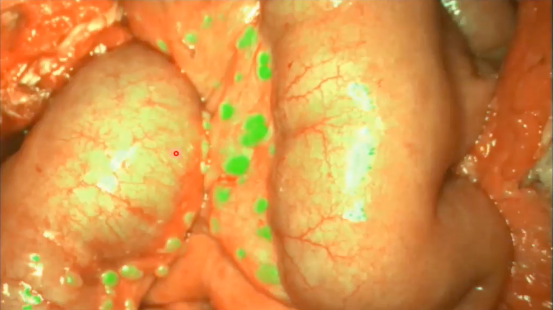
Low: [Over the years] we’ve designed a number of different tumor-specific targeting molecules; we call them ligands. These are molecules that have a high affinity for a receptor that might be expressed on cancer cells, but not unhealthy cells. We then use the ligands as homing molecules to carry an attached bright fluorescent light bulb to cancer cells. Upon homing in on the cancer cells, these tumor-specific ligands bind and carry the attached bright fluorescent light bulbs into the cancer cells. For several weeks thereafter, the cancer cells glow brightly, and the adjacent healthy cells remain dark. This allows the surgeon with the aid of a fluorescent lamp to see the cancer cells very clearly against the dark background.
Pafolacianine utilized in cancer cells

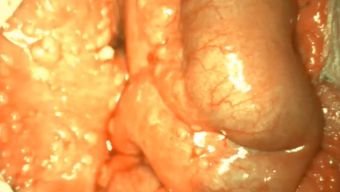
Cancer cells without the use of pafolacianine

How did you begin to develop this agent?
When I first developed this back in 2001, we tested it in mice, and it worked beautifully. I then went to a number of surgeons and said, ‘look at this capability, wouldn’t you like to have it during cancer surgery?’ The answer almost invariably was well, ‘we’re pretty good at finding cancer, so we don’t need this.’
I am frequently asked to give talks at scientific meetings, and I always put in a slide or 2 on the fluorescence guided surgery just to see if I can’t tease somebody into taking an interest, but for years it kept falling on deaf ears. Then finally, a [surgeon] in the Netherlands saw one of my talks and he said, ‘let’s do this’. He took our dye and he was able to obtain approval to test it in ovarian cancer patients in a phase 1 clinical trial in the Netherlands. He published these images and reported that he found 5 times more malignant lesions during surgeries with the aid of the fluorescent dye than without it. This was published in Nature Medicine, and suddenly, the whole world awakened to the opportunity. Many of these surgeons then started beating a pathway to our door to find out if they could get involved in the clinical trials. Now, in all fairness, the technology still hasn’t been seen by [many] of them. It’ll take a while, but I hope we can get the word out so that cancer patients everywhere can benefit.
What was the rationale for creating this agent for use during surgery for ovarian cancer?
We had been developing these tumor-homing molecules for use in delivering cancer killing drugs that can cause a patient’s hair to fall out, their guts to bleed, and their immune systems to become suppressed, rendering them susceptible to opportunistic infections and all sorts of other bad things that can happen when you put potent drugs into healthy cells. We decided the only solution for these problems was to selectively target these nasty drugs to cancer cells, and we developed a number of very specific and potent targeting ligands that would do that job.
We then realized that we had a resource that could be applied for a lot of other uses, and one of those that we envisioned was the highlighting, or illumination, of cancer cells so the surgeon would find them more easily and be able to resect them. A good fraction of tumors recur after surgery and the only explanation for this unwanted outcome is that the surgeon didn’t remove them all. The ability to illuminate these lesions so that they glow brightly will hopefully reduce the probability of overlooking some of them and leaving them left behind after the surgery is completed.
How can a surgeon distinguish healthy cells from cancer cells?
I will say that in many cases, the healthy tissue and the cancer tissue cannot be distinguished. In general, a surgeon uses 2 tools, one is visual inspection and the other one is palpation, the use their fingers. [They use palpation] because the tumor tissue is harder. When they feel a hard lump or something like that, in many cases, they’ll simply resect it because they’re worried [about its potential to be a malignant lesion]. If it’s not a cancer, it may be some sort of a granuloma, cyst, or an undesirable mass of that sort. They feel and look around a lot during cancer surgeries.
What this [agent] does is it gives them a third tool. With many surgeons now moving towards endoscopic surgeries where they don’t make a big incision but instead perform the surgery through a little hole, they lose the ability to palpate suspicious tissue, since they can’t feel it anymore but instead must detect it with an optical scope, just a tube with a light on the end of it. Many surgeons have traditionally relied more on palpation than on visual inspection. [With endoscopic surgery], you still have the visual inspection. But when you lose palpation, you better find something else to replace it or you’re going to miss a lot of malignant lesions. As we move more towards robotic surgeries, this technology will become increasingly important.
Can you discuss the data that supported this approval? Are there any ongoing clinical trials?
The data that were collected for FDA approval were obtained in patients that were suspected of having ovarian cancer. The patient was injected, usually an hour or 2 before surgery with pafolacianine which is usually very innocuous. The surgeon then waits an hour or 2 as the dye circulates through the bloodstream and passes through all the body tissues, both healthy and malignant. If it’s not captured by a receptor on a cancer cell, it’s excreted very quickly. Within a couple of hours, it’s virtually all gone except for the amount retained in the cancer cells. Then the surgeon initiates the surgery, turns on the lamp when he or she wants to see the tumor, and removes any malignant lesions that he or she can find.
We’re now moving forward [with future trials] aimed at identifying additional indications for pafolacianine. In fact, we just finished phase 3 clinical trials in lung cancer.
Are there plans to treat any other cancers than you have mentioned?
There are still other cancers that have the folate receptor for pafolacianine, like kidney cancer, triple-negative breast cancer, and endometrial cancer, [to name a few]. It turns out roughly 40% of human cancers overexpress the folate receptor, so it can be useful for [many diseases]. But at the same time, you need to understand that 60% of cancers don’t [express it]. We need to find a few other targeted fluorescent dyes that will illuminate those other cancers that don’t express a folate receptor. In fact, we have one that just finished phase 1 clinical trials that targets prostate-specific membrane antigen that’s overexpressed on almost all prostate cancers. That dye is currently looking very good. We also have 2 more dyes that when combined with pafolacianine will cover essentially 100% of all cancers. In the end, we intend to develop a cocktail of maybe 3 to 4 tumor-targeting fluorescent dyes that it will enable visualization of essentially all human cancers. That’s the plan, the science looks promising for that.
What should clinicians or patients know ahead of its use?
One of the problems associated with developing an entirely new technology is the task of informing the patient population of its capability and availability. If my wife were to come down with ovarian cancer, I wouldn’t hesitate to have this dye used in her surgery. It would be just an absolute must for me, but I doubt I would know about it if I weren’t involved in its development. Therefore, I think it will be important to make the availability and the potential power of this new technology known to not only patients, but also surgeons as quickly as possible. Just a small handful of patients have participated in these clinical trials and as far as I know, [the surgeons] are all very enthusiastic about it. Until the rest of the world finds out about it and asks their hospital to purchase a camera that is required for imaging these malignant lesions, it won’t be available for everybody. [Purchasing a camera is required for this usage]; we don’t sell the camera, and we don’t even make it. It’s going to be produced by other companies that are designing cameras for our dyes. The hospital will need to purchase a camera, and then we will need to train their surgeons on how to use it. It usually doesn’t take very much time, just a short visit, but we’ll send a professional to train each surgeon interested in the use of the fluorescent dye.
Reference
FDA approves new imaging drug to help identify ovarian cancer legions. News Release. FDA. November 29, 2021. Accessed December 17, 2021. https://bit.ly/3IYf3F9
Late Hepatic Recurrence From Granulosa Cell Tumor: A Case Report
Granulosa cell tumors exhibit late recurrence and rare hepatic metastasis, emphasizing the need for lifelong surveillance in affected patients.