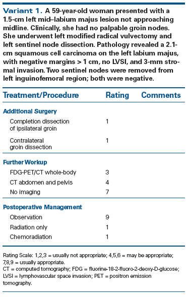
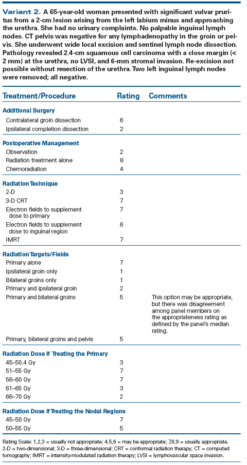
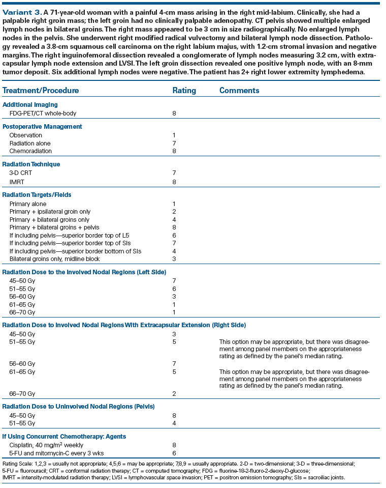
These American College of Radiology consensus guidelines were formed from an expert panel on the appropriate use of adjuvant therapy in vulvar cancer after primary treatment with surgery. The American College of Radiology Appropriateness Criteria® are evidence-based guidelines for specific clinical conditions that are reviewed every 3 years by a multidisciplinary expert panel. The guideline development and review include an extensive analysis of current medical literature from peer-reviewed journals and the application of a well-established consensus methodology (modified Delphi) to rate the appropriateness of imaging and treatment procedures by the panel. In those instances where evidence is lacking or not definitive, expert opinion may be used to recommend imaging or treatment. The panel reviewed the pertinent literature in vulvar cancer and voted on three variants to establish appropriate use of imaging, adjuvant radiation, including dose, fields, and technique, as well as adjuvant chemotherapy. This report will aid clinicians in selecting appropriate patients for adjuvant treatment and will provide guidelines for the optimal delivery of adjuvant radiation therapy and chemotherapy.
Summary of Literature Review
Introduction/Background
Vulvar carcinoma is a rare gynecologic malignancy in the United States; there were an estimated 4,850 new cases and 1,030 deaths from the disease in 2014.[1] Approximately 80% of vulvar cancer patients are diagnosed at an early stage, when the disease is still confined to the perineum without nodal metastases.[2] The mainstay of management for these localized cases of vulvar cancer is surgical resection and lymph node assessment, which entail a good prognosis. However, in cases with surgical margins that are close or positive or in cases found to have positive nodes, the patient’s prognosis is compromised.[3-5] In these less favorable situations, there is an overall survival (OS) benefit with adjuvant therapy.[6,7] Margin and nodal status are the two most common indications for adjuvant therapy.[4,8] Adjuvant therapy would also be considered in the presence of other pathologic risk factors, including lymphovascular space invasion (LVSI), deep invasion of the primary, or a large primary.[4]
The cornerstone of management for most vulvar cancers is lymph node assessment. This is typically carried out by lymph node dissection in patients with a primary lesion deeper than 1 mm. More recently, publication of prospective studies showing the benefit of sentinel lymph node dissection has led to increased utilization of this method in vulvar cancer.[9-11] Adjuvant therapy typically includes radiation. In some cases, adjuvant chemoradiation is offered; however, the evidence for this is limited, and adding chemotherapy in the adjuvant setting is still controversial.[12,13] Given the scarcity of vulvar cancer cases, there are a limited number of prospective trials evaluating the management of vulvar cancer patients. At present, the optimal method of adjuvant therapy is yet to be established (see Variant 1).
Recommendations for Staging
Vulvar cancer most commonly arises from the labia majora and labia minora. This malignancy typically spreads from the vulva to the ipsilateral superficial inguinofemoral lymph nodes, followed by the deep inguinofemoral lymph nodes, then into the pelvis. It would be atypical for contralateral nodes or pelvic lymph nodes to be involved without the involvement of ipsilateral nodes. However, the risk of contralateral lymph node metastases increases as the lesion approaches midline. Direct involvement of certain midline structures, including the glans clitoris and urethra, could have direct spread to the pelvic nodes. Approximately 30% to 40% of patients with clinically negative lymph nodes can have positive lymph nodes. Therefore, pathologic staging of the lymph nodes at risk is critical. Lymph nodes are pathologically assessed in patients with primary tumors that are deeper than 1 mm. Homesley et al[14] identify tumor thickness, lymphovascular invasion, age above 55 years, and poor differentiation as risk factors for nodal involvement. Typically, ipsilateral nodes are assessed in all of these patients. Additional contralateral assessment is completed when patients have tumors approaching midline or clinically concerning nodes. The risk of contralateral nodal involvement increases with each additional risk factor that is present.
For completion of International Federation of Gynecology and Obstetrics (FIGO) staging, the only imaging study that is required to complete a metastatic workup for vulvar cancer is imaging of the chest. Although additional imaging studies of the pelvis are not indicated for FIGO staging, they are commonly done to better delineate the extent of the tumor and to identify suspicious lymph nodes. However, randomized trials have shown a low nodal failure risk with negative sentinel nodes. The likelihood of detecting abnormal lymph nodes in the setting of a small primary lesion with negative sentinel nodes is low. Imaging studies are likely most helpful in patients with advanced lesions. Computed tomography (CT) pelvis and/or magnetic resonance imaging (MRI) can delineate the tumor and aid in treatment planning for patients with advanced lesions. Fluorine-18-2-fluoro-2-deoxy-D-glucose positron emission tomography (FDG-PET)/CT can provide further information to help guide management of vulvar cancer. Many of the studies assessing the usefulness of a PET scan are small retrospective studies. They consistently suggest a good positive predictive value of PET scans in this disease, particularly for nodal metastases. One retrospective analysis by Peiro et al[15] reviewed 13 preoperative PET/CT studies in patients with biopsy-proven vulvar cancer and compared them with histopathologic findings at the time of surgery. All pathological lymph nodes detected on PET/CT were confirmed after surgery. No new lesions were found during surgery that were not noted on PET/CT. A second review by Kamran et al[16] similarly reviewed preoperative PET/CT scans and correlated findings with histologic findings. This study also endorsed 100% specificity but 50% sensitivity. The positive predictive value was 100% and the negative predictive value was only 57%. Therefore, although PET/CT can aid in treatment planning prior to surgery, it cannot be used to rule out nodal metastases, and it does not replace surgical evaluation of the lymph nodes.
Prognosis
The prognosis for vulvar cancer is drastically different for tumors confined to the vulva vs node-positive vulvar cancer and cancer that has spread to distant organs. Based on Surveillance, Epidemiology, and End Results (SEER) data, the 5-year OS for cancer limited to the vulva is approximately 86%. When the patient presents with regional nodes involved, the 5-year OS is reduced to 54%. In the setting of metastatic disease, survival falls further, to approximately 16%. Overall, approximately 23% of patients have a local recurrence at 5 years. Local relapse strongly predicts for cancer-related death. After a patient’s cancer has recurred, the risk of a cancer-related death at 1 year is approximately 60% and at 3 years is 70% (see Variant 2).[17]
Implications of Surgical Margin Status
In the era of conservative surgery, including wide local excision and modified radical vulvectomy, the extent of surgical margins becomes an important prognostic factor for local recurrence.[4] With close or positive margins, re-excision should be considered to obtain negative surgical margins whenever possible. However, when re-excision leads to excessive morbidity, then adjuvant radiation is considered. In a series by Heaps et al,[4] a cohort of 135 patients who were treated with surgical resection of the primary lesion with no adjuvant therapy was reviewed. There were 21 vulvar recurrences. Factors that correlated with local recurrence included depth of invasion > 5 mm, presence of LVSI, increasing keratinization, > 10 mitoses/10 high-power fields (HPF), and surgical margin < 8 mm. In fact, the strongest predictor of local recurrence was surgical margin < 8 mm (P = .0001). All 21 vulvar recurrences had a tumor-free margin of < 8 mm. Of note, 8 mm is measured in formalin-fixed tissue, which corresponds to a margin of approximately 1 cm in fresh tissue. Other studies looking at adjuvant radiation in patients with close or positive margins used 8 mm as the cutoff for a close margin.[6,18] One retrospective study that compared patients with close and positive margins who received radiation vs those who did not showed that adjuvant radiation led to a significant decrease in local recurrence, from 58% to 16%. Furthermore, local recurrence in vulvar cancer was a significant predictor of death, with a 2-year actuarial survival of 25% following relapse.[6]
Additional analysis of the Heaps et al[4] study suggests that although recurrences occurred up to a margin of 8 mm, the most predictive value was associated with a margin of 4.8 mm, which correctly identified 91% of patients who did not have recurrences and 62% of those with a recurrence. In one of the largest retrospective studies (N = 205) to date, Viswanathan et al[19] evaluated margin status as a predictor of local recurrence. Patients were stratified into negative margins, close margins (defined as < 1 cm), and positive margins. In this study, although vulvar recurrences were seen in surgical margins up to 9 mm, the risk for vulvar recurrence was significantly higher in those patients with margins < 5 mm.
Current literature suggests that patients with negative lymph nodes and surgical margins > 8 mm for the primary tumor can generally be observed after surgery. However, some may opt for a less stringent margin status and may use 5 mm as an appropriate cutoff for observing a patient postoperatively without adjuvant therapy.
Radiation Dose
Although many studies have established that adjuvant radiation reduces the risk of local recurrence in patients with positive margins, few have studied the dose necessary to achieve this advantage. Vulvar cancer studies have generally shown that doses range from approximately 45 Gy to 64 Gy. Viswanathan et al[19] published one study that specifically analyzed the relationship between dose and local recurrence. The median dose to the vulva was 45 Gy for women who experienced a vulvar recurrence and 50.4 Gy for those who did not have a recurrence. More specifically, the rate of vulvar recurrence was 21% (4/19) in women treated with doses ≥ 56 Gy and 34% (11/32) in those women treated with doses ≤ 50.4 Gy. These data suggest that for women who have an indication for adjuvant radiation, like the patient in Variant 2, the dose to the vulva should reach at least 56 Gy when feasible (see Variant 3).
Lymph Node Management
Lymph node involvement is a negative prognostic factor in vulvar cancer. In a study by Farias-Eisner et al[3] that reviewed 74 patients who underwent resection of the primary lesion and lymphadenectomy, the OS for patients with negative and positive nodes was 98% and 45%, respectively. Multiple studies have looked into whether it was beneficial to give adjuvant radiation to patients with a single positive lymph node.[18,20,21] These studies showed no benefit and suggested that adjuvant radiation should be recommended only to those with multiple positive lymph nodes or to those with limited lymph node dissections in which < 12 nodes are removed. In a SEER analysis of 208 patients with a single positive inguinal lymph node, 92% underwent radical vulvectomy with a unilateral or bilateral lymph node dissection. The median number of lymph nodes resected was 13. Approximately 50% received adjuvant radiotherapy (RT). In the cohort overall, the 5-year disease-specific survival with adjuvant RT was 77% vs 61% with no RT (P = .02). There was also an improvement in OS; however, this was not statistically significant. Only after stratifying the patients by the extent of their lymph node dissection was it established that there was a statistically significant improvement in 5-year OS with adjuvant radiation if < 12 lymph nodes were removed (77% vs 55%). If > 12 lymph nodes were removed, then there was a trend towards improved 5-year OS with adjuvant radiation, but this was not statistically significant.[21] Most recently, a retrospective exploratory multicenter cohort study, the AGO-CaRE-1 study, analyzed the advantage of adjuvant radiation in patients with positive nodes. Of the 1,249 patients who were diagnosed with vulvar cancer, 447 had positive nodes, and 244 of these patients received adjuvant radiation. The majority of these patients had one or two positive nodes. This study showed a statistically significant benefit in progression-free survival and again a trend toward better survival with adjuvant radiation. The overall 3-year progression-free survival and OS for node-positive patients were 35.2% and 56.2% compared to 75.2% and 90.2% for node-negative patients. The progression-free survival for those node-positive patients who received adjuvant therapy was 39.6% compared to 25.9% in those who did not receive further therapy. The benefit was consistent regardless of the number of positive nodes, grade, or depth of invasion. Given the poor outcomes in a patient with nodal recurrence, this paper strongly supports the use of adjuvant radiation in a patient with one or more positive nodes.[5] In another review by Woelber et al[22] that looked at 157 patients, OS declined with each additional positive lymph node. At 2 years, OS was 88% in node-negative patients and 60%, 43%, and 29% with one, two, and more than two affected nodes, respectively. A subset analysis of patients who received adjuvant radiation found that adjuvant radiation eliminated these differences in survival. The most significant difference in survival with adjuvant radiation was noted in those patients who had only a single positive lymph node. Although adjuvant radiation for a single positive node is controversial, there appears to be sufficient evidence to offer adjuvant radiation in this setting.
Certain pathologic features of lymph nodes also predict worse outcomes, including extracapsular extension and > 50% involvement of a lymph node.[23] Gynecologic Oncology Group (GOG) 37, a prospective study, was conducted to determine the proper management of patients found to have node-positive vulvar cancer after inguinofemoral lymph node dissection. They randomized these patients to undergo either pelvic nodal resection or groin and pelvic radiation. At 6 years, the cumulative incidence of cancer-related death was 29% for the radiation group compared with 51% for the pelvic node resection group. Cancer-related death directly correlated with inguinal relapse, and adjuvant radiation reduced the number of inguinal recurrences.[24]
Summary of Recommendations
- Adjuvant treatment is recommended when surgical margins of the primary are < 8 mm or when positive lymph nodes are identified.
- Sentinel lymph node biopsy is an adequate pathologic assessment of the lymph nodes for patients with clinically negative lymph nodes and primary vulvar tumors < 4–6 cm.
- When treating for close or positive margins, radiation dose to the primary tumor bed should reach 56 Gy when tolerable.
- When groin nodes are involved, the involved nodal region and the pelvic nodal region should be treated. Radiation dose should be approximately 45–50 Gy if no extranodal extension is noted. With extranodal extension, the nodal region should be treated with > 56 Gy.
- Consider adjuvant chemotherapy with bulky nodal disease and extranodal extension, although toxicity may be increased compared to radiation therapy alone.
There is some controversy over whether a groin dissection is entirely necessary or whether it can be replaced with groin radiation. GOG 88 was designed to answer this question by randomizing patients to either bilateral inguinofemoral lymph node dissection or bilateral groin irradiation alone. In this study, the rate of groin recurrence was 18% in the radiation-alone arm vs 0% in the dissection arm.[25] This study closed early due to the failures seen only in the radiation arm. However, it is important to note that 20% of the patients in the surgical arm were found to have positive lymph nodes and subsequently received ipsilateral groin and pelvic radiation. Furthermore, in the radiation-alone arm, a dose of 50 Gy was prescribed to a depth of 3 cm. Koh et al[26] performed a study in which they analyzed the pretreatment CT scans of 50 patients with cervical or vulvar cancer and found that the depth of femoral vessels from the skin can range from 2 to 18.5 cm. Deep nodes are known to be in close proximity to these vessels. There was also a clear correlation between increasing body mass index and increasing depth of femoral vessels. Therefore, in GOG 88, in which 50% of patients had a body mass index > 28 (consistent with obesity), the radiation was not designed to cover all lymph nodes with 50 Gy. In the five patients who had groin failures on the radiation arm of GOG 88, the failures were in regions dosed with < 47 Gy. Three of these patients were underdosed by > 30%. Since then, a number of retrospective trials have looked at the same question and have shown no difference in recurrence-free survival between inguinal irradiation and lymph node dissection.[27,28]
Sentinel Lymph Nodes
In an attempt to reduce the significant morbidity of groin node dissection, many clinicians now use a sentinel lymph node biopsy (SLNB) to assess nodal involvement. There are two prospective trials that evaluated SLNB in patients who were clinically node-negative, with primary tumors that were limited to the vulva, < 6 cm, and of squamous histology. These trials established the feasibility and reliability of SLNBs, and many clinicians would argue that SLNB is now the standard of care for lymph node evaluation in this group of patients. GOG 173 was a prospective study of 452 patients who underwent unilateral or bilateral SLNB based on the location of the original tumor, followed by groin dissection. The sensitivity of SLNB was 91.7%. Of the patients who had a negative SLNB, 11 were found to have lymph node metastases on dissection, suggesting a low false-negative rate of 2% in patients with primary tumors of < 4 cm.[9] Another prospective study, GROINSS-V, recently reported long-term outcomes of patients who were found to have a negative SLNB and were observed without further intervention. At a median follow-up of 58.3 months, 3 out of 57 patients (5.2%) had a groin recurrence.[10] Based on these studies, SLNBs are a feasible option for nodal staging; however, patients must be informed that there is a risk of false-negatives. It is unclear, however, whether there is a difference in quality of life. Data on this matter are limited. However, less aggressive surgical management with sentinel node dissection should decrease the acute and late morbidity, including recurrent erysipelas, lymphedema, and wound dehiscence. One small study assessed quality of life of 62 patients who underwent SLNB or groin dissection using surveys. Although there was an increase in lymphedema in those patients who underwent dissection, there was no overall difference in quality of life between the two groups.[29]
Rationale for Chemotherapy
Chemotherapy has not been systematically studied in the adjuvant setting for vulvar cancer and is not routinely used as adjuvant therapy.[8,9,30] There are some retrospective studies that have included small subsets of patients who received adjuvant chemotherapy and provide some insight into its use. Han et al[12] conducted a review of 54 patients who received radiation therapy, 20 who received chemoradiation and 34 who received radiation alone. Of the 20 patients who underwent chemoradiation therapy, 14 were treated for primary or recurrent disease and 6 were treated after radical vulvectomy for high-risk disease. These patients received fluorouracil (5-FU) and mitomycin-C. In comparing the 6 patients who received adjuvant chemoradiation with those who received adjuvant radiation, there was no statistically significant difference in survival. Although there was a trend towards improved survival, there were too few patients to provide power to this analysis.
Other retrospective studies evaluating patients who received concurrent chemoradiation using 5-FU and mitomycin-C or cisplatin have shown significant morbidity and mortality associated with the addition of chemotherapy. There have been reports of grade 4 neutropenia, neutropenic sepsis, severe enterocolitis, grade 4 skin toxicity, bowel perforation, colovaginal fistula formation, pelvic fracture, and vaginal stenosis.[13,31] In cases with positive tumor margins, bulky nodal disease, or extranodal extension, consideration can be given to adding chemotherapy by extrapolating from cervical cancer data that suggest adjuvant chemotherapy can provide a benefit. However, it must be given cautiously, given its morbidity in combination with radiation.
Radiation Technique
In many of the prior studies discussed above that reviewed adjuvant radiation, including GOG 37, radiation targeted the pelvic and groin nodes and did not include the vulva. This was based on the rationale that surgery was an adequate treatment for the primary site and that vulvar recurrences are more safely and successfully salvaged than a nodal relapse. For example, in Homesley et al,[7] node-positive vulvar cancer patients who underwent radical vulvectomy and bilateral groin lymphadenectomy were prospectively randomized to subsequently undergo either ipsilateral pelvic lymphadenectomy or radiation to the groins and pelvis without direct radiation to the vulva. Yet the rates of vulvar recurrences in both groups were low and comparable (8.5% in the RT arm vs 9.1% in the surgery arm). Similarly, in GOG 37, the rate of vulvar recurrence at the median follow-up of 6 years was 8.8% in the pelvic lymphadenectomy arm and 7.4% in the arm that received groin and pelvic radiation. In Dusenbery et al,[32] a cohort of 27 patients was given adjuvant radiation to the nodes with a midline block in place to avoid radiation to the vulva. Of these patients, 17 relapsed (63%); 13 of these had vulvar relapses. Based on this study, there is an unacceptably high rate of vulvar relapse when the primary site is excluded from the radiation field. Of note, the margin status of the primary vulvar lesion in this study was not classified as per the Heaps criteria, contributing to the higher rate of relapses seen. Thus, whether to routinely treat the vulvar primary when treating the groins remains controversial.
Intensity-modulated radiation therapy (IMRT) is commonly used for many gynecologic and pelvic malignancies to decrease the dose to the perineal skin, bowel, and bladder. Beriwal et al[33] describes the advantages of IMRT for vulvar cancer. For a malignancy that requires a complicated plan encompassing the primary, bilateral groins, and pelvis, IMRT provides more conformal and homogeneous dose delivery, with less skin, bowel, bladder, and rectum toxicity. In order to delineate an adequate clinical target volume that covers lymph nodes at risk, Kim et al[34] reviewed 22 cases of pelvic malignancies that involved inguinal lymph nodes and noted where PET-avid lymph nodes were in relation to the femoral vessels. They found that most nodes were medial and anteromedial to the vessels. To cover at least 90% of the disease, the field needed to extend > 2–3 cm in most directions from the vessel. Anatomic borders can be defined laterally by the medial border of the iliopsoas muscle, medially by the lateral border of the adductor longus or medial end of the pectineus, posterolaterally by the iliopsoas muscle and the anterior aspect of the pectineus muscle, and anteromedially by the anterior edge of the sartorius muscle.
The American College of Radiology seeks and encourages collaboration with other organizations on the development of the ACR Appropriateness Criteria® through society representation on expert panels. Participation by representatives from collaborating societies on the expert panel does not necessarily imply individual or society endorsement of the final document.
Financial Disclosure: The authors have no significant financial interest in or other relationship with the manufacturer of any product or provider of any service mentioned in this article.
Copyright © 2015 American College of Radiology. Reprinted with permission of the American College of Radiology.
Supporting Documents
For additional information on the ACR Appropriateness Criteria® methodology and other supporting documents, refer to www.acr.org/ac.
References:
1. Siegel R, Ma J, Zou Z, Jemal A. Cancer statistics, 2014. CA Cancer J Clin. 2014;64:9-29.
2. Beller U, Quinn MA, Benedet JL, et al. Carcinoma of the vulva. FIGO 26th Annual Report on the Results of Treatment in Gynecological Cancer. Int J Gyn Obstet. 2006;95(suppl 1):S7-S27.
3. Farias-Eisner R, Cirisano FD, Grouse D, et al. Conservative and individualized surgery for early squamous carcinoma of the vulva: the treatment of choice for stage I and II (T1-2N0-1M0) disease. Gyn Oncol. 1994;53:55-8.
4. Heaps JM, Fu YS, Montz FJ, et al. Surgical-pathologic variables predictive of local recurrence in squamous cell carcinoma of the vulva. Gyn Oncol. 1990;38:309-14.
5. Mahner S, Jueckstock J, Hilpert F, et al. Adjuvant therapy in lymph node-positive vulvar cancer: the AGO-CaRE-1 study. J Natl Cancer Inst. 2015;107:dju426.
6. Faul CM, Mirmow D, Huang Q, et al. Adjuvant radiation for vulvar carcinoma: improved local control. Int J Radiat Oncol Biol Phys. 1997;38:381-9.
7. Homesley HD, Bundy BN, Sedlis A, Adcock L. Radiation therapy versus pelvic node resection for carcinoma of the vulva with positive groin nodes. Obstet Gyn. 1986;68:733-40.
8. Gaffney DK, Du Bois A, Narayan K, et al. Patterns of care for radiotherapy in vulvar cancer: a Gynecologic Cancer Intergroup study. Int J Gyn Cancer. 2009;19:163-7.
9. Levenback CF, Ali S, Coleman RL, et al. Lymphatic mapping and sentinel lymph node biopsy in women with squamous cell carcinoma of the vulva: a gynecologic oncology group study. J Clin Oncol. 2012;30:3786-91.
10. Robison K, Roque D, McCourt C, et al. Long-term follow-up of vulvar cancer patients evaluated with sentinel lymph node biopsy alone. Gyn Oncol. 2014;133:416-20.
11. Van der Zee AG, Oonk MH, De Hullu JA, et al. Sentinel node dissection is safe in the treatment of early-stage vulvar cancer. J Clin Oncol. 2008;26:884-9.
12. Han SC, Kim DH, Higgins SA, et al. Chemoradiation as primary or adjuvant treatment for locally advanced carcinoma of the vulva. Int J Radiat Oncol Biol Phys. 2000;47:1235-44.
13. Mulayim N, Foster Silver D, Schwartz PE, Higgins S. Chemoradiation with 5-fluorouracil and mitomycin C in the treatment of vulvar squamous cell carcinoma. Gyn Oncol. 2004;93:659-66.
14. Homesley HD, Bundy BN, Sedlis A, et al. Prognostic factors for groin node metastasis in squamous cell carcinoma of the vulva (a Gynecologic Oncology Group study). Gyn Oncol. 1993;49:279-83.
15. Peiro V, Chiva L, Gonzalez A, et al. [Utility of the PET/CT in vulvar cancer management]. Revista espanola de medicina nuclear e imagen molecular. 2014;33:87-92.
16. Kamran MW, O’Toole F, Meghen K, et al. Whole-body [18F]fluoro-2-deoxyglucose positron emission tomography scan as combined PET-CT staging prior to planned radical vulvectomy and inguinofemoral lymphadenectomy for squamous vulvar cancer: a correlation with groin node metastasis. Eur J Gyn Oncol. 2014;35:230-5.
17. Rouzier R, Haddad B, Plantier F, et al. Local relapse in patients treated for squamous cell vulvar carcinoma: incidence and prognostic value. Obstet Gyn. 2002;100:1159-67.
18. Chan JK, Sugiyama V, Pham H, et al. Margin distance and other clinico-pathologic prognostic factors in vulvar carcinoma: a multivariate analysis. Gyn Oncol. 2007;104:636-41.
19. Viswanathan AN, Pinto AP, Schultz D, et al. Relationship of margin status and radiation dose to recurrence in post-operative vulvar carcinoma. Gyn Oncol. 2013;130:545-9.
20. Fons G, Groenen SM, Oonk MH, et al. Adjuvant radiotherapy in patients with vulvar cancer and one intra capsular lymph node metastasis is not beneficial. Gyn Oncol. 2009;114:343-5.
21. Parthasarathy A, Cheung MK, Osann K, et al. The benefit of adjuvant radiation therapy in single-node-positive squamous cell vulvar carcinoma. Gyn Oncol. 2006;103:1095-9.
22. Woelber L, Eulenburg C, Choschzick M, et al. Prognostic role of lymph node metastases in vulvar cancer and implications for adjuvant treatment. Int J Gyn Cancer. 2012;22:503-8.
23. van der Velden J, van Lindert AC, Lammes FB, et al. Extracapsular growth of lymph node metastases in squamous cell carcinoma of the vulva. The impact on recurrence and survival. Cancer. 1995;75:2885-90.
24. Kunos C, Simpkins F, Gibbons H, et al. Radiation therapy compared with pelvic node resection for node-positive vulvar cancer: a randomized controlled trial. Obstet Gyn. 2009;114:537-46.
25. Stehman FB, Bundy BN, Thomas G, et al. Groin dissection versus groin radiation in carcinoma of the vulva: a Gynecologic Oncology Group study. Int J Radiat Oncol Biol Phys. 1992;24:389-96.
26. Koh WJ, Chiu M, Stelzer KJ, et al. Femoral vessel depth and the implications for groin node radiation. Int J Radiat Oncol Biol Phys. 1993;27:969-74.
27. Hallak S, Ladi L, Sorbe B. Prophylactic inguinal-femoral irradiation as an alternative to primary lymphadenectomy in treatment of vulvar carcinoma. Int J Oncol. 2007;31:1077-85.
28. Petereit DG, Mehta MP, Buchler DA, Kinsella TJ. A retrospective review of nodal treatment for vulvar cancer. Am J Clin Oncol. 1993;16:38-42.
29. Oonk MH, van Os MA, de Bock GH, et al. A comparison of quality of life between vulvar cancer patients after sentinel lymph node procedure only and inguinofemoral lymphadenectomy. Gyn Oncol. 2009;113:301-5.
30. Woelber L, Kock L, Gieseking F, et al. Clinical management of primary vulvar cancer. Eur J Cancer. 2011;47:2315-21.
31. Mak RH, Halasz LM, Tanaka CK, et al. Outcomes after radiation therapy with concurrent weekly platinum-based chemotherapy or every-3-4-week 5-fluorouracil-containing regimens for squamous cell carcinoma of the vulva. Gyn Oncol. 2011;120:101-7.
32. Dusenbery KE, Carlson JW, LaPorte RM, et al. Radical vulvectomy with postoperative irradiation for vulvar cancer: therapeutic implications of a central block. Int J Radiat Oncol Biol Phys. 1994;29:989-98.
33. Beriwal S, Heron DE, Kim H, et al. Intensity-modulated radiotherapy for the treatment of vulvar carcinoma: a comparative dosimetric study with early clinical outcome. Int J Radiat Oncol Biol Phys. 2006;64:1395-400.
34. Kim CH, Olson AC, Kim H, Beriwal S. Contouring inguinal and femoral nodes; how much margin is needed around the vessels? Pract Radiat Oncol. 2012;2:274-8.


