Cancer Management Chapter 10: Gastric cancer
Gastric cancer is more common than esophageal cancer in Western countries but is less fatal. More than 21,130 new cases of gastric cancer will be diagnosed in the United States in the year 2009, with 10,620 deaths expected. Worldwide, gastric cancer represents approximately 930,000 new cases and accounts for more than 700,000 deaths. The incidence and mortality of gastric cancer have been declining in most developed countries, including the United States; the age-adjusted risk (world estimate) fell 5% from 1985 to 1990.
Gastric cancer is more common than esophageal cancer in Western countries but is less fatal. More than 21,130 new cases of gastric cancer will be diagnosed in the United States in the year 2009, with 10,620 deaths expected. Worldwide, gastric cancer represents approximately 930,000 new cases and accounts for more than 700,000 deaths. The incidence and mortality of gastric cancer have been declining in most developed countries, including the United States; the age-adjusted risk (world estimate) fell 5% from 1985 to 1990.
Gastric cancer is defined as any malignant tumor arising from the region extending between the gastroesophageal (GE) junction and the pylorus. It may not be possible to determine the site of origin if the cancer involves the GE junction itself, a situation that has become more common in recent years.
Epidemiology
Gender Gastric cancer occurs more frequently in men, with a male-to-female ratio of 2.3:1.0; mortality is approximately doubled in men.
Age The incidence of gastric cancer increases with age. In the United States, most cases occur between the ages of 65 and 74 years, with a median age of 70 for males and 74 for females.
Race Gastric cancer occurs 2.2 times more frequently in American blacks than whites; in black males, it tends to occur at a younger age (68 years).
Geography Evidence of an association between environment and diet and gastric cancer comes from the profound differences in incidence seen in various parts of the world. Almost 40% of cases occur in China, where it is the most common cancer, but age-adjusted incidence rates are highest in Korea.
Survival Most patients still present with advanced disease, and their survival remains poor. From 1989 to 1995, only 20% of patients with gastric cancer presented with localized disease. The relative 5-year survival rate for gastric cancer of all stages is 24%.
Incidence Significant increases in age-adjusted incidence rates for tumors arising in the gastric cardia have been seen in males. Rates for other gastric adenocarcinomas either have not significantly changed (black males) or have declined (white males). Overall, rates of gastric adenocarcinoma rose for white females between 1974 and 1994.
Etiology, risk factors, and prevention
Diet and environment Studies of immigrants have demonstrated that high-risk populations (eg, Koreans) have a dramatic decrease in the risk of gastric carcinoma when they migrate to the West and change their dietary habits. Low consumption of vegetables and fruits and high intake of salts, nitrates, and smoked or pickled foods have been associated with an increased risk of gastric carcinoma. Conversely, the increasing availability of refrigerated foods has contributed to the decline in incidence rates. Recent laboratory data from Japan suggest that oolong tea may contain a substance that can kill stomach cancer cells.
Occupational exposure in coal mining and processing of nickel, rubber, and timber has been reported to increase the risk of gastric carcinoma. Cigarette smoking may also increase the risk.
Intestinal metaplasia, a premalignant lesion, is common in locations where gastric cancer is common and is seen in 80% of resected gastric specimens in Japan. Individuals with blood group A may have a greater risk of gastric carcinoma than individuals with other blood groups. The risk appears to be for the infiltrative type of gastric carcinoma (rather than the exophytic type).
Gastric resection Although reports have suggested that patients undergoing gastric resection for benign disease (usually peptic ulcer disease) are at increased risk of subsequently developing gastric cancer, this association has not been definitely proven. Gastric resection may result in increased gastric pH and subsequent intestinal metaplasia in affected patients.
Pernicious anemia Although it has been widely reported that pernicious anemia is associated with the subsequent development of gastric carcinoma, this relationship also has been questioned.
Genetic abnormalities The genetic abnormalities associated with gastric cancer are still poorly understood. Abnormalities of the tumor-suppressor gene TP53 (alias p53) are found in over 60% of gastric cancer patients and the adenomatous polyposis coli (APC) gene, in over 50%. The significance of these findings is not clear at present.
Overexpression, amplification, and/or mutations of oncogenes c-Ki-ras, HER-2/neu(aka c-erb-b2), and c-myc most likely play a role in the development of some gastric neoplasms. A high S-phase fraction has been associated with an increased risk of relapse as well.
Hereditary diffuse gastric cancer occurs due to mutations in the E-cadherin (CDH1) gene. Studies of prophylactic gastrectomy in carriers demonstrate high rates of occult cancers, approximating 80%.
Family history Family members of a patient with gastric cancer have a twofold to threefold higher risk of stomach cancer than the general population.
Prevention Recent studies strongly implicate cyclo-oxygenase 2 (COX-2) in the development of many human cancers, including gastric cancer. Potential mechanisms of oncogenesis include stimulation of tumor angiogenesis, inhibition of apoptosis, immune suppression, and enhancement of invasive potential. Furthermore, COX-2 inhibitors have been shown to decrease the size of gastric adenomas in mice. This information strongly suggests that COX-2 inhibitors may play a role in the prevention, and even treatment, of GI tumors. However, trials with COX-2 inhibitors in other advanced GI malignancies, such as colon cancer, have not been positive, so patients should not be treated with these agents outside the setting of a clinical trial.
Helicobacter pylori infection is associated with gastric lymphomas and adenocarcinomas. The overall risk of developing malignancy in the presence of infection is low; however, more than 40% to 50% of gastric cancers are linked with H pylori. The bacterium has been designated a class I carcinogen. Antibiotics alone can cure localized, node-negative MALT (mucosa-associated lymphoid tissue) lymphomas in about 50% of patients. With regard to gastric adenocarcinoma, H pylori infection is associated with a 2.8-fold increase in the relative risk of the disease compared with uninfected controls. Data from Japan and China suggest that H pylori infection can lead to chronic atrophic gastritis. This condition appears to be a major risk factor for gastric cancer. Eradication of H pylori should thus decrease the incidence of gastric cancer by preventing it, and pilot trials assessing this strategy have been undertaken in Central and South America.
Signs and symptoms
Most gastric cancers are diagnosed at an advanced stage. Presenting signs and symptoms are often nonspecific and typically include pain, weight loss, vomiting, and anorexia.
Hematemesis is present in 10% to 15% of patients.
Physical findings Peritoneal implants to the pelvis may be palpable on rectal examination (Blumer’s shelf). Extension of disease to the liver may be appreciated as hepatomegaly on physical examination. Nodal metastases can be found in the supraclavicular fossa (Virchow’s node), axilla, or umbilical region. Ascites can accompany advanced intraperitoneal spread of disease.
Screening and diagnosis
Routine screening for gastric cancer is generally not performed in Western countries because the disease is so uncommon. However, screening appears more effective in high-incidence areas. Mass screening, as has been practiced in Japan since the 1960s, has probably contributed to the 2.5-fold improvement in long-term survival compared with Western countries, though differences in biology may also play a role.
Endoscopy and barium x-rays The diagnosis of gastric cancer in a patient presenting with any constellation of the symptoms previously described revolves around the use of upper endoscopy or double-contrast barium x-rays. The advantage of endoscopy is that it allows for direct visualization of abnormalities and directed biopsies. Barium x-rays do not facilitate biopsies but are less invasive and may provide information regarding motility.
CT scan Once a diagnosis has been established and careful physical examination and routine blood tests have been performed, a CT scan of the chest, abdomen, and pelvis should be obtained to help assess tumor extent, nodal involvement, and metastatic disease. CT may demonstrate an intraluminal mass arising from the gastric wall or focal or diffuse gastric wall thickening. It is not useful in determining the depth of tumor penetration unless the carcinoma has extended through the entire gastric wall. Direct extension of the gastric tumor to the liver, spleen, or pancreas can be visualized on CT, as can metastatic involvement of celiac, retrocrural, retroperitoneal, and porta hepatis nodes. Ascites, intraperitoneal seeding, and distant metastases (liver, lungs, bone) can also be detected.
Endoscopic ultrasonography (EUS) is a staging technique that complements information gained by CT. Specifically, depth of tumor invasion, including invasion of nearby organs, can be assessed more accurately by EUS than by CT. Furthermore, perigastric regional nodes are more accurately evaluated by EUS, whereas regional nodes farther from the primary tumor are more accurately evaluated by CT. Specific ultrasonographic features may aid in the diagnosis and staging of patients with gastric lymphomas.
Capsule video endoscopy A capsule containing a tiny camera is swallowed by the patient. Two pictures per second are taken. Once clinical studies demonstrated that the procedure was safe and effective, the FDA cleared this device for clinical use in 2001. The capsule can be especially helpful in imaging the small intestine.
Laparoscopy Laparoscopy is particularly suited to detect small-volume visceral and peritoneal metastases missed on a CT scan. It should be performed prior to curative-intent locoregional therapy or preoperative chemoradiation therapy.
Bone scan A bone scan should be obtained if the patient has bony pain or an elevated alkaline phosphatase level.
PET scan PET scanning may be used to show distant and metastatic disease and may also be helpful in assessing response to neoadjuvant therapy. In the latter setting, PET response correlates with better survival.
Pathology
Adenocarcinoma is the predominant form of gastric cancer, accounting for approximately 95% of cases. Histologically, adenocarcinomas are classified as intestinal or diffuse; mixed types occur but are rare. Intestinal-type cancers are characterized by cohesive cells that form glandlike structures and are often preceded by intestinal metaplasia. Diffuse-type cancers are composed of infiltrating gastric mucous cells that infrequently form masses or ulcers.
Primary lymphoma of the stomach is increasing in frequency and, occasionally, may be difficult to distinguish from adenocarcinoma.
Stromal tumors GI stromal tumors (GISTs) are mesenchymal tumors of the GI tract, most commonly arising from the stomach. These tumors share an ancestor with, or arise from, the interstitial cells of Cajal (the pacemaker cells of the gut). GISTs commonly express KIT (CD117), but this is not required for diagnosis. Most GISTs have mutations in either the c-kit or PDGFR (platelet-derived growth factor receptor) genes.
Other histologic types Infrequently, other histologic types are found in the stomach, such as squamous cell carcinomas, small-cell carcinomas, and carcinoid tumors.
Metastatic spread of disease from primaries in other organs (eg, breast cancer and malignant melanoma) is also seen occasionally.
Gastric carcinomas spread by direct extension (lesser and greater omentum, liver and diaphragm, spleen, pancreas, transverse colon); regional and distant nodal metastases; hematogenous metastases (liver, lungs, bone, brain); and peritoneal metastases. Multicentricity characterizes up to 20% of gastric cancers.
Staging and prognosis
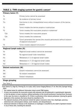
At present, epithelial gastric cancers are most commonly staged by the TNM system. Stromal tumors, lymphomas, carcinoids, and sarcomas are not covered by these TNM criteria. The most recent update of this staging system (Table 1) allows for a more precise nodal classification based on the number of lymph nodes involved.

A more detailed Japanese staging system has been shown to have prognostic importance in gastric cancer. However, these results have not yet been duplicated in the United States, and this system is not widely used around the world. Resected GISTs can be assigned a numeric risk of recurrence based on tumor size, mitotic rate, and location of the primary.
Prognostic factors Aneuploidy may predict a poor prognosis in patients with adenocarcinoma of the distal stomach. High plasma levels of vascular endothelial growth factor (VEGF) and the presence of carcinoembryonic antigen (CEA) in peritoneal washings predict poor survival in surgically resected patients. As with colorectal cancer, intratumoral levels of dihydropyrimidine dehydrogenase (DPD) may be prognostic of gastric cancer; low levels appear to predict better response to fluorouracil (5-FU)–based chemotherapy and longer survival. The prognostic implications of tumor-suppressor genes and oncogenes are an area of active investigation. Patients with cancers of the diffuse type fare worse than those with intestinal-type lesions.
It has long been known that survival rates for patients with gastric cancers are higher in Asian nations than in the United States, but it was not clear whether this represented results from superior surgical techniques or racial differences in underlying biology. Recent analysis of outcomes in Asian-Americans versus white patients suggests true differences exist in tumor biology, though the specifics have not yet been identified.
Treatment
PRIMARY TREATMENT OF LOCALIZED DISEASE
Management of gastric cancer relies primarily on surgical resection of the involved stomach, with reconstruction to preserve intestinal continuity, as resection provides the only chance for cure. Radiotherapy and chemotherapy now have better roles as adjuncts to surgery and in patients with unresectable tumors. Perioperative chemotherapy and chemoradiation therapy remain active areas of current investigation.
Surgery
The objectives of operative treatment for potentially curable gastric cancers are confirmation of resectability, performance of a complete resection, facilitation of appropriate pathologic staging, and reestablishment of GI continuity and function.
Confirmation of resectability Laparoscopy has emerged as an excellent tool to assess the extent of disease and resectability before the surgeon performs an open laparotomy. Laparoscopy adds to the accuracy of preoperative imaging primarily in cases of peritoneal spread or small liver metastases. As a result, morbidity, hospital stay, and costs have been reduced significantly in patients with unresectable lesions. In addition, peritoneal washings can be obtained.
The initial experience with laparoscopic ultrasonography has shown that its value lies in identifying lesions with a high risk of recurrence (T2b or >, N+), for which a preoperative chemotherapy protocol may be available.
As this Handbook went to press the American Joint Committee on Cancer (AJCC) published an updated edition of the AJCC Cancer Staging Manual. Changes were made to the staging system for gastric cancer. These included the following: T and N categories were modified; stage groupings were changed; Esophagogastric junction tumors or tumors less than or equal to 5 cm from the esophagogastric junction and crossing into the esophagogastric junction are staged using the TNM system for esophageal adenocarcinoma. For additional information see Edge SB et al: AJCC Cancer Staging Manual, 7th edition, chapter 11. Springer-Verlag, New York, 2010. Watch also for updates online at www.cancernetwork.com/cancer-management.
Extent of resection The extent of gastric resection depends on the site and extent of the primary cancer. Subtotal gastrectomy is preferred over total gastrectomy, because it leads to comparable survival but lower morbidity. A 5-cm margin of normal stomach appears to be sufficient in proximal and distal resections. For lesions of the GE junction or the proximal third of the stomach, proximal subtotal gastrectomy can be performed. If total gastrectomy is necessary, transection of the distal esophagus and proximal duodenum is required, and omentectomy is performed. In Japan, there is a growing experience with more limited resections of early-stage gastric cancer. This trend includes endoscopic mucosal resection (EMR) of nonulcerated T1 N0 lesions and pylorus-preserving gastrectomy. Laparoscopic resections are also being performed more frequently.
Extent of lymphadenectomy The extent of lymph node resection, including the number removed at the time of gastrectomy, continues to be controversial. Preferably, lymphadenectomy includes the lymphatic chains along the celiac, left gastric, splenic, and hepatic arteries, which allows for more precise lymph node staging. The exact level designation of lymph nodes varies with the site and intragastric location of the primary tumor. Based on the TNM staging criteria, 15 or more lymph nodes should be obtained as minimum and examined for an accurate N classification. Removal of lymph nodes immediately adjacent to the stomach (paracardial, paragastric at the lesser or greater curvature, parapyloric) has been termed D1 dissection. A more extensive D2 dissection would also remove retroperitoneal “second echelon” lymph nodes along the celiac trunk, left gastric artery, hepatic artery, splenic artery, and splenic hilus.
Circumstances and extent of surgical therapy of gastric cancer continue to be discussed. A recent analysis of 27,420 gastrectomy patients from the NCOB links superior postoperative mortality and long-term survival to high-volume hospitals
(Bilimoria KY et al: J Clin Oncol 26:4626–4633, 2008)
. Adding a para-aortic lymphadenectomy to a standard D2 dissection does not improve recurrence-free survival based on a prospective randomized trial
(Sasako M et al: N Engl J Med 359:453–462, 2008)
. A growing experience with laparoscopy or robotic-assisted distal gastrectomy reports low morbidity and improved recovery
(Kim W et al: Ann Surg 248:793–799, 2008; Kim YW et al: Ann Surg 248:721–727, 2008; Song J et al: Ann Surg 249:927–932, 2009)
. While validation in Western patients is pending, early selected patient series appear complementary
(Strong VE et al: Ann Surg Oncol 16:1507–1513, 2009; Guzman EA et al: Ann Surg Oncol 16:2218–2223, 2009
).
Improved long-term survival rates for Japanese patients had been attributed to the extended lymphadenectomies routinely performed in this country. Because the improvement in survival after gastrectomy during recent decades was usually associated with the performance of extended lymph node dissections (D2 dissections or greater), this practice appeared to be sensible if performed with acceptable complication rates. Retrospective data had shown that D2 lymphadenectomy is safe and does not increase morbidity.
On the other hand, two European randomized trials showed no significant differences in overall long-term survival between D1 and D2 dissection groups. Both studies found higher postoperative morbidity and mortality in the D2 (extended) group, largely due to a higher rate of splenectomy and/or partial pancreatectomy performed with those dissections. When a subset of patients with N2 disease were studied in long-term follow-up in the Dutch randomized trial, a survival advantage was shown with D2 dissection. Gastrectomy with extended lymphadenectomy should primarily be performed in specialized centers by experienced surgeons, and splenectomy and pancreatectomy should be avoided; for adequate staging, at least 15 lymph nodes should be removed and analyzed. Recent population data reviews have linked higher total lymph node numbers (up to 40) to superior survival.
A recent phase III trial attempted to address this issue for distal esophageal and GE junction adenocarcinomas. In this study, patients with T3 or T4 adenocarcinoma of the gastroesophageal junction were randomized to receive 2.5 cycles of induction chemotherapy with cisplatin, 5-FU, and leucovorin followed by surgery vs 2 cycles of the same regimen followed by chemoradiotherapy with cisplatin and etoposide and then by surgery. Although the study closed early due to poor accrual, there was a significant improvement in pathologic complete response rate and the rate of tumor-free lymph nodes in the radiation-containing regimen. There was also a trend for improved survival in the group receiving chemoradiotherapy, but this did not reach significance. These results suggest there is an advantage to radiotherapy in neoadjuvant regimens
(Stahl M, et al: J Clin Oncol 27:851–856, 2009)
.
Reconstruction methods After distal gastrectomy, Billroth I gastroduodenostomy or, more commonly, Billroth II gastrojejunostomy methods are acceptable for reconstruction. Reflux esophagitis is a common problem when the gastric reservoir is too small. After total or subtotal gastrectomy, a Roux-en-Y esophagojejunostomy is usually performed.
Resection of extragastric organs may be required to control T4 disease. Such a resection can be associated with long-term survival. Splenectomy should be avoided unless it is indicated by direct tumor extension, because it significantly increases the rate of complications.
Neoadjuvant Therapy
Prompted by the promising results and acceptable toxicity of preoperative (neoadjuvant) chemoradiation therapy in other parts of the GI tract (eg, esophagus, rectum), there is growing interest in neoadjuvant therapy for gastric cancer. Neoadjuvant treatment may be performed in an attempt to convert an initially unresectable cancer to resectable status (so-called conversion therapy), or it may be used in advanced but resectable disease.
The MAGIC trial investigated perioperative treatment with epirubicin, cisplatin, and 5–fluorouracil versus surgery alone, demonstrating improvements in progression-free and overall survival rates with the addition of chemotherapy. In resectable patients, perioperative chemotherapy added to surgery is now a standard of care in many parts of the world.
Adjuvant Therapy
The 5-year survival rate after “curative resection” for gastric cancer is only between 30% and 40% (Table 2). Treatment failure stems from a combination of local or regional recurrence and distant metastases. Investigators have studied adjuvant therapy in the hope of improving treatment results. A North American Intergroup trial randomizing resected patients (stages IB–IV[M0]) to receive chemoradiation therapy or observation showed significant improvement in median disease-free (median 19 vs 30 months) and overall (26 vs 35 months) survival with adjuvant therapy, and the use of postoperative chemoradiation therapy, usually with continuous infusion of 5-FU, is the standard of care in the United States.
A recent review of the SEER database found that receipt of adjuvant therapy is associated with an increase in survival in patients undergoing surgical resection for gastric cancer
(Kozak KRet al: Int J Radiat Oncol Biol Phys 72:517–521, 2008)
. However, as many as 60% of patients who are eligible for adjuvant therapy do not receive it, suggesting that adjuvant therapy is underutilized or limited by a reduced postoperative performance status
(Sherr DL et al: 2009 Gastrointestinal Cancers Symposium: abstract 78, 2009)
Radiotherapy
Radiotherapy can decrease the rate of locoregional failure but has not been shown to improve survival as a single postoperative modality. Postoperative radiotherapy may be appropriate in patients who are not candidates for chemotherapy.
Chemotherapy
Chemotherapy alone as a surgical adjunct does not have a defined role in the United States. Individual randomized trials of chemotherapy plus surgery vs resection alone have showed no definite survival advantage, with the possible exception of patients with widespread nodal involvement and older patients who may do better with chemotherapy. Meta-analyses of postoperative chemotherapy plus surgery vs resection alone have tended to show minor reductions in death rates, but no specific regimen can be recommended.
Intraperitoneal (IP) chemotherapy is theoretically attractive in adjuvant treatment of resected gastric cancer, given the usual pattern of recurrence. Kang and associates performed a randomized phase III study testing mitomycin and doxifluridine with or without IP cisplatin plus mitomycin, in potentially curatively resected patients with gastric cancers grossly involving the serosa. Rates of postoperative complications were similar in both groups. Those getting IP chemotherapy had significantly improved relapse-free and overall survival rates. As it is not clear whether the IP chemotherapy itself, or just the earlier start of chemotherapy in general, was responsible for the benefits seen, more study is necessary. This approach is not ready for wholesale adoption
(Kang Y, et al: J Clin Oncol 26: abstract LBA4571, 2008)
.
Chemoradiation therapy
Patients with T3–T4 any N M0 tumors are at highest risk of locoregional recurrence after potentially curative surgery (surgery in which all macroscopic tumor has been resected with no evidence of metastatic disease) for gastric cancer. Even patients with node-negative disease (T3 N0) have a gastric cancer-related mortality of about 50% within 5 years. Mortality is significantly worse in patients with node-positive disease or in those with incomplete (R1, R2) resection.
In the North American intergroup trial mentioned in the section on “Adjuvant therapy,” patients were randomized to receive chemoradiation therapy or observation following resection of stages IB–IV (M0) adenocarcinoma of the stomach. Chemoradiation therapy following resection of these high-risk patients significantly improved both disease-free and overall survival rates. Because of the apparent benefit of reducing locoregional recurrences, but not distant recurrences, it is possible that more routine use of D2 lymphadenectomy may modify this recommendation in the future. D2 lymphadenectomy was performed in only 10% of the patients in this trial. Subgroup analysis revealed that outcome did not differ based upon the type of lymphadenectomy (P = .80). Still, since only a small percentage of patients underwent the recommended D2 dissection, further research is necessary before firm conclusions can be made in this area. A 10-year update of this trial was recently presented at the 2009 American Society of Clinical Oncology meeting. Patients who were offered chemoradiation continued to enjoy improved survival (HR = 1.32) and disease-free survival (HR = 1.51).
In RTOG 0114, a randomized Phase II trial of two paclitaxel-containing chemoradiation regimens in patients with com-pletely resected gastric cancer, 78 patients with resected IB–IIIB gastric cancer received either paclitaxel and cisplatin for 2 cycles followed by 45 Gy with concurrent cisplatin and paclitaxel vs paclitaxel, cisplatin, and 5-FU for 2 cycles followed by 45 Gy with concurrent paclitaxel and cisplatin. Grade 3 or higher GI toxicity occurred in 59% of those on the 5-FU–paclitaxel arm requiring closure of that arm at interim analysis. The 2-year disease-free survival for the paclitaxel-cisplatin arm was 52%, less than the predefined lower boundary, suggesting that paclitaxel-containing regimens did not improve outcomes over those achieved on INT0116
(Schwartz GK, et al: J Clin Oncol 27:1956–1962, 2009)
.
Despite this trial, significant controversy regarding the need for adjuvant treatment persists and is perhaps growing. Many studies support the contention that aggressive, formal D2 resection may obviate the need for adjuvant treatment in many cases. Other studies and subgroup analyses support the recommendations for adjuvant treatment as concluded in the North American trial. These conflicting results, as well as distinct differences in results between Eastern and Western nations, suggest that this issue may take many years to resolve. In the interim, it is appropriate to recommend adjuvant chemoradiotherapy to those patients in North America who undergo initial gastrectomy.
GISTs have a high risk of recurrence, even following complete resection. The American College of Surgical Oncologists performed a phase III trial of surgery alone vs surgery followed by 1 year of adjuvant imatinib (Gleevec) in patients with resected intermediate or high-risk GISTs. Accrual to the trial was halted early when it was apparent those given postoperative imatinib had a markedly lower 1-year relapse-free survival rate than did those treated with surgery alone (97% vs 83%; P = .0000014). Interestingly, there was no difference in overall survival, leaving open the question of whether patients should be treated immediately following surgery or should wait for relapse and use imatinib in the salvage setting. Most North American GIST experts favor immediate imatinib, and the drug recently received FDA approval in this setting.
Unresectable tumors
Patients with unresectable gastric cancers and no evidence of metastatic disease can be expected to survive approximately 6 months without any treatment.
Palliative resection Palliative resection, bypass, and/or stenting may be appropriate for some patients with obstructive lesions. Palliative resection may also be suitable for patients with bleeding gastric cancers that are not resectable for cure. Generally, resection appears to offer better palliative results than bypass.
Radiotherapy Radiation therapy alone can provide palliation in patients with bleeding or obstruction who are not operative candidates. Radiotherapy may convert unresectable cancers to resectable tumors.

Chemoradiation therapy Patients with locally advanced disease may be appropriately treated with chemoradiation therapy. This approach can provide relatively long-lasting palliation and may render some unresectable cancers resectable. Older studies have shown that postoperative chemoradiation therapy can reduce relapse rates and prolong survival in patients with incompletely resected stomach cancer.
Medical treatment of advanced gastric cancer
When possible, all newly diagnosed patients with disseminated gastric cancer should be considered candidates for clinical trials, and those with good performance status should be offered systemic therapy. Even though cure is not expected with chemotherapy, such treatment may provide palliation in selected patients and sometimes durable remissions. Several randomized chemotherapy trials have suggested improvement in survival and probably quality of life vs best supportive care alone across first and second lines of therapy.
A recent phase II trial of bevacizumab (Avastin), added to modified docetaxel (Taxotere), cisplatin, and 5-FU (DCF) reported extraordinarily high response rates, as well as progression-free survival (PFS) rates approaching the best results seen to date. Jhawer and colleagues gave patients with untreated metastatic gastroesophageal adenocarcinomas the chemobiologic regimen detailed, aiming for an improvement in 6-month PFS from 43% to 63%. Preliminary results in 42 patients showed the combination was well tolerated (rates of neutropenia were lower than those previous reported for DCF with bevacizumab). Six-month PFS was 83% (95% CI: 68%–93%), and median survival was not reached. This regimen needs validation in a phase III trial, but it is extremely promising
(Jhawer M, et al: 2009 Gastrointestinal Cancers Symposium: abstract 10, 2009)
.
Single-agent therapy
Several agents have established activity in gastric cancer: 5-FU, platinums (cisplatin, oxaliplatin [Eloxatin], and carboplatin), mitomycin, etoposide, some anthracyclines (doxorubicin and epirubicin), taxanes (paclitaxel and docetaxel [Taxotere]), irinotecan, antimetabolites (pemetrexed [Alimta], methotrexate, trimetrexate (Neutrexin), and oral fluoropyrimidines (uracil and tegafur [UFT], S-1, capecitabine [Xeloda]). 5-FU and cisplatin have been used most commonly. The responses seen with single-agent chemotherapy have been traditionally partial and mostly short-lived, with little, if any, impact on overall survival.
Novel agents recently tested in patients with advanced gastric cancer include the epidermal growth factor receptor inhibitor cetuximab (Erbitux) and the VEGF inhibitor bevacizumab (Avastin). Bevacizumab appears promising when given with chemotherapy.
Trastuzumab (Herceptin) is a monoclonal antibody against HER2 that has been demonstrated to improve survival in patients with HER2-positive breast cancer. A phase III trial reported at ASCO 2009 by Van cutsem and associates (the ToGa study) has evaluated the efficacy and safety of trastuzumab in the treatment of HER2-positive advanced gastric cancer. The study included 584 patients who were randomly assigned to treatment with trastuzumab plus standard chemotherapy (fluorouracil or capecitabine and cisplatin), every 3 weeks for 6 cycles, or standard chemotherapy alone. Patients who received trastuzumab had a median overall survival of 13.8 months, compared with 11.1 months in the standard chemotherapy group (
P
= .0046). They also had a significantly higher overall response rate (47.3% vs 34.5%,
P
= .0017). Importantly, the two groups did not differ with respect to symptomatic congestive heart failure. Significant but asymptomatic reductions in left ventricular ejection fraction were reported in 4.6% of the trastuzumab group and in 1.1% of the standard chemotherapy group
(Van Cutsem E et al: J Clin Oncol 27[18S]:abstract LBA4509, 2009)
.
Combination chemotherapy
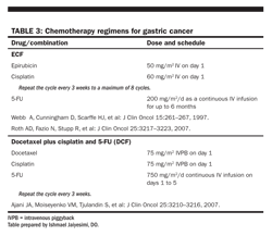
Response rates are consistently higher when combination chemotherapy regimens are used in gastric cancer. Combination therapy has been generally preferred over single agents (Table 3).
In the 1980s, the combination of 5-FU, doxorubicin, and mitomycin (FAM) was considered the standard regimen in the treatment of advanced gastric cancer. However, the NCCTG randomly compared this regimen with 5-FU plus doxorubicin and single-agent 5-FU and found no difference in survival among the patients treated with the three regimens.
Several different regimens, including FAMTX (5-FU, doxorubicin, and methotrexate) and ELF (etoposide, leucovorin, and 5-FU), have been tested. Most regimens show markedly better response rates and longer survival in early trials than in phase III studies. No combination has been confirmed as superior. However, ECF (epirubicin, cisplatin, 5-FU) approaches standard of care in Canada and in some parts of Europe. This regimen has proved superior to FAMTX in terms of objective response rate and survival and superior to the mitomycin, cisplatin, 5-FU (MCF) regimen in terms of toxicity. A combination of docetaxel, cisplatin, and 5-FU was shown to offer superior survival rates vs cisplatin and 5-FU; however, grade 3/4 toxic events occurred in 81% of patients treated with triple therapy. Modifications of this regimen have been proposed and await further testing. The search for the optimal combination regimen continues, with the promising newer agents usually being introduced in combination regimens.
S-1, an oral fluoropyrimidine, was especially promising in the treatment of advanced gastric cancer, based on phase II studies. Results from FLAGS, a phase III study of S-1 plus cisplatin vs 5-FU plus cisplatin in 1,029 patients with advanced untreated gastric or gastroesophageal adenocarcinoma were recently reported. Median overall survival was 8.6 months in patients receiving S-1 vs 7.9 months in the control arm (
P
= .1983). The S-1–containing arm did have lower rates of severe or greater neutropenia and treatment-related deaths. Differing doses of cisplatin on the two arms may have contributed to some of the differences, and make interpretation of results difficult. Nonetheless, the lack of improved efficacy is disappointing
(Ajani JA, et al: 2009 Gastrointestinal Cancers Symposium: abstract 8, 2009)
.
Newer agents with somewhat similar mechanisms of action to those of classic drugs have been tried in patients with advanced gastric cancer. Disappointing results were recently seen, however, in a randomized trial (see sidebar). Oxaliplatin- and cisplatin-containing regimens have been compared, with the former being less toxic and perhaps more efficacious.
Gastric cancer patients should be encouraged to participate in well-designed clinical trials. Outside experimental regimens, the recommended therapy for patients with good performance status is a 5-FU- or cisplatin–based regimen.
References:
SUGGESTED READING
Ajani JA, Moiseyenko VM, Tjulandin S, et al: Clinical benefit with docetaxel plus fluorouracil and cisplatin compared with cisplatin and fluorouracil in a phase III trial of advanced gastric or gastroesophageal cancer adenocarcinoma: The V-325 Study Group. J Clin Oncol 25:3205â3209, 2007.
Bennett JJ, Gonen M, D’Angelica M, et al: Is detection of asymptomatic recurrence after curative resection associated with improved survival in patients with gastric cancer? J Am Coll Surg 201:503â510, 2005.
Blanke CD, Demetri GD, von Mehren M, et al: Long-term results from a randomized phase II trial of standard-versus higher-dose imatinib mesylate for patients with unresectable or metastatic gastrointestinal stromal tumors expressing KIT. J Clin Oncol 26:620â625, 2008.
Blanke CD, Rankin C, Demetri GD, et al: Phase III randomized, intergroup trial assessing imatinib mesylate at two dose levels in patients with unresectable or metastatic gastrointestinal stromal tumors expressing the kit receptor tyrosine kinase: S0033. J Clin Oncol 26:626â632, 2008.
Cunningham D, Starling N, Rao S, et al: Capecitabine and oxaliplatin for advanced esophagogastric cancer. N Engl J Med 358:36â46, 2008.
Dematteo RP, Ballman KV, Antonescu CR, et al: Adjuvant imatinib mesylate after resection of localised, primary gastrointestinal stromal tumour: A randomised, double-blind, placebo-controlled trial. Lancet 373:1097â1104, 2009.
Heinrich MC, Owzar K, Corless CL, et al: Correlation of kinase genotype and clinical outcome in the North American Intergroup Phase III Trial of imatinib mesylate for treatment of advanced gastrointestinal stromal tumor: CALGB 150105 Study by Cancer and Leukemia Group B and Southwest Oncology Group. J Clin Oncol 26:5360â5367, 2008.
Kim W, Song KY, Lee HJ, et al: The impact of comorbidity on surgical outcomes in laparoscopy-assisted distal gastrectomy: A retrospective analysis of multicenter results. Ann Surg 248:793â799, 2008.
Kozak KR, Moody JS: The survival impact of the intergroup 0116 trial on patients with gastric cancer. Int J Radiat Oncol Biol Phys 72:517â521, 2008.
Sakuramoto S, Sasako M, Yamaguchi T, et al: Adjuvant chemotherapy for gastric cancer with S-1, an oral fluoropyrimidine. N Engl J Med 357:1810â1820, 2007.
Sasako M, Sano T, Yamamoto S, et al: D2 lymphadenectomy alone or with para-aortic nodal dissection for gastric cancer. N Engl J Med 359:453â462, 2008.
Stahl M, Walz MK, Stuschke M, et al: Phase III comparison of preoperative chemotherapy compared with chemoradiotherapy in patients with locally advanced adenocarcinoma of the esophagogastric junction. J Clin Oncol 27:851â856, 2009.
Abbreviations in this chapter
MAGIC = Medical Research Council Adjuvant Gastric Infusional Chemotherapy; NCCTG = North Central Cancer Treatment Group
Progress Reported in Advanced Esophageal Cancer
June 7th 2010Meeting podcast: Listen to nationally regarded GI cancer specialist and clinical researcher, Peter C. Enzinger, MD, Assistant Professor of Medicine, Dana-Farber Cancer Institute, discuss new developments and trends in the management of esophageal cancer.