Cancer Management Chapter 16: Urothelial and kidney cancers
In the year 2009, an estimated 70,980 new cases of bladder cancer will be diagnosed in the United States, and approximately 14,330 patients will die of this disease.Urothelial cancers encompass carcinomas of the bladder, ureters, and renal pelvis; these cancers occur at a ratio of 50:3:1, respectively. Cancer of the urothelium is a multifocal process. Patients with cancer of the upper urinary tract have a 30% to 50% chance of developing cancer of the bladder at some time in their lives. On the other hand, patients with bladder cancer have a 2% to 3% chance of developing cancer of the upper urinary tract. The incidence of renal pelvis tumors is decreasing.
UROTHELIAL CANCER
In the year 2009, an estimated 70,980 new cases of bladder cancer will be diagnosed in the United States, and approximately 14,330 patients will die of this disease.
Urothelial cancers encompass carcinomas of the bladder, ureters, and renal pelvis; these cancers occur at a ratio of 50:3:1, respectively. Cancer of the urothelium is a multifocal process. Patients with cancer of the upper urinary tract have a 30% to 50% chance of developing cancer of the bladder at some time in their lives. On the other hand, patients with bladder cancer have a 2% to 3% chance of developing cancer of the upper urinary tract. The incidence of renal pelvis tumors is decreasing.
Epidemiology
Gender
Urothelial cancers occur more commonly in men than in women (3:1) and have a peak incidence in the seventh decade of life.
Race
Cancers of the urothelial tract are also more common in whites than in blacks (2:1).
Etiology and risk factors
Cigarette smoking
The major cause of urothelial cancer is cigarette smoking. A strong correlation exists between the duration and amount of cigarette smoking and cancers at all levels of the urothelial tract. This association holds for both transitional cell and squamous cell carcinomas.
Analgesic abuse
Abuse of compound analgesics, especially those containing phenacetin, has been associated with an increased risk of cancers of the urothelial tract. This risk appears to be greatest for the renal pelvis, and cancer at this site is usually preceded by renal papillary necrosis. The risk associated with analgesic abuse is seen after the consumption of excessive amounts (5 kg).
Chronic urinary tract inflammation
Chronic urinary tract inflammation also has been associated with urothelial cancers. Upper urinary tract stones are associated with renal pelvis cancers. Chronic bladder infections can predispose patients to cancer of the bladder, usually squamous cell cancer.
Occupational exposure
Occupational exposure to toxins has been associated with an increased risk of urothelial cancers. Workers exposed to arylamines in the organic chemical, rubber, and paint and dye industries have an increased risk of urothelial cancer similar to that originally reported for aniline dye workers.
Balkan nephropathy
An increased risk of cancer of the renal pelvis and ureters occurs in patients with Balkan nephropathy. This disorder is a familial nephropathy of unknown cause that results in progressive inflammation of the renal parenchyma, leading to renal failure and multifocal, superficial, low-grade cancers of the renal pelvis and ureters.
Genetic factors
There are reports of families (eg, Lynch syndrome) with a higher risk of urothelial carcinoma of the urothelium, but the genetic basis for this familial clustering remains undefined.
Signs and symptoms
Hematuria
Blood in the urine is the most common symptom in patients presenting with urothelial tract cancer. It is most often painless, unless obstruction due to a clot or tumor and/or deeper levels of tumor invasion have already occurred.
Urinary voiding symptoms
Urinary voiding symptoms of urgency, frequency, and/or dysuria are also seen in patients with cancers of the bladder or ureters but are uncommon in patients with cancers of the renal pelvis.
Vesical irritation without hematuria
Vesical irritation without hematuria can be seen, especially in patients with carcinoma in situ of the urinary bladder.
Symptoms of advanced disease
Pain is usually a symptom of more advanced disease, as is edema of the lower extremities secondary to lymphatic obstruction.
Diagnosis
Initial work-up
The initial evaluation of a patient suspected of having urothelial cancer consists of excretory urography (CT, MRI or IVP) followed by cystoscopy. Retrograde pyelography can better define the exact location of upper tract lesions. Definitive urethroscopic examination and biopsy can be accomplished utilizing rigid or flexible instrumentation.
At the time of cystoscopy, urine is obtained from both ureters for cytology, and brush biopsy is obtained from suspicious lesions of the ureter. Brush biopsies significantly increase the diagnostic yield over urine cytology alone. Also, at the time of cystoscopy, a bimanual examination is performed to determine whether a palpable mass is present and whether the bladder is mobile or fixed.
Evaluation of a primary bladder tumor
In addition to biopsy of suspicious lesions, evaluation of a bladder primary tumor includes biopsy of selected mucosal sites to detect possible concomitant carcinoma in situ. Biopsies of the primary lesion must include bladder wall muscle to determine whether there is invasion of muscle by the overlying carcinoma.
CT
For urothelial cancers of the upper tract or muscle invasive bladder cancers, a CT scan of the abdomen/pelvis is performed to detect local extension of the cancer, involvement of the abdominal/pelvic lymph nodes, or systemic metastases. The CT imaging usually consists of an abdominal/pelvic CT scan with contrast (usually with delayed images to assess the entire urinary tract).
Bone scan
For patients with bone pain or an elevated alkaline phosphatase level, a radioisotope bone scan is performed.
A chest x-ray
A chest x-ray completes the staging evaluation.
Pathology
Transitional cell carcinomas
These constitute 90% to 95% of urothelial tract cancers.
Squamous cell cancers
These malignancies account for 3% to 7% of urothelial carcinomas and are more common in the renal pelvis and ureters.
Adenocarcinomas
These tumors account for a small percentage (< 3%) of bladder malignancies and are predominantly located in the trigone region. Adenocarcinomas of the bladder that arise from the dome are thought to be urachal in origin.
Carcinoma in situ
In approximately 30% of newly diagnosed bladder cancers, there are multiple sites of bladder involvement, most commonly with carcinoma in situ. Although carcinoma in situ can occur without macroscopic cancer, it most commonly accompanies higher disease stages.
When carcinoma in situ is associated with superficial tumors, rates of recurrence and disease progression (development of muscle invasion) are higher (50%–80%) than when no such association is present (10%). Carcinoma in situ involving the bladder diffusely without an associated superficial tumor is also considered an aggressive disease. Most patients with this type of cancer will develop invasive cancers of the bladder.
Staging and prognosis
Staging system
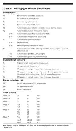
Urothelial tract cancers are staged according to the AJCC TNM classification system (Table 1). Superficial bladder cancer includes papillary tumors that involve only the mucosa (Ta) or submucosa

(T1) and flat carcinoma in situ (Tis). The natural history of superficial bladder cancer is unpredictable, and recurrences are common. Most tumors recur within 6 to 12 months and are of the same stage and grade, but 10% to 15% of patients with superficial cancer will develop invasive or metastatic disease.
Prognostic factors
For carcinomas confined to the bladder, ureters, or renal pelvis, the most important prognostic factors are T stage and differentiation pattern. The impact of associated carcinoma in situ on Ta and T1 lesions was discussed previously (see section on “Pathology”). Less-differentiated Ta–T1 lesions also are associated with higher recurrence and disease progression rates. Patients with well-differentiated Ta lesions without carcinoma in situ have a 95% survival rate, whereas those with high-grade T1 lesions have a 10-year survival rate of 50%. The presence of lymphovascular invasion within the surgical specimen appears to be independently associated with overall survival, cause-specific survival, as well as local and distant recurrence in patients with node-negative bladder cancer at the time of cystectomy. As such, the presence of lymphovascular invasion should be included in the pathologic assessment of bladder cancer.
Muscle invasive carcinoma carries a 5-year survival rate of 20% to 50%. When regional lymph nodes are involved, the 5-year survival rate is 0% to 20%.
A retrospective series of patients with no viable tumor at the time of radical cystectomy (pT0) would suggest that up to 9% of these patients may develop a recurrence following surgery. On multivariate analysis, the presence of lymphovascular invasion and concomitant carcinoma in situ on the transurethral bladder tumor resection specimen predicted poorer overall and recurrence-free survival, respectively (
Kassouf W et al: Eur Urol, 52:769–774, 2007).
Treatment
Localized disease
Surgical approaches to superficial bladder cancer
Transurethral resection Most patients with superficial bladder cancer can be treated adequately with transurethral resection (TUR). Such procedures preserve bladder function, entail minimal morbidity, and can be performed repeatedly. Survival rates > 70% at 5 years are expected.Although TUR removes existing tumors, it does not prevent the development of new lesions. Patients should be followed closely.
Laser The neodymium:yttrium-aluminum-garnet (Nd:YAG) laser has achieved good local tumor control when used in the treatment of superficial bladder tumors. However, it has not been adopted for general use because of its limitations in obtaining material for staging and grading of tumors.
Partial cystectomy is an infrequently utilized treatment option for patients whose tumors are not accessible or amenable to TUR but are solitary in location and away from the trigone.
Radical cystectomy is generally not used for the treatment of superficial bladder tumors. The indications for radical cystectomy include:
• Unusually large tumors that are not amenable to complete TUR, even on repeated occasions
• Some high-grade tumors
• Multiple tumors or frequent recurrences that make TUR impractical
• Symptomatic diffuse carcinoma in situ (Tis) that proves unresponsive to intravesical therapy
• Prostatic stromal involvement.
Intravesical therapy The indications for intravesical therapy include:
• Stage T1 tumors, especially if multiple
• Multifocal papillary Ta lesions, especially grade 2 or 3
• Diffuse Tis
• Rapidly recurring Ta, T1, or Tis disease.
In the United States, four intravesical agents are commonly used: thiotepa, an alkylating agent; bacillus Calmette-Gurin (BCG), an immune modulator/stimulator; and mitomycin and doxorubicin, both antibiotic chemotherapeutic agents. The dose of BCG varies with the strain (50 mg [Tice] or 60 mg [Connaught]). Mitomycin doses range from 20 to 40 mg. Although all four agents reduce the tumor recurrence rate, BCG is the most effective particularly for high-grade disease. For the treatment of papillary Ta and T1 lesions, BCG and mitomycin have the greatest efficacy (complete response rate: approximately 50%). For the treatment of Tis, BCG is extremely effective.
Following radical cystectomy, patients remain at risk of upper tract recurrence, with a previous report estimating the ?incidence of upper tract recurrence at 2.5%. Only urethral tumor involvement was predictive of upper tract recurrence. Despite routine surveillance of the upper urinary tracts, 78% of these recurrences were detected only upon development of symptoms, with the median survival following recurrence being only 1.7 years. Furthermore, the ?detection of asymptomatic upper tract recurrences via routine surveillance strategies did not predict lower pathologic stage, absence of nodal metastasis, or the improved survival in patients at time of nephroureterectomy (
Sanderson KM et al: J Urol 177:2088–2094, 2007).
In a meta-analysis comparing intravesical BCG and chemotherapy (mitomycin, epirubicin, doxorubicin, or sequential mitomycin/doxorubicin), intravesical BCG was superior in reducing the risk of short- and long-term treatment failure for Tis. Therefore, intravesical BCG appears to be the agent of choice for Tis.
Surgical approaches to invasive bladder cancer Radical cystectomy The most standardized treatment for invasive bladder cancer (stage II or higher) is radical cystectomy. Candidates for radical cystectomy include:
• Patients with muscle-invasive tumor
• Patients with high-grade, invasive, lamina propria tumors with evidence of lymphovascular invasion, with or without Tis
• Patients with diffuse Tis or recurrent superficial cancer who do not respond to intravesical therapy.
In men, radical cystectomy with pelvic lymph node dissection and removal of the bladder, seminal vesicles, and prostate. In women, radical cystectomy entails pelvic lymph node dissection and anterior exenteration, including both ovaries, fallopian tubes, uterus, cervix, anterior vaginal wall, bladder, and urethra.
Partial cystectomy is an infrequently utilized treatment option that should only be considered when there is a solitary lesion in the dome of the bladder and when random biopsy results from remote areas of the bladder and prostatic urethra are negative.
Urethrectomy is routinely included in the anterior exenteration performed in female patients. Urethrectomy in male patients is performed if the tumor grossly involves the prostatic urethra or if prior TUR biopsy results of the prostatic stroma are positive. Delayed urethrectomy for positive urethral cytology or biopsy is required in about 10% of male patients.
Urinary reconstruction may involve any one of the following: intestinal conduits (eg, ileal, jejunal, or colonic), continent cutaneous diversion (eg, Indiana or Kock pouch), or orthotopic reconstruction (in both male and female patients).
Surgical approaches to ureteral and renal pelvic tumors
Optimal surgical management of urothelial malignancies of the ureter and renal pelvis consists of nephroureterectomy with excision of a bladder cuff. Some tumors may respond well to local resection, and tumor specifics may allow for a more conservative intervention particularly in low-grade tumors.
Upper ureteral and renal pelvic tumors (because of similar tumor behavior and anatomic aspects) may be considered as a group, whereas lower ureteral tumors may be considered as a separate group.
Upper ureteral and renal pelvic tumors are best treated with nephroureterectomy. Solitary, low-grade upper tract tumors may be considered for segmental excision or ureteroscopic surgery if close surveillance is feasible. Care should be exercised, however, as multicentricity is more probable, and the risk of recurrence is greater than for lower ureteral lesions.
Lower ureteral lesions may be managed by nephroureterectomy, segmental resection, and neovesical reimplantation or by endoscopic resection. A 15% recurrence rate is seen after segmental resection or endoscopic excision. Careful follow-up is mandatory. Disease progression, the development of a ureteral stricture precluding periodic surveillance, and poor patient compliance are indications to abandon conservative management and perform nephroureterectomy.
Radiation therapy
Radiation therapy for bladder cancer Primary radiation or chemoradiation therapy Radiation therapy, either alone or in conjunction with chemotherapy, is the modality of choice for patients whose clinical condition precludes surgery, either because of extensive disease or poor overall status. Trials have shown that patients treated with irradiation and cisplatin with or without fluorouracil (5-FU) have improved local control as compared with patients treated with irradiation alone.
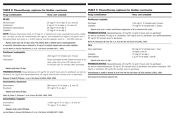
The most frequently utilized systemic chemotherapy regimens for urothelial carcinoma are shown in

. Other studies suggest that TUR followed by radiation therapy combined with cisplatin and 5-FU chemotherapy, with cystectomy reserved for salvage, provides a survival equivalent to that achieved with initial radical cystectomy while allowing for bladder preservation in many patients. The extent of TUR and the absence of hydronephrosis are important prognostic factors in studies of bladder-conserving treatment. Updates from institutions in Europe and the United States on over 600 patients with long-term follow-up support the durability of outcomes previously reported.
A randomized phase III study of bladder preservation with or without neoadjuvant chemotherapy following TUR, conducted by the RTOG, revealed no advantage to the use of MCV (methotrexate, cisplatin, and vinblastine) before radiation therapy and concurrent cisplatin. The favorable outcome without neoadjuvant chemotherapy may make bladder preservation a more acceptable option for a wider range of patients.
Radiation dose and technique Initially, a pelvic field is treated to 4,000–4,500 cGy utilizing a three-dimensional conformal technique, with daily or twice-daily fractionation. A cystoscopy is performed with biopsies. If a complete response is confirmed, the bladder tumor site is then boosted to a total dose of 6,480 cGy utilizing multifield techniques.
Radiation therapy for renal pelvic and ureteral cancers
In patients with renal pelvic and ureteral lesions who have undergone nephroureterectomy, postoperative local-field irradiation is offered if there is periureteral, perirenal, or peripelvic extension or lymph node involvement. A dose of approximately 4,500 to 5,400 cGy is delivered utilizing multifield techniques.
Palliative irradiation
Palliative radiation therapy is effective in controlling pain from local and metastatic disease and in providing hemostatic control. A randomized study comparing 3,500 cGy in 10 fractions and 2,100 cGy in three hypofractionated treatments revealed high rates of relief of hematuria, frequency, dysuria, and nocturia with either regimen. In selected cases of bladder cancer, aggressive palliation to approximately 6,000 cGy may be warranted to provide long-term local tumor control. Concurrent chemotherapy should be considered.
Neoadjuvant/adjuvant chemotherapy
Perioperative chemotherapy to reduce the risk of recurrence before or after cystectomy is a debated topic. Multiple randomized trials of different designs have given various chemotherapy regimens before and/or after cystectomy. Many of these trials had inadequate power or methodologic flaws that limited interpretation. In general, there is likely a small (5% to 10%) reduction in the risk of recurrence for perioperative chemotherapy given to high-risk (T3 and/or N+) patients.
Chemotherapy for advanced disease
Treatment of advanced metastatic urothelial cancer is generally considered to be palliative. Response rates are high with cisplatin-containing regimens (50% to 60%), but the duration of response is short, and median survival is 12 to 14 months. A small subset of patients (5% to 10%; usually with only lymph node metastases) can have a complete response to chemotherapy. A randomized trial showed an advantage for a regimen of M-VAC (methotrexate, vinblastine, Adriamycin [doxorubicin], and cisplatin) over cisplatin alone with regard to progression-free and overall survival. In another randomized trial, the combination of gemcitabine (Gemzar) and cisplatin exhibited equivalent survival to M-VAC in metastatic bladder cancer but was clinically better tolerated. Thus, cisplatin plus gemcitabine has become a common standard of care. Carboplatin can be substituted if a contraindication to cisplatin exists (eg, neuropathy, poor renal function).
KIDNEY CANCER
Approximately 57,760 new cases of renal tumors will be diagnosed in the year 2009 in the United States, with an associated 12,980 deaths. There has been a steady increase in the incidence of renal cell carcinoma that is not explained by the increased use of diagnostic imaging procedures. Mortality rates have also shown a steady increase over the past 2 decades.
Epidemiology
Gender and age
This malignancy is twice as common in men as in women. Most cases of renal cell carcinoma are diagnosed in the fourth to sixth decades of life, but the disease has been reported in all age groups.
Ethnicity
Renal cell carcinoma is more common in persons of northern European ancestry than in those of African or Asian descent.
Etiology and risk factors
Renal cell carcinoma occurs most commonly as a sporadic form and rarely (2%) as a familial form. The exact etiology of sporadic renal cell carcinoma has not been determined. However, smoking, obesity, and renal dialysis have been associated with an increased incidence of the disease.
Genetic factors
von Hippel-Landay disease (VHL)
VHL, an autosomal-dominant disease, is associated with retinal angiomas, CNS hemangioblastomas, and renal cell carcinoma.
Chromosomal abnormalities
Deletions of the short arm of chromosome 3 (3p) occur commonly in renal cell carcinoma associated with VHL disease. In the rare familial forms of renal cell carcinoma, translocations affecting chromosome 3p can be present. Sporadic renal cell carcinoma of the clear cell is also associated with VHL-gene silencing.
Associated malignancy
Two studies from large patient databases have reported a higher-than-expected incidence of both renal cell cancer and lymphoma. No explanation for this association has been found.
Signs and symptoms
Renal cell carcinoma has been associated with a wide array of signs and symptoms. The classic triad of hematuria, flank mass, and flank pain occurs in only 10% of patients and is usually associated with a poor prognosis. With the routine use of CT scanning for various diagnostic reasons, renal cell carcinoma is being diagnosed more frequently as an incidental finding.
Hematuria
More than half of patients with renal cell carcinoma present with hematuria, either gross or microscopic.
Other common signs/symptoms
Other commonly associated signs and symptoms of renal cell carcinoma include normocytic/normochromic anemia, fever, and weight loss.
Less common signs/symptoms
Less frequently occurring, but often described, signs and symptoms include polycythemia, hepatic dysfunction not associated with hepatic metastasis which is termed Stauffer’s syndrome, and hypercalcemia. Although not a common finding at the time of diagnosis of renal cell carcinoma, hypercalcemia ultimately occurs in up to 25% of patients with metastatic disease.
Diagnosis
Pre- and post-contrast-enhanced CT scanning
This technique has virtually replaced excretory urography and renal ultrasonography in the evaluation of suspected renal cell carcinoma. In most cases, CT imaging can differentiate cystic from solid masses and also supplies information about lymph nodes and renal vein/inferior vena cava (IVC) involvement.
Ultrasonography
Ultrasound is useful in evaluating questionable cystic renal lesions if CT imaging is inconclusive.
Venography and MRI
When IVC involvement by tumor is suspected, either IVC venography or MRI is needed to evaluate its extent. MRI is currently the preferred imaging technique for assessing IVC involvement at most centers. Transesophageal echocardiography is occasionally obtained pre- or intraoperatively to determine the proximal extent of the IVC thrombus particularly in those suspected to be beyond the diaphragm.
Renal arteriography
Renal arteriography is not used as frequently now as it was in the past in the evaluation of suspected renal cell carcinoma. In patients with small, indeterminate lesions, arteriography may be helpful. It is also used by the surgeon as part of the preoperative evaluation and management of a large renal neoplasm.
Evaluation of extra-abdominal disease sites
This includes a chest x-ray or CT imaging of the chest. A bone scan is required if a patient has symptoms suggestive of bone metastasis and/or an elevated alkaline phosphatase level.
Brain CT or MRI
A CT or MRI of the brain is indicated if neurologic signs or symptoms occur or if needed for staging before systemic therapy is given.
Pathology
Renal cell carcinoma arises from the proximal renal tubular epithelium. Histologically, renal cell carcinoma can be of various cellular types: clear cell (70% to 80%), papillary (10% to 15%), and chromophobe (5%). Oncocytoma is a benign renal tumor. Approximately 10% to 20% of renal cell carcinomas have sarcomatoid features (spindled cells that can occur in any subtype), which is a more aggressive malignancy with a worse prognosis.
Staging and prognosis
Staging system
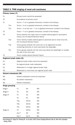
The preferred staging system for renal cell carcinoma is the TNM classification (Table 3).

Prognostic factors
The natural history of renal cell carcinoma is highly variable. However, approximately 30% of patients present with metastatic disease at diagnosis, and one-third of the remainder will develop metastasis during follow-up.
Five-year survival rates after nephrectomy for tumors confined to the renal parenchyma (T1/2) are > 80%. Renal vein involvement without nodal involvement does not affect survival. Lymph node involvement and/or extracapsular spread is associated with a 5-year survival of 10% to 25%. Patients with metastatic disease have a median survival of 2 years.
Several prognostic schemes have been developed for both localized and metastatic renal cell carcinomas. In general, factors such as tumor stage and grade, performance status, hemoglobin value, calcium, and lactate dehydrogenase levels, and time interval to development of metastatic disease are important.
Treatment
Surgery
Surgical resection (radical or partial nephrectomy) is the established therapy for localized renal cell carcinoma. When performing a radical nephrectomy, the kidneys, adrenal gland, and perirenal fat (structures bound by Gerota’s fascia) are removed. Also, limited regional lymph node dissection can be performed for staging purposes. Partial nephrectomy is standard in patients with smaller tumors (eg, < 4 cm) or in whom radical nephrectomy would unacceptably compromise overall renal function.
Because complete resection is the only known cure for renal cell carcinoma, even in locally advanced disease, surgery is considered if the involved adjacent structures can be safely removed. In patients with metastatic renal cell carcinoma, two randomized, controlled trials have shown a survival benefit of 6 months (combined analysis) with a debulking nephrectomy prior to interferon α immunotherapy, as compared with immunotherapy alone. However, patients must be carefully selected prior to the nephrectomy and should have a performance status of 0 to 1 according to the WHO criteria. The performance status of a patient prior to treatment is an important determinant of disease-related outcome and should be considered in making treatment decisions.
Radiation therapy for renal cell carcinoma
Primary radiation therapy
Radiation therapy may be considered for palliation as the primary therapy for renal cell carcinoma in patients whose clinical condition precludes surgery, either because of extensive disease or poor overall condition. A dose of 4,500 cGy is delivered, with consideration of a boost up to 5,500 cGy.
Postoperative radiation therapy
This modality has not been shown to prevent recurrence.
Palliation
Radiation therapy is commonly used for palliation for metastatic and local disease.
Systemic therapy for advanced disease
Metastatic renal cell carcinoma is resistant to chemotherapeutic agents. An extensive review of currently available agents concluded that the overall response rate to chemotherapy is 6%.
There is no standard adjuvant therapy for renal cell carcinoma after surgical resection regardless of recurrence risk. Multiple agents, including hormone therapy, radiation, immune therapy, and chemotherapy, have been tried, and none have produced benefit in the adjuvant setting. Thus, observation is the current standard of care. There are ongoing trials testing targeted agents in the adjuvant setting.
Interleukin-2 (IL-2, aldesleukin [Proleukin])
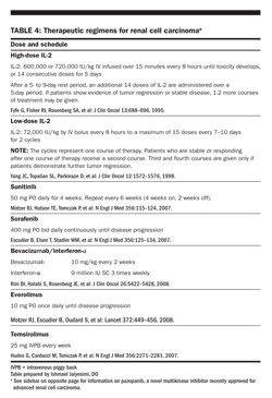
The first FDA-approved treatment for metastatic renal cell carcinoma was high-dose IL-2 (Table 4).

High-dose regimen High-dose IL-2 (720,000 IU/kg IV piggy back every 8 hours for 14 doses, repeated once after a 9-day rest) results in a 15% remission rate (7% complete responses, 8% partial responses). The majority of responses to IL-2 are durable, with a median response duration of 54 months.
The major toxicity of high-dose IL-2 is a sepsis-like syndrome, which includes a progressive decrease in systemic vascular resistance and an associated decrease in intravascular volume due to a “capillary leak.” Management includes judicious use of fluids and vasopressor support to maintain blood pressure and intravascular volume and at the same time to avoid pulmonary toxicity due to noncardiogenic pulmonary edema from the capillary leak. This syndrome is totally reversible.
Other doses and schedules Because of the toxicity of high-dose IL-2, other doses and schedules have been and are being evaluated. Several trials of low-dose IL-2 (3–18 × 106 IU/d), either alone or combined with interferon-α, have reported outcomes similar to those achieved with high-dose IL-2.
Biologic agents Four oral multikinase inhibitors have been approved by the FDA for the treatment of advanced kidney cancer. In addition, a monoclonal antibody has been tested extensively in treating the disease and, in combination with interferon-α, was FDA approved to treat metastatic renal cell carcinoma.
Sorafenib (Nexavar) targets several serine/threonine and receptor tyrosine kinases, especially vascular endothelial growth factor, thought to be integral to the biology of renal cell carcinoma. A phase III, placebo-controlled trial was conducted in 769 patients with advanced renal cell carcinoma who had received prior systemic treatment. The recommended oral dose of sorafenib (400 mg twice daily) was used. The median progression-free survival was 5.5 months in the sorafenib group versus 2.8 months in the placebo group. Toxic effects associated with sorafenib included reversible skin rashes in 40% and hand-foot skin reactions in 30% of patients. Notably, the incidence of treatment-emergent cardiac ischemia/infarction events was higher with sorafenib (2.9% vs 0.4%).
A report by Sternberg et al at ASCO 2009 supports the efficacy of pazopanib (Votrient), a multikinase angiogenesis inhibitor, in the treatment of advanced renal cell carcinoma (RCC). In this phase III study, 435 patients with advanced RCC were randomly assigned to treatment with pazopanib (800 mg/d orally) or placebo. Progression-free survival was significantly better in the pazopanib group than in the placebo group (hazard ratio [HR], 0.46,
P
< .0000001). This benefit was observed for both treatment-nave patients (HR, 0.40,
P
< .0000001) and for those who had previously received one cytokine-based treatment (HR, 0.54,
P
< .001). The response rate was 30% with pazopanib, compared with 3% with placebo, and the median duration of response was 58.7 weeks. Most of the adverse events associated with this therapy were grade 1 or 2. The most common laboratory abnormality was an elevation in alanine aminotransferase (
Sternberg CN et al: J Clin Oncol 27[15S]:abstract 5021, 2009
). FDA approved pazopanib for advanced renal cell carcinoma in October 2009.
Sunitinib (Sutent) targets several receptor tyrosine kinases. Initial phase II trials of sunitinib, given as 50 mg once daily PO for 4 weeks followed by 2 weeks off, to 169 metastatic renal cell cancer patients who had failed prior cytokine-based therapy demonstrated an investigator-assessed objective response rate of 45%, a median duration of response of 11.9 months, and a median progression-free survival of 8.4 months. A phase III trial of sunitinib versus interferon-α in 750 untreated metastatic renal cell cancer patients demonstrated a significant advantage in an independently assessed objective response rate (31%; 95% confidence interval [CI] = 26%–36% vs 6%; P < .001) and progression-free survival (11 months vs 5 months). Sunitinib-treated patients had a median overall survival of 26.4 versus 21.8 months for interferon-α-treated patients (P = .051).
Temsirolimus (Torisel) and bevacizumab (Avastin) were added to the NCCN Kidney Cancer Guidelines as options for first-line treatment of relapsed or medically unresectable stage IV renal cell carcinoma with predominant clear cell histology and, in the case of temsirolimus, non-clear cell histology. The recommendations were based on the results of large randomized trials. Temsirolimus significantly prolonged median overall survival in a phase III trial of 626 patients (P = .0078) and received FDA approval for first-line as well subsequent treatment of advanced renal cell carcinoma.
Bevacizumab (Avastin) Two separate, multicenter, international studies have established bevacizumab-based therapy as robust in the front-line setting. One phase III trial randomized 649 untreated patients with metastatic renal cell carcinoma to treatment with interferon α-2a, recombinant (Roferon) plus placebo infusion or to interferon α-2a plus bevacizumab infusion 10 mg/kg every 2 weeks. A significant advantage for bevacizumab plus interferon α-2a was observed for objective response rate (31% vs 13%, respectively; P < .0001) and progression-free survival (10.2 months versus 5.4 months; P < .0001). The hazard ratio (HR) for progression in the bevacizumab plus interferon α-2a arm was 0.63 (95% confidence interval [CI] = 0.52–0.75; P = .0001).
Everolimus is an oral inhibitor of mTOR kinase that has shown activity in metastatic renal cell carcinoma in phase II studies. Motzer and colleagues recently presented results of a planned interim analysis of a phase III study assessing the clinical benefit of everolimus in patients who had progressive metastatic renal cell carcinoma _after receiving sunitinib, sorafenib, or both. The primary endpoint was progression-free survival. A total of 410 patients were randomized to receive everolimus, 10 mg, or placebo in a 2:1 ratio. Prior bevacizumab and/or cytokine treatment was allowed. The results showed a significant improvement in progression-free survival for everolimus vs placebo (progression-free survival: = 4.0 months for everolimus vs 1.9 months for placebo,
P
< .001) (
Motzer RJ et al: Lancet 372:449-456, 2008
).
A second multicenter, phase III trial conducted in the United States and Canada through the CALGB was nearly identical in design, except that it lacked a placebo infusion and did not require prior nephrectomy. The median progression-free survival was 8.5 months in patients receiving bevacizumab plus interferon α-2a (95% CI =7.5–9.7) vs 5.2 months for patients using interferon α-2a monotherapy ((95% CI = 3.1–5.6; P < .0001). The HR for progression for patients receiving bevacizumab plus interferon α-2a after adjusting for stratification factors was 0.71 (P < .0001). Also, among patients with measurable disease, the objective response rate was higher in patients treated with bevacizumab plus interferon α-2a (25.5%) than for those given interferon α-2a monotherapy (13.1%, P = .0001).
In the AVOREN trial, bevacizumab significantly increased progression-free survival (10.2 months vs 5.4 months; P = .0001) of patients with metastatic renal cell carcinoma when administered in combination with interferon-α 2a.
As always, patients should be encouraged to participate in ongoing clinical trials for metastatic renal cell cancer.
References:
SUGGESTED READING
ON UROTHELIAL CANCER
Barton Grossman H, Natale RB, Tangen CM, et al: Neoadjuvant chemotherapy plus cystectomy compared with cystectomy alone for locally advanced bladder cancer. N Engl J Med 349:859â866, 2003.
Duchesne GM, Bolger JJ, Griffiths GD, et al: A randomized trial of hypofractionated schedules of palliative radiotherapy in the management of bladder carcinoma: Results of Medical Research Council Trial BA09. Int J Radiat Oncol Biol Phys 47:379â388, 2000.
Hudson MA, Herr HW: Carcinoma in situ of the bladder. J Urol 153:564â572, 1995.
Hussain MH, Glass TR, Forman J, et al: Combination cisplatin, 5-fluorouracil, and radiation therapy for locally advanced unresectable or medically unfit bladder cancer cases: A Southwest Oncology Group study. J Urol 165:56â60, 2001.
Lotan Y, Gupta A, Shariat SF, et al: Lymphovascular invasion is independently associated with overall survival, cause-specific survival, and local and distant recurrence in patients with negative lymph nodes at radical cystectomy. J Clin Oncol 23:6533â6539, 2005.
Milosevic M, Gospodarowicz M, Zietman A, et al: Radiotherapy for bladder cancer. Urology 69:80â92, 2007.
Sylvester RJ, van der Meijden AP, Witjes JA, et al: Bacillus calmette-guerin versus chemotherapy for the intravesical treatment of patients with carcinoma in situ of the bladder: A meta-analysis of the published results of randomized clinical trials. J Urol 174:86â92, 2005.
Von der Maase H, Hansen SW, Roberts JT, et al: Gemcitabine and cisplatin versus methotrexate, vinblastine, doxorubicin, and cisplatin in advanced or metastatic bladder cancer: Results of a large, randomized, multinational, multicenter, phase III study. J Clin Oncol 17:3068â3077, 2000.
ON KIDNEY CANCER
Dutcher JP, Szczylik C, Tannir N, et al: Correlation of survival with tumor histology, age, and prognostic risk group for previously untreated patients with advanced renal cell carcinoma receiving temsirolimus or interferon-alpha. J Clin Oncol 25:5033, 2007.
Escudier B, Eisen T, Stadler WM, et al: Sorafenib in advanced clear-cell renal-cell carcinoma. N Engl J Med 356:125â134, 2007.
Escudier B, Koralewski P, Pluzanska A, et al: A randomized, controlled, double-blind phase III study (AVOREN) of bevacizumab/interferon-α 2a vs placebo/interferon-α 2a as first-line therapy in metastatic renal cell carcinoma. J Clin Oncol 25:3, 2007.
Escudier B, Pluzanska A, Koralewski P, et al. Bevacizumab plus interferon alfa-2a for treatment of metastatic renal cell carcinoma: a randomised, double-blind phase III trial. Lancet 370:2103â2111, 2007.
Flanigan RC, Salmon SE, Blumenstein BA, et al: Nephrectomy followed by interferon alfa-2b compared with interferon alfa-2b alone for metastatic renal cell cancer. N Engl J Med 345:1655â1659, 2001.
Hudes G, Carducci M, Tomczak P, et al: Temsirolimus, interferon alfa, or both for advanced renal-cell carcinoma. N Engl J Med 356:2271â2281, 2007.
Mickisch GH, Garin A, van Poppel H, et al: Radical nephrectomy plus interferon-alfa-based immunotherapy compared with interferon alone in metastatic renal-cell carcinoma: A randomized trial. Lancet 358:966â970, 2001.
Motzer RJ, Dror Michaelson MD, Redman BG, et al: Activity of SU11248, a multitargeted inhibitor of vascular endothelial growth factor receptor and platelet-derived growth factor receptor, in patients with metastatic renal cell carcinoma. J Clin Oncol 24:16â24, 2006.
Motzer RJ, Hutson TE, Tomczak P, et al: Sunitinib versus interferon alfa in metastatic renal-cell carcinoma. N Engl J Med 356:115â124, 2007.
Motzer RJ, Rini BI, Bukowski RM, et al: Sunitinib in patients with metastatic renal cell carcinoma. JAMA 295:2516â2524, 2006.
Parkinson DR, Sznol M: High-dose interleukin-2 in the therapy of metastatic renal cell carcinoma. Semin Oncol 22:61â66, 1995.
Rabinovitch RA, Zelefsky MJ, Gaynor JJ, et al: Patterns of failure following surgical resection of renal cell carcinoma: Implications for adjuvant local and systemic therapy. J Clin Oncol 12:206â212, 1994.
Rini BI, Halabi S, Rosenberg JE, et al. Bevacizumab plus interferon alfa compared with interferon alfa monotherapy in patients with metastatic renal cell carcinoma: CALGB 90206. J Clin Oncol 26:5422â5428, 2008.
Stodlen WM, Vogelzang NJ: Low-dose interleukin-2 in the treatment of metastatic renal cell carcinoma. Semin Oncol 22:67â73, 1995.
Zisman A, Pantuck AJ, Dorey F, et al: Improved prognostication of renal cell carcinoma using an integrated staging system. J Clin Oncol 19:1649â1657, 2001.
Abbreviations in this chapter
AJCC = American Joint Committee on Cancer; AVOREN = Avastin for Renal Cell Cancer; CALGB = Cancer and Leukemia Group B; FDA = US Food and Drug Administration; NCCN = National Comprehensive Cancer Network; RTOG = Radiation Treatment Oncology Group; WHO = World Health Organization