Hodgkin lymphoma is a unique disease entity characterized by a low number of neoplastic tumor cells surrounded by an inflammatory microenvironment composed of dysfunctional immune cells. Recent molecular and genetic studies have revealed that upregulation of the immune checkpoint pathway programmed death 1/programmed death ligand 1 is a key oncogenic driver of Hodgkin lymphoma. Corroborating these mechanistic studies, early-phase clinical trials using the checkpoint inhibitors nivolumab and pembrolizumab in treatment regimens for relapsed and/or refractory Hodgkin lymphoma have demonstrated impressive response rates, a promising durability of response, and a favorable side-effect profile. Given its targeted mechanism of action, acceptable safety, and clinically meaningful activity, the checkpoint inhibitor nivolumab was recently approved by the US Food and Drug Administration as therapy for classical Hodgkin lymphoma that has relapsed or progressed after autologous stem cell transplantation (ASCT) and post-ASCT consolidation therapy with brentuximab vedotin. In this article we review the scientific rationale, preclinical evidence, and most recent clinical data for the use of checkpoint inhibitor therapy in patients with relapsed Hodgkin lymphoma.
Introduction
With more than 9,000 new cases of Hodgkin lymphoma diagnosed annually in the United States, patients with relapsed disease remain a significant clinical challenge.[1] Classical Hodgkin lymphoma is characterized by the presence of less than 1% multinucleated giant cells, called Hodgkin and Reed-Sternberg (HRS) cells, within a vast reactive milieu of immune cells that include lymphocytes, histiocytes, eosinophils, macrophages, plasma cells, and fibroblasts.[2] This tumor microenvironment is supported by autocrine and/or paracrine production of inflammatory cytokines that promote tumor evasion from host growth control and immune surveillance, and that underlie the constitutional inflammatory symptoms associated with Hodgkin lymphoma.[3]
The initial treatment for Hodgkin lymphoma patients is based on the stage and tumor burden at presentation. For patients with advanced disease, risk status is traditionally stratified based on the presence or absence of seven independent prognostic factors-ie, by the International Prognostic Score, called IPS-7: male sex, age ≥ 45 years, stage IV disease, hemoglobin level < 105 g/L, white blood cell count ≥ 15 × 109/L, lymphocyte count < 0.6 × 10⁹/L or < 8% of differential, and albumin level < 40 g/L.[4,5] More recently, a streamlined IPS-3 has been proposed, comprising three clinical factors: age, disease stage, and hemoglobin level.[6] Patients with early-stage, good-risk disease are usually treated with either single-modality cytotoxic chemotherapy (ie, doxorubicin, bleomycin, vinblastine, and dacarbazine [ABVD]) or combined-modality therapies that include abbreviated courses of ABVD followed by involved-field radiation therapy. Patients with advanced-stage and/or poor-risk disease usually receive a prolonged or more intense course of chemotherapy consisting of either ABVD or a regimen of bleomycin, etoposide, doxorubicin, cyclophosphamide, vincristine, procarbazine, and prednisone (BEACOPP), with the occasional inclusion of radiation treatment to sites of tumor bulk.[7] For patients with relapsed or refractory disease, salvage chemotherapy followed by high-dose chemotherapy and autologous stem cell transplantation (ASCT) remains the standard of care, and offers the highest chance for long-term disease control and cure.[7,8] Additional therapeutic options for patients who are ineligible for ASCT or in whom ASCT has failed include consolidation therapy with brentuximab vedotin (BV), an antibody-drug conjugate targeting CD30; palliative chemotherapy; targeted therapies, such as inhibitors of the mammalian target of rapamycin (mTOR) pathway and histone deacetylase; allogeneic stem cell transplant (SCT); and participation in a clinical trial.[8] The checkpoint inhibitor nivolumab, a humanized monoclonal antibody, is now approved by the US Food and Drug Administration (FDA) for patients who have relapsed after BV and allogeneic SCT.
Two major challenges facing clinicians caring for patients with Hodgkin lymphoma are the minimization of long-term toxicities of therapy, and the improvement in salvage strategies for patients with relapsed and refractory disease. Longitudinal epidemiological studies have demonstrated a persistent risk of secondary malignancy for up to 40 years after curative treatment for Hodgkin lymphoma.[9,10] In addition, the risk of premature coronary artery disease in patients who receive radiation treatment that encompasses the cardiac field increases 10 years post exposure.[11] In terms of salvage therapy, despite the progress made in recent years, including the incorporation of BV and other novel targeted therapies such as inhibitors of the mTOR pathway, the cure rate for relapsed disease is still less than 50%. Allogeneic SCT can potentially provide cure to a small subset of relapsed patients but is associated with considerable morbidity and mortality.[12]
Under normal physiological conditions, the host utilizes a plethora of immunologic inhibitory pathways, including the checkpoint blockade, to maintain self-tolerance and to modulate the duration and amplitude of the physiological immune response.[13] In solid tumors, the concept of checkpoint inhibitor–based therapy derives from the understanding that most solid tumors have myriad genetic and epigenetic alterations that provide a diverse set of neoantigens used by the immune system to distinguish tumor cells from normal cells, and that tumor cells use this mechanism to evade host immune surveillance.[14]
Antibodies have been developed that target ligands and receptors in immune checkpoint pathways, and these new agents are proving to be promising therapeutics for treatment of both solid tumors and Hodgkin lymphoma. Antibodies to cytotoxic T-lymphocyte–associated antigen 4 (CTLA-4) were the first immune checkpoint inhibitors to receive FDA approval for the treatment of malignant melanoma, based on a survival benefit of 30% over 3 years reported in phase III studies.[15] More recently, checkpoint inhibitor antibodies against the protein programmed death 1 (PD-1) have been approved for the treatment of several solid tumors, including melanoma and non–small-cell lung cancer.[15] To date, development of these therapies has been slower in hematologic malignancies, with the exception of Hodgkin lymphoma.[16] This may be due to lower mutational burden in hematologic malignancies, resulting in a lower level of neoantigens.[17] However, Hodgkin lymphoma stands out among all lymphomas, with its high responsiveness to PD-1 blockade (> 70% overall response rate [ORR]) and significant clinical benefit in patients with relapsed and refractory disease.[18-20]
Targeting Immune Systems in Hodgkin Lymphoma Pathogenesis
In many patients with Hodgkin lymphoma, deficiencies of host immune functions, such as T cell–mediated viral clearance, have been noted, sometimes even predating a diagnosis of Hodgkin lymphoma.[21] Interestingly, in some patients this immune deficiency may persist years after successful treatment of their disease.[22] Supporting this notion, it has long been appreciated that patients with human immunodeficiency virus infection or autoimmune disease have an increased incidence and prevalence of both Hodgkin and non-Hodgkin lymphoma.[23]
In 2008, Yamamoto et al showed for the first time that HRS cell lines and primary tumor samples overexpressed programmed death ligand 1 (PD-L1).[24] Most importantly, PD-1 itself was markedly overexpressed in the tumor-infiltrating CD4 and CD8 T cells of 3 patients with Hodgkin lymphoma.[24] Artificial blocking of the PD-1 pathway restored the production of interferon gamma. These data suggested that activation of the PD-1 pathway in T cells induces “T-cell exhaustion,” a chronic state of effector T-cell dysfunction resulting from the expression of inhibitory receptors and decreased production of activating cytokines. Overexpression of PD-L1 on HRS cells and of PD-1 on tumor-infiltrating lymphocytes creates a potent inhibitory signal that helps to maintain the immunosuppressive Hodgkin lymphoma microenvironment, and allows HRS evasion of immune surveillance.[24] These findings were confirmed in a large set of primary Hodgkin lymphoma tumor samples, in which HRS cells were found to express high levels of PD-L1 and programmed death ligand 2 (PD-L2) compared with cell samples from other aggressive types of Hodgkin lymphoma (nodular lymphocyte–predominant disease) or non-Hodgkin lymphoma (Burkitt lymphoma and diffuse large B-cell lymphoma, not otherwise specified).[25] A subsequent immunohistochemistry study of tumor specimens from patients with primary Hodgkin lymphoma confirmed a significantly higher level of PD-L1 expression in classical Hodgkin lymphoma compared with primary mediastinal B-cell lymphomas or diffuse large B-cell lymphoma.[26] Results of an earlier study by Muenst et al supported the role of PD-1/PD-L1 pathway activation in the pathogenesis of Hodgkin lymphoma; the authors used tissue microarray technology to evaluate 189 cases of classical Hodgkin lymphoma and found that an increased number of PD-1–positive tumor-infiltrating lymphocytes was a stage-independent negative prognostic factor for overall survival.[27]
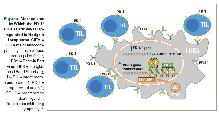
Several mechanisms have been proposed to account for the upregulation of the PD-1 pathway in Hodgkin lymphoma. Genomics-based approaches that combine high-resolution copy number data with transcriptional profiles have identified selective amplification of PD-L1 and PD-L2 genes on chromosome 9p24.1 in HRS cells.[28] In addition, expression of the Janus kinase 2 (JAK2) gene is amplified, leading to JAK2 protein overexpression and subsequent transcriptional activation of PD-L1.[28] Most recently, the prognostic significance of PD-L1 and PD-L2 genetic alterations has been demonstrated using fluorescence in situ hybridization analysis of 108 primary samples from patients with Hodgkin lymphoma; 97% of the patients had concordant alterations of the PD-L1 and PD-L2 loci on 9p24.1, and this amplification was associated with a more advanced stage of disease and worsened progression-free survival (PFS).[29]
Several groups subsequently found additional mechanisms that contributed to the upregulation of PD-L1 and PD-L2. Steidl et al utilized whole-transcriptome paired-end sequencing in Hodgkin lymphoma cell lines and identified a highly expressed chromosomal fusion gene involving CIITA, which encodes a protein called the major histocompatibility complex class II transcriptional activator, leading to downregulation of surface human leukocyte antigen class II expression and overexpression of PD-L1 and PD-L2.[30] Constitutive activation of the JunB-enriched activator protein-1 transcriptional complex through an enhancer element in the PD-L1 gene in HRS cells has also been described.[31] In addition, infection with Epstein-Barr virus causes increased expression of the PD-L1 gene and PD-L1 protein, mediated by latent membrane protein 1, and associated with increased levels of JAK/signal transducers and activators of transcription-dependent promoter and enriched activator protein-1.[31] However, an analysis of 87 cases of primary Hodgkin lymphoma samples found no correlation between Epstein-Barr virus–encoded small RNA (EBER) expression and PD-1/PD-L1 expression.[32] By overexpressing PD-L1 and PD-L2 on their surface, HRS cells are able to shut down the immune response, thus evading immune surveillance. The Figure summarizes the mechanisms by which the PD-1/PD-L1 pathway is upregulated in the tumor microenvironment of Hodgkin lymphoma.
Clinical Studies of Checkpoint Inhibition in Hodgkin Lymphoma
Anti–CTLA-4 therapy
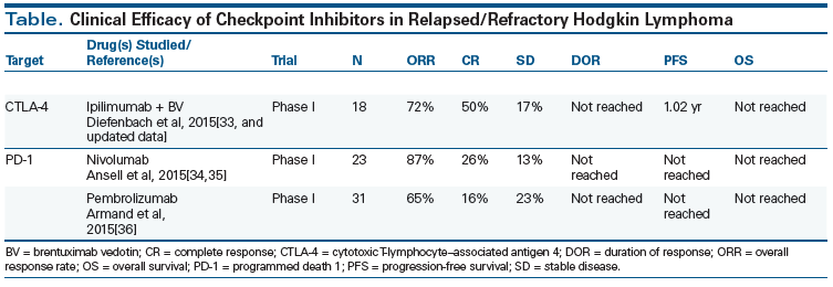
The safety and preliminary efficacy of the combination of BV and ipilimumab, an FDA-approved CTLA-4 antibody, in a phase I study of 23 patients with relapsed and refractory Hodgkin lymphoma was presented at the 2015 American Society of Hematology (ASH) Annual Meeting.[33] Ipilimumab was administered at a dose of 1 mg/kg or 3 mg/kg, following the typical induction-maintenance regimen, and BV was dosed at 1.8 mg/kg every 3 weeks. The regimen was well tolerated, with only manageable immune-related adverse events and no dose-limiting toxicities. Among 18 evaluable patients, the ORR was 72% and the complete response (CR) rate was 50%. This study demonstrates that the combination of BV and the checkpoint inhibitor ipilimumab appears safe, has promising activity in patients with relapsed Hodgkin lymphoma, and suggests a proof of concept for the combination of checkpoint inhibitors and antibody-drug conjugate or cytotoxic therapy platforms.
Anti–PD-1 therapy
The CheckMate-039 study of the anti–PD-1 checkpoint inhibitor nivolumab in relapsed and refractory Hodgkin lymphoma was published in 2015. In this landmark phase I trial in 23 patients with relapsed/refractory, heavily pretreated disease (78% treated with BV, ASCT, or both), nivolumab was administered at a starting dose of 1 mg/kg, with the dose increased up to 3 mg/kg for up to 2 years. An 87% ORR and 20% CR rate were observed.[34] The drug was well tolerated, with no significant dose-limiting toxicities. Immunohistochemistry confirmed the high level of PD-L1 expression in HRS cells in this study. An update of the trial was presented at the 2015 ASH Annual Meeting. After an extended follow-up of 101 weeks, the median duration of response and median PFS had yet to be reached, showing promising remission durability in this traditionally difficult-to-treat patient population.[35] The overall survival rate was 91% at 1 year and was 83% at 1.5 years. Most importantly, this follow-up analysis included long-term data for the cohort of patients who were treated with nivolumab until confirmed CR, or for up to 2 years if they had a partial response (PR) or stable disease, and then were followed for 1 year after treatment discontinuation. At the time of data cutoff, of the 20 responding patients, 3 remained on nivolumab treatment with ongoing responses and 13 had discontinued treatment without progressive disease; 5 of these 13 patients underwent allogeneic SCT, 4 developed progressive disease following initial response, and 1 patient was re-treated post-progression and achieved a second CR.[35] Based on these results, the FDA approved nivolumab for treatment of relapsed/refractory classical Hodgkin lymphoma after ASCT and BV.
TO PUT THAT INTO CONTEXT
[[{"type":"media","view_mode":"media_crop","fid":"52872","attributes":{"alt":"","class":"media-image","id":"media_crop_4620514871393","media_crop_h":"0","media_crop_image_style":"-1","media_crop_instance":"6572","media_crop_rotate":"0","media_crop_scale_h":"0","media_crop_scale_w":"0","media_crop_w":"0","media_crop_x":"0","media_crop_y":"0","style":"height: 158px; width: 144px;","title":" ","typeof":"foaf:Image"}}]]
Alex F. Herrera, MD
City of Hope National Medical Center
Duarte, CaliforniaWhat Is the Role of Immune Checkpoint Inhibitors in Hodgkin Lymphoma?The majority of patients with classical Hodgkin lymphoma will be cured with standard therapy. However, a significant minority of patients will have relapsed or refractory disease that does not respond to second-line therapy and/or autologous stem cell transplantation, and outcomes in these patients have historically been poor. In recent years, we have learned that alterations of the programmed death 1 (PD-1) pathway are nearly universal in classical Hodgkin lymphoma and central to its pathogenesis. The overwhelming success of checkpoint inhibitors, particularly PD-1 inhibitors, in treating relapsed and refractory classical Hodgkin lymphoma is truly a practice-changing development. In addition to providing effective treatment for patients in whom options were previously limited, there is an opportunity to utilize these well-tolerated agents in earlier lines of therapy and potentially alter the course of the disease while minimizing the toxicity of classical Hodgkin lymphoma treatment.What Key Questions Remain to Be Answered?Despite the tremendous potential for immune checkpoint inhibitors to improve outcomes for patients with classical Hodgkin lymphoma, there are still important unanswered questions about their role. Not all patients with classical Hodgkin lymphoma benefit from treatment with checkpoint inhibitors, and few experience a complete response. Combinations of checkpoint inhibitors with other agents may be required to increase response rates and deepen remissions. The identification of biomarkers of both response and subsequent treatment failure will be critical in selecting candidates for therapy and optimizing the sequencing of novel therapies. Given the durable responses to checkpoint inhibitor therapy observed in classical Hodgkin lymphoma patients who have not achieved complete response, a better understanding of the composition of residual hypermetabolic lesions after checkpoint therapy should be sought. In light of the durability of remissions, as well as the emerging signal of toxicity in previously anti–PD-1–treated patients who undergo allogeneic stem cell transplantation, clarity is needed concerning the respective roles of these therapies in classical Hodgkin lymphoma.The next phase of investigation of checkpoint inhibitors in classical Hodgkin lymphoma is underway, with ongoing trials evaluating earlier introduction of PD-1 inhibitors and novel therapeutic combinations. The potential of these powerful agents is great, and through these studies we will learn whether their effectiveness can be augmented without increasing toxicity.Financial Disclosure: Dr. Herrera receives research funding from Genentech-Roche, Immune Design, Pharmacyclics, and Seattle Genetics.Acknowledgment of Research Support: Dr. Herrera is supported by the following grants from the National Institutes of Health (NIH): NIH 2K12CA001727-21 and NIH 5P50CA107399-10. The content is solely the responsibility of Dr. Herrera and does not necessarily represent the official views of the NIH.
Similar efficacy and safety results were reported for KEYNOTE-013, a phase Ib multicenter, multicohort trial that evaluated the anti–PD-1 humanized monoclonal antibody pembrolizumab.[36] In this study, 31 patients with classical Hodgkin lymphoma unresponsive to treatment with BV (100%) and ASCT (71%) received 10 mg/kg of pembrolizumab every 2 weeks for up to 2 years. At the time this trial was presented at the 2015 ASH Annual Meeting, the ORR was 65%, with a CR rate of 16%. In terms of best responses, 90% of patients had a diameter reduction of ≥ 50% in their target lesions compared with baseline. Eighty percent of responses occurred by week 12, and 71% of patients had responses with a duration of at least 24 weeks. The PFS rate at 24 weeks was 69%. Only one immune-related adverse event was noted, and 3 patients went on to receive allogeneic SCT as consolidation. Immunohistochemistry of pretreatment tumor tissue showed that most were positive for PD-L1/PD-L2 expression. In April 2016, the FDA granted a Breakthrough Therapy Designation to pembrolizumab for the treatment of relapsed and refractory classical Hodgkin lymphoma. The Table summarizes clinical efficacy results from key trials of checkpoint inhibitors in Hodgkin lymphoma.
Following the FDA approval of nivolumab, the 2016 National Comprehensive Cancer Network guidelines recommended that nivolumab be considered as a treatment option in patients with classical Hodgkin lymphoma whose disease had relapsed or progressed despite ASCT and BV maintenance therapy.[37] At present, the use of checkpoint inhibitors prior to ASCT and/or treatment with BV, in combination with other chemotherapy or targeted agents, or in earlier lines of therapy, should only occur in the context of a clinical trial.
Checkpoint inhibition in the setting of allogeneic SCT
The FDA-approved anti–CTLA-4 antibody ipilimumab was investigated in a phase I study of 29 patients with advanced hematologic malignancies (including 14 with Hodgkin lymphoma) who experienced relapse after undergoing allogeneic SCT.[38] Given the concern about immunotherapy possibly worsening chronic graft-vs-host disease (GVHD) in these patients, a wide dose range of 0.1- to 3.0-mg/kg ipilimumab was used. Dose-limiting toxicity was not reached and no worsening GVHD or graft rejection occurred. Two patients with Hodgkin lymphoma achieved a CR, and a PR was observed in a patient with refractory mantle cell lymphoma. Ipilimumab was well tolerated. Organ-specific immune adverse events (grade 3 arthritis, grade 2 hyperthyroidism, and recurrent grade 4 pneumonitis) occurred in 4 patients.
Preliminary results of the expansion cohort of this study were reported at the 2015 ASH Annual Meeting. Repeated dosing of ipilimumab at 3 mg/kg and 10 mg/kg was carried out according to a schedule for induction and maintenance therapy.[39] A total of 28 patients with relapsed hematologic malignancies after allogeneic SCT were enrolled, including 7 with Hodgkin lymphoma. In 21 evaluable patients, the ORR was 33%, including 5 of 12 patients with acute myeloid leukemia who achieved CR, and 1 patient with Hodgkin lymphoma who achieved a PR. Five GVHD-related dose-limiting toxicities and 4 immune-related events were reported. These studies suggested that anti–CTLA-4 therapy is reasonably safe following allogeneic SCT for patients without evidence of GVHD, particularly at a low dose. The unexpectedly high response rate for the cohort with acute myeloid leukemia also supports the notion that immune checkpoint inhibition may play a role in augmenting the graft-vs-tumor effect.
The safety and optimal timing of checkpoint inhibitor therapy prior to or following allogeneic SCT remains unknown. The desired graft-vs-tumor effect of allogeneic SCT induces generalized immune stimulation and inhibition of normal checkpoint function.[40] Tumor relapse after allogeneic SCT invariably involves escape from immune surveillance by mechanisms such as increased PD-1/PD-L1 expression on CD8-positive T cells.[41] Checkpoint inhibition carries a theoretical risk of exacerbating GVHD. In a murine model of acute GVHD, both PD-L1 and PD-L2 expression were increased at the baseline, and blockade of PD-1/PD-L1 signaling worsened acute GVHD and enhanced the lethality of treatment.[42] Moreover, in PD-L1–deficient hosts, donor T cells exhibit increased levels of aerobic glycolysis and oxidative phosphorylation, leading to their increased proliferation and activation; this higher rate of metabolic activity may contribute to acute GVHD.[42] Similar findings have also been demonstrated in a murine model of chronic GVHD.[43]
The available clinical data, while limited, suggest that careful investigation of potential graft-vs-tumor effects from immune checkpoint blockade in cancer treatment is warranted. The anti–CTLA-4 antibody ipilimumab appears safe to use at low to intermediate doses (1 mg/kg and 3 mg/kg) in patients who have undergone allogeneic SCT, but at the highest dose (10 mg/kg) a worsening of chronic GVHD was observed.[33] The safety and efficacy of nivolumab treatment for relapsed Hodgkin lymphoma after allogeneic SCT was presented at the 2015 ASH Annual Meeting, in a single-institution retrospective analysis of 12 patients. The patients were without a history of grade 3/4 acute or chronic GVHD and were required to be off immunosuppression for more than 4 weeks. Nivolumab was given at a dosage of 3 mg/kg every 2 weeks, and patients received a median of 4 cycles of therapy. The ORR was 87.5%, with three CRs and four PRs. Two cases of grade 3/4 acute GVHD developed, necessitating dose delay or dose reduction.[44] Similarly, pembrolizumab was used successfully in 2 patients with Hodgkin lymphoma who relapsed after allogeneic SCT and were maintained on low-dose prednisone. Both patients responded without evidence of acute GVHD.[45] However, case reports of severe and fatal GVHD have also been described in patients receiving anti–PD-1 therapy after allogeneic SCT for Hodgkin lymphoma.[46,47]
Although the safety of allogeneic SCT following checkpoint inhibitor therapy remains unknown, there is at least a theoretical risk of acute GVHD in this setting due to the long half-life of these antibodies. Not yet reported in the medical literature but described in the nivolumab prescribing information are significant complications, including fatal events, that have occurred in patients who underwent allogeneic SCT at various intervals after receiving treatment with nivolumab.[48] In total, data for 17 patients have been described; 15 underwent reduced-intensity conditioning and 2 underwent myeloablative conditioning. The median age at time of allogeneic SCT was 33 years, and a median of 9 doses of nivolumab were administered (range, 4 to 16). Of 17 patients, 6 (35%) died from complications of allogeneic SCT after nivolumab. Five deaths occurred in the setting of severe or refractory GVHD. Grade 3 or higher acute GVHD was reported in 5 of the 17 patients (29%). Hyperacute GVHD, a steroid-requiring febrile syndrome, encephalitis, and hepatic veno-occlusive disease have also been reported.[48]
Checkpoint Inhibition and the Future of Therapy for Hodgkin Lymphoma
High priorities in Hodgkin lymphoma clinical research continue to be reduction of long-term toxicity of treatment for patients with low-risk Hodgkin lymphoma and improvement in the efficacy of treatment for high-risk patients. The high response rate, favorable side effect profile, and potential durability of checkpoint inhibitor therapy suggest that this approach holds great promise as a cornerstone of therapy for relapsed and refractory Hodgkin lymphoma-as a single agent and potentially in novel combination treatment platforms with antibody-drug conjugates or other cytotoxic chemotherapy regimens. A variety of clinical trials are underway to test these hypotheses, with the goal of identifying rational combinations that can increase rates of CR and durable responses in these patients. In addition, given the persistent risk of toxicity from long-term therapy with these agents, especially for high-risk Hodgkin lymphoma patients who have undergone extensive prior treatment with alkylator-based therapy, the activity of checkpoint inhibitors in the relapsed setting suggests their potential as a component of upfront therapy.
Financial Disclosure:Dr. Diefenbach receives research funding and honoraria from, and is a consultant to, Bristol Myers-Squibb and Seattle Genetics. Dr. Lin has no significant financial interest or other relationship with the manufacturers of any products or providers of any service mentioned in this article.
References:
1. Ansell SM. Hodgkin lymphoma: 2016 update on diagnosis, risk-stratification, and management. Am J Hematol. 2016;91:435-42.
2. Haluska FG, Brufsky AM, Canellos GP. The cellular biology of the Reed-Sternberg cell. Blood. 1994;84:1005-19.
3. Steidl C, Connors JM, Gascoyne RD. Molecular pathogenesis of Hodgkin’s lymphoma: increasing evidence of the importance of the microenvironment. J Clin Oncol. 2011;29:1812-26.
4. Hasenclever D, Diehl V. A prognostic score for advanced Hodgkin’s disease. International Prognostic Factors Project on Advanced Hodgkin’s Disease. N Engl J Med. 1998;339:1506-14.
5. Moccia AA, Donaldson J, Chhanabhai M, et al. International Prognostic Score in advanced-stage Hodgkin’s lymphoma: altered utility in the modern era. J Clin Oncol. 2012;30:3383-8.
6. Diefenbach CS, Li H, Hong F, et al. Evaluation of the International Prognostic Score (IPS-7) and a Simpler Prognostic Score (IPS-3) for advanced Hodgkin lymphoma in the modern era. Br J Haematol. 2015;171:530-8.
7. Johnson P, McKenzie H. How I treat advanced classic Hodgkin’s lymphoma. Blood. 2015;125:1717-23.
8. Montanari F, Diefenbach C. Relapsed Hodgkin’s lymphoma: management strategies. Curr Hematol Malig Rep. 2014;9:284-93.
9. Schaapveld M, Aleman BM, van Eggermond AM, et al. Second cancer risk up to 40 years after treatment for Hodgkin’s lymphoma. N Engl J Med. 2015;373:2499-511.
10. Swerdlow AJ, Barber JA, Hudson GV, et al. Risk of second malignancy after Hodgkin’s disease in a collaborative British cohort: the relation to age at treatment. J Clin Oncol. 2000;18:498-509.
11. Heidenreich PA, Schnittger I, Strauss HW, et al. Screening for coronary artery disease after mediastinal irradiation for Hodgkin’s disease. J Clin Oncol. 2007;25:43-9.
12. Sureda A, Domenech E, Schmitz N, Dreger P; Lymphoma Working Party of the European Group for Stem Cell Transplantation. The role of allogeneic stem cell transplantation in Hodgkin’s lymphoma. Curr Treat Options Oncol. 2014;15:238-47.
13. Pardoll DM. The blockade of immune checkpoints in cancer immunotherapy. Nat Rev Cancer. 2012;12:252-64.
14. Hanahan D, Weinberg RA. Hallmarks of cancer: the next generation. Cell. 2011;144:646-74.
15. Postow MA, Callahan MK, Wolchok JD. Immune checkpoint blockade in cancer therapy. J Clin Oncol. 2015;33:1974-82.
16. Armand P. Immune checkpoint blockade in hematologic malignancies. Blood. 2015;125:3393-400.
17. Alexandrov LB, Nik-Zainal S, Wedge DC, et al; Australian Pancreatic Cancer Genome Initiative; ICGC Breast Cancer Consortium; ICGC MMML-Seq Consortium; ICGC PedBrain. Signatures of mutational processes in human cancer. Nature. 2013;500:415-21.
18. Novakovic BJ. Checkpoint inhibitors in Hodgkin’s lymphoma. Eur J Haematol. 2016;96:335-43.
19. Villasboas JC, Ansell S. Checkpoint inhibition: programmed cell death 1 and programmed cell death 1 ligand inhibitors in Hodgkin lymphoma. Cancer J. 2016;22:17-22.
20. Hawkes EA, Grigg A, Chong G. Programmed cell death-1 inhibition in lymphoma. Lancet Oncol. 2015;16:e234-e245.
21. Kennedy-Nasser AA, Hanley P, Bollard CM. Hodgkin’s disease and the role of the immune system. Pediatr Hematol Oncol. 2011;28:176-86.
22. Björkholm M, Holm G, Mellstedt H. Persisting lymphocyte deficiencies during remission in Hodgkin’s disease. Clin Exp Immunol. 1977;28:389-93.
23. Robert NJ, Schneiderman H. Hodgkin’s disease and the acquired immunodeficiency syndrome. Ann Intern Med. 1984;101:142-3.
24. Yamamoto R, Nishikori M, Kitawaki T, et al. PD-1-PD-1 ligand interaction contributes to immunosuppressive microenvironment of Hodgkin lymphoma. Blood. 2008;111:3220-4.
25. Chen BJ, Chapuy B, Ouyang J, et al. PD-L1 expression is characteristic of a subset of aggressive B-cell lymphomas and virus-associated malignancies. Clin Cancer Res. 2013;19:3462-73.
26. Menter T, Bodmer-Haecki A, Dirnhofer S, Tzankov A. Evaluation of the diagnostic and prognostic value of PDL1 expression in Hodgkin and B-cell lymphomas. Hum Pathol. 2016;54:17-24.
27. Muenst S, Hoeller S, Dirnhofer S, Tzankov A. Increased programmed death-1+ tumor-infiltrating lymphocytes in classical Hodgkin lymphoma substantiate reduced overall survival. Hum Pathol. 2009;40:1715-22.
28. Green MR, Monti S, Rodig SJ, et al. Integrative analysis reveals selective 9p24.1 amplification, increased PD-L1 ligand expression, and further induction via JAK2 in nodular sclerosing Hodgkin lymphoma and primary mediastinal large B-cell lymphoma. Blood. 2010;116:3268-77.
29. Roemer MG, Advani RH, Ligon AH, et al. PD-L1 and PD-L2 genetic alterations define classical Hodgkin’s lymphoma and predict outcome. J Clin Oncol. 2016 Apr 11. [Epub ahead of print]
30. Steidl C, Shah SP, Woolcock BW, et al. MHC class II activator CIITA is a recurrent gene fusion partner in lymphoid cancers. Nature. 2011;471:377-81.
31. Green MR, Rodig S, Juszczynski P, et al. Constitutive AP-1 activity and EBV infection induce PD-L1 in Hodgkin lymphomas and posttransplant lymphoproliferative disorders: implications for targeted therapy. Clin Cancer Res. 2012;18:1611-8.
32. Paydas S, Baqir E, Seydaoglu G, et al. Programmed death-1 (PD-1), programmed death-ligand 1 (PD-L1), and EBV-encoded RNA (EBER) expression in Hodgkin lymphoma. Ann Hematol. 2015;94:1545-52.
33. Diefenbach CS, Hong F, Cohen JB, et al. Preliminary safety and efficacy of the combination of brentuximab vedotin and ipilimumab in relapsed/refractory Hodgkin’s lymphoma: a trial of the ECOG-ACRIN Cancer Research Group (E4412). Blood. 2015;126(suppl):abstr 585.
34. Ansell SM, Lesokhin AM, Borrello I, et al. PD-1 blockade with nivolumab in relapsed or refractory Hodgkin’s lymphoma. N Engl J Med. 2015;372:311-9.
35. Ansell S, Armand P, Timmerman JM, et al. Nivolumab in patients (pts) with relapsed or refractory classical Hodgkin lymphoma (R/R cHL): clinical outcomes from extended follow-up of a phase I study (CA209-039). Blood. 2015;126(suppl):abstr 583.
36. Armand P, Shipp MA, Ribrag V, et al. PD-1 blockade with pembrolizumab in patients with classical Hodgkin lymphoma after brentuximab vedotin failure: safety, efficacy, and biomarker assessment. Blood. 2015;126(suppl):abstr 584.
37. National Comprehensive Cancer Network. NCCN guidelines for Hodgkin lymphoma. Version 1.2016. https://www.nccn.org/professionals/physician_gls/f_guidelines.asp. Accessed September 7, 2016.
38. Bashey A, Medina B, Corringham S, et al. CTLA4 blockade with ipilimumab to treat relapse of malignancy after allogeneic hematopoietic cell transplantation. Blood. 2009;113:1581-8.
39. Davids MS, Kim HT, Costello CL, et al. A multicenter phase I/Ib study of ipilimumab for relapsed hematologic malignancies after allogeneic hematopoietic stem cell transplantation. Blood. 2015;126(suppl):abstr 860.
40. Mosaad YM. Immunology of hematopoietic stem cell transplant. Immunol Invest. 2014;43:858-87.
41. Norde WJ, Maas F, Hobo W, et al. PD-1/PD-L1 interactions contribute to functional T-cell impairment in patients who relapse with cancer after allogeneic stem cell transplantation. Cancer Res. 2011;71:5111-22.
42. Saha A, Aoyama K, Taylor PA, et al. Host programmed death ligand 1 is dominant over programmed death ligand 2 expression in regulating graft-versus-host disease lethality. Blood. 2013;122:3602-73.
43. Fujiwara H, Maeda Y, Kobayashi K, et al. Programmed death-1 pathway in host tissues ameliorates Th17/Th1-mediated experimental chronic graft-versus-host disease. J Immunol. 2014;193:2565-73.
44. Herbaux C, Gauthier J, Brice P, et al. Nivolumab is effective and reasonably safe in relapsed or refractory Hodgkin’s lymphoma after allogeneic hematopoietic cell transplantation: a study from the Lysa and SFGM-TC. Blood. 2015;126(suppl):abstr 3979.
45. Villasboas JC, Ansell SM, Witzig TE. Targeting the PD-1 pathway in patients with relapsed classic Hodgkin lymphoma following allogeneic stem cell transplant is safe and effective. Oncotarget. 2016;7:13260-4.
46. Mori S, Ahmed W, Patel RD, Dohrer AL. Steroid refractory acute liver GvHD in a Hodgkin’s patient after allogeneic stem cell transplantation following treatment with anti PD-1 antibody, nivolumab, for relapsed disease. Biol Blood Marrow Transplant. 2016;22(suppl):S392-S393.
47. Singh AK, Porrata F, Aljitawi O, et al. Fatal GvHD induced by PD-1 inhibitor pembrolizumab in a patient with Hodgkin’s lymphoma. Bone Marrow Transplant. 2016 Apr 25. [Epub ahead of print]
48. Opdivo full prescribing information. http://www.accessdata.fda.gov/drugsatfda_docs/label/2016/125554s019lbl.pdf. Accessed September 6, 2016.


