Ductal Carcinoma in Situ
A 57-year-old patient presents for evaluation of right nipple discomfort. The patient has family history of a mother and sister with premenopausal breast cancer.
Clinical History
A 57-year-old patient presents for evaluation of right nipple discomfort. The patient has family history of a mother and sister with premenopausal breast cancer.
Findings
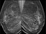
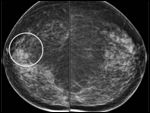
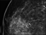
Digital mammography shows area of calcifications (Figure 1a-b). Magnification views demonstrate intraductal pleomorphic microcalcifications in the right 11:00 area (Figure 1c).
No noted findings on physical exam or ultrasound.
Diagnosis
Vacuum assisted stereotactic needle core biopsy of the right breast 11:00 microcalcifications reveals ductal carcinoma in situ.


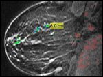
Discussion
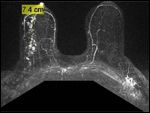
This case demonstrates the benefit of multimodality imaging including digital mammography and breast magnetic resonance imaging (MRI) in this patient, which led to a diagnosis of ductal carcinoma in situ (DCIS). There has been some conflicting information in the literature regarding MRI and its usefulness in the evaluation of DCIS. For this patient, the MRI revealed the extensive area of involvement more so than mammography. This information was critical to the surgeon for surgical planning.
This article was originally published at Diagnostic Imaging


