Management of Mycosis Fungoides: Part 2. Treatment
Mycosis fungoides is a low-grade lymphoproliferative disorder ofskin-homing CD4+ lymphocytes that may produce patches, plaques,tumors, erythroderma, and, ultimately, systemic dissemination. Treatmentselection is generally guided by institutional experience, patientpreference, and toxicity profile, as data from phase III clinical trials arelimited. Effective topical treatments currently include mechlorethamine(Mustargen), carmustine (BCNU, BiCNU), corticosteroids, bexarotene(Targretin, a novel rexinoid), psoralen plus ultraviolet A, ultraviolet B,and total-skin electron-beam radiotherapy. Effective systemic treatmentsinclude interferon, retinoids, bexarotene, denileukin diftitox(Ontak), extracorporeal photopheresis, chemotherapy, and high-dosechemotherapy with allogeneic bone marrow transplant. Each of thesetreatments is discussed in detail, followed by specific recommendationsfor each stage of mycosis fungoides.
ABSTRACT: Mycosis fungoides is a low-grade lymphoproliferative disorder of skin-homing CD4+ lymphocytes that may produce patches, plaques, tumors, erythroderma, and, ultimately, systemic dissemination. Treatment selection is generally guided by institutional experience, patient preference, and toxicity profile, as data from phase III clinical trials are limited. Effective topical treatments currently include mechlorethamine (Mustargen), carmustine (BCNU, BiCNU), corticosteroids, bexarotene (Targretin, a novel rexinoid), psoralen plus ultraviolet A, ultraviolet B, and total-skin electron-beam radiotherapy. Effective systemic treatments include interferon, retinoids, bexarotene, denileukin diftitox (Ontak), extracorporeal photopheresis, chemotherapy, and high-dose chemotherapy with allogeneic bone marrow transplant. Each of these treatments is discussed in detail, followed by specific recommendations for each stage of mycosis fungoides.
Mycosis fungoides is a lowgrade lymphoproliferative disorder of skin-homing CD4+ lymphocytes that may produce patches, plaques, tumors, erythroderma, and, ultimately, result in systemic dissemination. Details regarding epidemiology, clinical presentation, histopathology, molecular profile, staging, and prognosis were discussed in the last issue of ONCOLOGY. This part of the article will focus on the treatment of mycosis fungoides, providing an overview of major topical and systemic therapies followed by a discussion of specific treatment recommendations for each stage of mycosis fungoides.
Topical Therapies
Mechlorethamine
Topical mechlorethamine hydrochloride (Mustargen), also known as nitrogen mustard, is an alkylating agent with proven activity in the treatment of mycosis fungoides patches and plaques. Typically, a 10 to 20 mg percent solution of mechlorethamine is applied once daily to all skin surfaces if diffuse skin involvement is present, and only to involved sites if skin disease is limited. For patients with slow response, the frequency of application may be increased to twice daily or the concentration may be increased to 30 to 40 mg percent. Therapy is typically continued for at least 6 months after complete skin clearance.
Cutaneous intolerance, manifested by erythema and pruritus, occurs in roughly 50% of patients treated with aqueous mechlorethamine[1] but is reduced to less than 10% in patients treated with mechlorethamine dissolved in an ointment such as Aquaphor.[2] Options for patients with cutaneous intolerance include changing from an aqueous to an ointment-based preparation, reducing the concentration of topical mechlorethamine to 1 mg percent followed by gradual dose titration, applying concomitant topical steroids, and attempting systemic desensitization. Other cutaneous side effects of mechlorethamine may include xerosis, hyperpigmentation, and, rarely, bullous reactions, urticaria, and Stevens-Johnson syndrome.[1]
Bone marrow suppression is not observed due to minimal systemic absorption. Mechlorethamine monotherapy has not been associated with an appreciable risk of secondary skin cancers,[ 2] but use of the drug in combination with other topical therapies such as total-skin irradiation or psoralen plus ultraviolet A (PUVA) may increase the risk of cutaneous basal and squamous cell carcinomas.[3]
Carmustine
Topical carmustine (BCNU, BiCNU), another alkylating agent with activity similar to mechlorethamine, is typically applied to all skin surfaces once daily at doses of 10 to 20 mg/d for 4 to 8 weeks. Due to systemic absorption that results in bone marrow suppression, the maximum duration of treatment is limited. Cutaneous hypersensitivity is uncommon (7% in one series) but chronic skin telangiectasis may occur.[4]
Topical Steroids
Glucocorticoids are an important component in the treatment of hematologic malignancies, likely due to their ability to induce apoptosis of neoplastic lymphoid cells. Class I (most potent) topical steroids, such as 0.05% clobetastol propionate, 0.05% diflorasone diacetate, and 0.05% halobetasol propionate (Ultravate), effectively treat most mycosis fungoides patches and plaques. These agents are applied twice daily to involved skin for 2 to 3 months before treatment efficacy is assessed.[5] Patients with steroid-responsive disease typically continue maintenance therapy for at least 1 month after clearing. Toxicity includes reversible depression of serum cortisol (in 10% to 15% of patients) and skin atrophy.[5]
Topical Rexinoids
Bexarotene (Targretin) belongs to a new class of agents called rexinoids that bind to the retinoid X receptor, resulting in transcription of various genes that control cellular differentiation and proliferation.[6] Promising phase I/II data suggest that 1.0% topical bexarotene gel applied twice daily is well-tolerated and effective in patients with stage IA-IIA mycosis fungoides.[7] Toxicity appears limited to skin irritation. As with systemic retinoids, bexarotene in both its topical and systemic forms should be avoided in pregnant women because of possible teratogenic effects.
PUVA and Ultraviolet B
Ultraviolet radiation effectively treats a variety of skin disorders, although the exact mechanism of action remains unclear. Ultraviolet light is divided into three classes-UVA, UVB, UVC, in order of increasing energy. UVA has a wavelength of 320 to 400 nm and activates the oral photochemotherapeutic agent 8- methoxypsoralen (8-MOP, Oxsoralen), resulting in DNA crosslinking and apoptotic cell death.[8] In contrast, UVB has a wavelength from 290 to 320 nm and effectively treats patches without the addition of an oral agent.
Contraindications to therapy with UVR include systemic lupus erythematosus, previous or current nonmelanoma skin cancer, pregnancy, porphyria, and genetic syndromes due to DNA repair defects.[9] In addition, ingestion of oral 8-methoxypsoralen should be avoided in patients with preexisting liver dysfunction, as liver toxicity is possible in this subgroup.
Patients treated with PUVA ingest 0.6 mg/kg of 8-methoxypsoralen 1.5 to 3 hours prior to receiving UVA irradiation.[10] The initial UVA dose may be as low as 0.5 J/cm2 and is increased by 0.5 to 1.0 J/cm2 every treatment session until complete response or tolerance dose is achieved.[9] Treatments are given two to three times per week until skin clearing and are then gradually tapered to once every 2 to 4 weeks for no more than 1 year.
Mycosis fungoides patches can be effectively treated by broadband UVB[11] or narrowband UVB (wavelength: 311-312 nm)[12,13] without psoralen ingestion. Therapy is typically initiated at doses that are 50% to 70% of the dose required to induce minimal skin erythema. Patients receive treatment 3 days per week with gradual increases in dose until patient tolerance and/or clinical response is achieved. Following maximal response, treatments continue for several months, but the frequency is gradually tapered to once every 2 weeks.[11] When selecting patients for UVB therapy, it is important to consider skin type, as UVB radiation may be less effective in people with darkly pigmented skin, given the ability of melanin to absorb ultraviolet radiation.[13]
Acute side effects of PUVA and UVB include skin erythema (which may be painful), hyperpigmentation, xerosis, pruritus, and blistering. Eye goggles are used to decrease the risk of cataract formation. PUVA may cause nausea and vomiting after ingestion of 8-methoxypsoralen, but this may be avoided by the substitution of 5-methoxypsoralen, which is currently available in Europe.[9] Long-term toxicity includes photoaging (PUVA ≈ UVB) and cutaneous carcinogenesis (PUVA > UVB).[14]
Total-Skin Electron Radiotherapy
TABLE 1

Guidelines for Total-Skin Electron-Beam Therapy
Total-skin electron-beam radiotherapy (TSEBT) is a technically challenging modality used for the treatment of T1-4 skin disease. Guidelines for TSEBT developed by the European Organization for Research and Treatment of Cancer (EORTC) Cutaneous Lymphoma Project Group are presented in Table 1.[15]
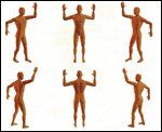
At our institution, these objectives are achieved with a mounted linear accelerator that emits 6-MeV electrons. The patient stands 3.8 meters from the gantry, and just behind a 3.2-mm thick Lexan polycarbonate screen that serves to attenuate and scatter the electron beam, resulting in an electron energy of 3.9 MeV at the skin surface. A total of six treatment positions are designated: anteroposterior, right and left anterior oblique, posteroanterior, and right and left posterior oblique (Figure 1). These positions maximize skin unfolding, thereby improving dose homogeneity in the lateral dimension. Each position is treated with two fields-upper and lower-to maximize dose homogeneity in the vertical dimension.
FIGURE 1

Treatment Positions Used in Total-Skin Electron-Beam Therapy
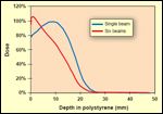
On treatment day 1, the anteroposterior, right posterior oblique, and left posterior oblique positions are treated. On treatment day 2, the posterior, right anterior oblique, and left anterior oblique positions are treated with the same dose. Over the course of a 48-hour treatment cycle, a patient will receive 2 Gy to the entire skin surface. This pattern continues, with patients receiving treatment 4 days a week for a total of 9 weeks, thereby delivering a total dose of 36 Gy to the skin surface. The depth-dose curves for both a single beam and all six beams are presented in Figure 2, which illustrates how this treatment arrangement satisfies the guidelines for skin dose and soft-tissue dose established by the Cutaneous Lymphoma Project Group.
FIGURE 2

Representative Depth-Dose Curves for Total-Skin Electron-Beam Therapy
A complex regimen of patch and boost treatments is utilized to ensure adequate dose delivery to all sites of cutaneous disease. For example, our institution uses 120-kV orthovoltage photons to deliver patch treatments to the soles of feet (14 Gy in 14 fractions, treatment days 1-7 and 30-36) and perineum (18 Gy in 18 fractions, treatment days 1-9 and 28-36). In addition, an electron reflector mounted above the patient's head increases the dose to the scalp. Patients with plaques or tumors that are bleeding, weeping, or painful also receive an upfront boost (2 Gy in 5 fractions) with either electrons or orthovoltage photons. Asymptomatic plaques and tumors that persist at the end of treatment receive a similar boost.
Acute and chronic morbidity is minimized by using small daily fractions and a complicated shielding regimen that protects the eyes, ears, lips, hands, and feet. Common acute toxicities from TSEBT include mild skin erythema, whole-body alopecia, possible loss of fingernails and toenails, hand and foot edema, and hypohidrosis. In general, TSEBT does not cause serious long-term complications, although permanent nail dystrophy, xerosis, partial scalp alopecia, and fingertip dysesthesias have been described.[15]
Systemic Therapies
Interferon
Interferon alpha-2a (Roferon-A) is an effective agent, particularly for patch and plaque disease, likely due to a direct antitumor effect and/or immunomodulation.[ 16] Interferonalpha has been used alone[16,17] or in combination with retinoids, PUVA,[18-20] extracorporeal photopheresis,[ 21] purine analogs,[22] or pentostatin (2′-deoxycoformycin, Nipent).[23] To date, no randomized trials have compared interferon-alpha plus another therapy with interferonalpha alone, although one prospective trial did show a benefit from combining interferon-alpha with PUVA as compared to interferonalpha plus a retinoid.[24]
Interferon-alpha may be administered subcutaneously or intramuscularly at a starting dose of 3 million units three times a week and may be gradually increased to 12 million units daily.[16] A phase I trial established 12 million IU/m2 three times a week as the maximal tolerated dose when interferon-alpha is combined with PUVA.[25] Toxicity may include flulike symptoms, psychiatric disturbances including depression and confusion, elevated transaminases, leukopenia, and thrombocytopenia.[19,20] Furthermore, when interferon-alpha was combined with fludarabine in a phase II trial, 2 of 35 (6%) patients developed persistent bone marrow aplasia.[22] Despite these side effects, in a recent phase II trial of interferonalpha and PUVA, only 8% of patients withdrew as a result of toxicity.[20]
Retinoids
Oral retinoids such as isotretinoin (Accutane) and acitretin (Soriatane) influence cell growth and differentiation and have demonstrated moderate activity in the treatment of patches and plaques. Retinoids can be safely combined with other therapies such as PUVA,[26] interferon-alpha, and TSEBT, and may permit reduction in PUVA dose, thereby reducing toxicity. Typical starting doses are 1 mg/kg/d for isotretinoin and 25 to 50 mg/d for acitretin.[27] Side effects include photosensitivity, xerosis, myalgias, arthralgias, headaches, impaired night vision, corneal opacities, teratogenicity, elevated transaminases, hyperlipidemia, and pancreatitis.[27]
Rexinoids
Oral bexarotene is administered once daily with food at a dose of 300 mg/m2/d and has been approved by the Food and Drug Administration for use in all stages of treatment-refractory mycosis fungoides.[28,29] Preliminary data suggest that bexarotene can be combined safely with other therapies including PUVA, extracorporeal photopheresis, interferon-alpha, and mechlorethamine. For patients who obtain a complete remission, bexarotene is typically continued indefinitely, though at a lower daily dose, provided that toxicities are manageable.[6]
Hypertriglyceridemia, the most common adverse event, occurs in 80% of treated patients and may result in reversible pancreatitis if triglyceride levels exceed 800 mg/dL.[6] Therefore, atorvastatin (Lipitor) or fenofibrate (Tricor) should be initiated if tryglyceride levels exceed 350 mg/dL. Gemfibrozil increases serum bexarotene concentrations, resulting in a paradoxical elevation of triglycerides, and should therefore be avoided.[6] Another side effect, central hypothyroidism, affects roughly 75% of patients but responds well to levothyroxine and resolves if bexarotene treatment is discontinued.[6] Other side effects include self-limited headaches, mild leukopenia, mild transaminase elevations, skin peeling, and pruritus. In the phase II/III trial of bexarotene for advanced, refractory cutaneous T-cell lymphoma, only 10% of patients receiving the optimal dose withdrew as a result of an adverse event.[28]
Denileukin Diftitox
Denileukin diftitox (Ontak) is a recombinant fusion protein that contains portions of interleukin-2 and diphtheria toxin and has proven activity in refractory mycosis fungoides (stages IB-IVA).[30] It selectively targets T cells that express the high-affinity interleukin-2 receptor (a complex of CD25, CD122, and CD132), resulting in endocytosis of diphtheria toxin, inhibition of protein synthesis, and cell death. Accordingly, denileukin diftitox should be considered only for mycosis fungoides patients with neoplastic T cells that express the highactivity interleukin-2 receptor. For example, the phase III clinical trial that led to the approval of denileukin diftitox required that at least 20% of the cells comprising the cutaneous lymphoid infiltrate express CD25.[30] Denileukin diftitox is typically administered by venous infusion for 5 consecutive days at a dose of 9 or 18 μ/kg/d for up to eight cycles and should be avoided in patients with poorly controlled hypertension, heart failure, liver disease, or renal impairment.
Despite pretreatment administration of acetaminophen and an antihistamine, toxicities were commonly encountered in the phase III clinical trial of denileukin diftitox. Acute hypersensitivity reactions including dyspnea, back pain, hypotension, and chest pain occurred in 60% of patients. Over 90% of patients experienced some degree of constitutional symptoms such as a flu-like illness or gastrointestinal discomfort. Furthermore, a vascular leak syndrome characterized by hypotension, hypoalbuminemia, and edema (at least two of the three) was encountered in 25% of patients. Administration of prophylactic intravenous corticosteroids and fluids reduces the risk of this toxicity. Other toxicities may include thrombotic events, infections, transaminase elevations, renal impairment, and lymphopenia. In total, 21% of the patients in this trial withdrew due to adverse events.[30]
Extracorporeal Photochemotherapy and Transimmunization
Extracorporeal photochemotherapy, also know as photopheresis, is a novel immune therapy that has shown activity in the treatment of erythrodermic mycosis fungoides, with or without blood involvement.[8,31] Peripheral blood leukocytes are harvested by leukapheresis, mixed with 8-methoxypsoralen, exposed to 2 J/cm2 of ultraviolet A, and then reinfused into the patient. This results in DNA crosslinking and gradual apoptotic death of the circulating mycosis fungoides cells that were exposed to UVA.
For unclear reasons, monocytes are resistant to the apoptotic effects of extracorporeal photochemotherapy. Instead, they are stimulated to become immature antigen-presenting dendritic cells via the physical process of leukapheresis. Therefore, once treated leukocytes are reinfused into the bloodstream, activated dendritic cells may phagocytose remnants of the apoptotic mycosis fungoides cells, present their antigens on major histocompatability class I, and stimulate expansion of antitumor CD8+ T cells.[32] Transimmunization is a variation of this theme, in which leukocytes treated with 8-methoxypsoralen and UVA are then incubated overnight under physiologic conditions in order to optimize loading of mycosis fungoides antigens onto antigen-presenting cells; this concept is now the subject of a clinical trial.[32]
Extracorporeal photochemotherapy is typically administered on 2 consecutive days every 4 weeks, although the frequency may be increased in patients with extensive disease.[33] For patients who achieve a complete response, therapy should be maintained for roughly 6 months and then gradually tapered. In general, this approach is well tolerated, although hypotension, arrhythmias, and heart failure may occur due to fluid shifts. Patients with a history of cardiac disease therefore require close monitoring.[ 31] Extracorporeal photochemotherapy has been safely combined with interferon-alpha and TSEBT.[21]
Chemotherapy
Systemic chemotherapy is typically reserved for refractory cutaneous disease, nodal involvement, visceral disease, or large-cell transformation. Both single-agent and combination regimens produce reasonable response rates and symptomatic improvement but are not curative. Numerous agents, either alone or in combination, have been studied, including methotrexate, trimetrexate (Neutrexin), fluorouracil, cyclophosphamide (Cytoxan, Neosar), doxorubicin, vincristine, prednisone, mechlorethamine, cisplatin, etoposide, bleomycin, vinblastine, gemcitabine (Gemzar), fludarabine (Fludara), 2-chlorodeoxyadenosine (Cladribine, Leustatin), pentostatin, and pegylated liposomal doxorubicin (Doxil).[ 22,23,34-39] A complete discussion of the contraindications and toxicity profiles of all these agents is beyond the scope of this review.
High-Dose Chemotherapy With Autologous Stem Cell Rescue or Allogeneic Bone Marrow Transplant
Investigators have reported only limited experience using high-dose chemotherapy with either autologous or allogeneic rescue in the treatment of advanced, refractory mycosis fungoides. Autologous stem-cell rescue has resulted in several complete responses; however, 15 of the 17 such patients reported in the literature relapsed within 1 year.[40-42] In contrast, high-dose chemotherapy with allogeneic bone marrow transplantation may result in prolonged diseasefree survival. Case reports have documented patients with advanced mycosis fungoides who are alive without recurrent disease at 15 months, 2 years, and 4.5 years after transplant.[43,44] A fourth patient whose disease recurred after allogeneic transplant developed a second complete response upon withdrawal of prophylactic cyclosporine, suggesting that allogeneic transplant may induce a graft-vs-tumor effect.[45] Thus, in contrast to conventional chemotherapy, high-dose chemotherapy with allogeneic bone marrow transplant may be potentially curative and should be considered for younger patients with good performance status, preferably as part of an institutional protocol.
Treatment by Stage
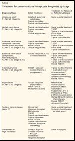
TABLE 2

Treatment Recommendations for Mycosis Fungoides by Stage
Treatment selection is largely determined by institutional expertise, patient preference, and toxicity profile, as rigorous phase III comparisons of different treatment modalities have not been conducted. The only major phase III trial, published in 1989, randomized 103 patients with stage IA-IVB mycosis fungoides to sequential topical therapy or combined TSEBT and systemic chemotherapy. Although patients in the combined-modality arm were more likely to obtain a complete response, no differences in long-term disease-free or overall survival were observed.[46] Therefore, this trial serves as justification for avoiding systemic chemotherapy and, instead, selecting the least morbid and most effective topical therapy to optimize control of cutaneous symptoms. Treatment recommendations for each stage are presented in Table 2.
Unilesional Patch
Patients with an isolated patch (T1, N0, M0; stage IA) experience longterm disease-free survival in excess of 85% when treated with superficial radiotherapy (electrons or orthovoltage) to a total dose of at least 30 Gy.[47-49] Local application of carmustine or mechlorethamine is also reasonable, although these therapies have not been formally compared to localized radiotherapy and published experience with them is limited.
Limited Patches and/or Plaques
For patients with limited skin involvement (T1, N0, M0; stage IA and T1, N1, M0; stage IIA), topical mechlorethamine is a reasonable firstline treatment, with a complete response rate of 65%, partial response rate of 28%, and 10-year diseasespecific survival of 95% in 203 patients followed longitudinally at Stanford.[2] Although retrospective data indicate that 10-year relapse-free survival is higher for patients who receive TSEBT as opposed to mechlorethamine (59% vs 45%), no difference in overall survival has been appreciated.[50] Thus, as TSEBT is far more costly and time consuming, we prefer to reserve TSEBT for patients with T1 disease refractory to other standard therapies.
Several other modalities are reasonable first-line treatment options for T1 disease. For patients with limited patches, UVB therapy results in a complete response rate of roughly 80% with a median response duration of 2 years.[11,12] PUVA is appropriate for both patches and plaques, producing complete responses rates of 80% to 100%. Although relapse is common, most patients will respond to additional PUVA.[51,52] Similar to mechlorethamine, topical carmustine produced a complete response rate of 86%, partial response rate of 12%, and 5-year relapse-free survival of 35% in a series of 49 patients.[4]
For T1 disease that recurs after mechlorethamine, carmustine, UVB, or PUVA therapy, retreatment with these agents may result in another response. Other options for recurrent or refractory T1 disease include TSEBT (complete response: 95%),[15] class I steroids (complete response: 63%, partial response: 31%),[5] interferonalpha monotherapy (complete response: 50%, partial response: 35%),[16] topical bexarotene (complete response: 21%, partial response: 42%, median duration of response: 99 weeks),[7] and oral bexarotene (complete response: 7%, partial response: 47%).[29]
Extensive Patches and/or Plaques
Relatively asymptomatic patients with extensive patches and/or minimally indurated plaques (T2, N0, M0; stage IB and T2, N1, M0; stage IIA) should receive treatment similar to that recommended for T1 disease. Again, topical mechlorethamine is an excellent choice for these patients, resulting in a complete response rate of 34%, partial response rate of 38%, and 10-year relapse-free survival rate of 20%.[2] Similarly, carmustine produced a complete response rate of 47%, partial response rate of 37%, and 5-year relapse-free survival rate of 10%.[4] Topical corticosteroids may also prove to be helpful for patients who have extensive patches, with recent data suggesting a complete response rate of 25% and partial response rate of 57%, including patients in whom other topical therapies have failed.[5]
As an alternative to topical chemotherapy, topical phototherapy may be considered for relatively asymptomatic patients who have extensive patch/plaque disease. UVB should be reserved for extensive patch disease only, due to its superficial penetration. In contrast, PUVA may be considered for patients with thin plaques and has resulted in complete response rates of 60% to 100%. Unfortunately, over half of these patients will relapse within 2 years.[5,51,52] Although randomized data are lacking, phase II trials suggest promising improvements in both complete response rate and duration of remission when interferon-alpha is added to PUVA for the treatment of T2 disease.[19,20,24]
For more symptomatic disease or deeply infiltrative plaques, we favor TSEBT as initial treatment, due to superior response rates that produce rapid palliation. For example, complete response rates for T2 disease range from 76% to 90%, compared with a complete response rate of 39% for topical mechlorethamine.[15,53] Retrospective data suggest that adjuvant PUVA or mechlorethamine following TSEBT may lengthen the disease-free interval.[53,54]
Patients with recurrent or refractory T2 disease may be considered for repeat TSEBT (complete response rates of 40% to 80%),[55,56] PUVA with interferon-alpha,[19] mechlorethamine, carmustine, or oral or topical bexarotene.[7,29]
Cutaneous Tumors
Radiotherapy is an important treatment modality for cutaneous tumors (T3, N0, M0; stage IIB) due to its ability to treat the full thickness of deeply infiltrative lesions. For the rare patient with asymptomatic tumors involving less than 10% of the skin, either topical mechlorethamine with local radiotherapy or TSEBT are reasonable first-line treatment options, both producing similar 5-year overall survival (roughly 50%).[53] However, most patients with T3 disease present with extensive, symptomatic tumors. Such patients often benefit from TSEBT as a first-line, palliative treatment, due to a superior complete response rate as compared with topical mechlorethamine plus localized radiotherapy (44%-54% vs 8%).[53,57]
Adjuvant therapy should be strongly considered for patients who achieve a complete response to TSEBT. Retrospective data suggest that adjuvant topical mechlorethamine may improve the duration of response, resulting in a 5-year relapse-free survival of 55%, compared to 30% with TSEBT alone.[53] Adjuvant photopheresis is another reasonable option, with retrospective data showing a 5-year overall survival of 100% for patients receiving this modality, compared with 50% in patients who did not receive adjuvant therapy.[58]
Patients with recurrent tumors should be considered for either repeat TSEBT[55,56] or localized radiotherapy to palliate cutaneous symptoms. In addition, oral bexarotene has shown efficacy in patients with relapsed or refractory stage IIB disease, producing an overall response rate of 57%.[28] Another option, denileukin diftitox, resulted in an overall response rate of 30% for patients with treatmentrefractory stage IB-IVA mycosis fungoides.[30]
Systemic chemotherapy should also be considered and may result in complete response rates of 20% to 60%, though the duration of response is often only several months. To date, no clear benefit of combination as opposed to single-agent chemotherapy has been determined.[35] Although still experimental, allogeneic bone marrow transplant may be considered for young patients with an excellent performance status.
Erythroderma
TSEBT is an appropriate initial therapy for erythrodermic mycosis fungoides (T4, N0, M0; stage III), given its ability to produce a rapid and sustained response, thereby ameliorating the severe cutaneous symptoms experienced by such patients. Retrospective data indicate that TSEBT produced a 74% complete response rate, median progression-free survival of 1 year, and 5-year progression-free survival of 36%.[59] Not surprisingly, patients without blood involvement are more likely to obtain a complete and durable response. Following TSEBT, adjuvant photopheresis should be strongly considered, as retrospective data suggest that such treatment improves progression- free and cause-specific survival.[60]
For patients without access to TSEBT, several other topical treatment options exist. For example, a series of 10 patients treated with PUVA reported a complete response rate of 70% and median progression-free survival of 5 months.[10] Although the addition of interferon-alpha to PUVA has not been studied in a randomized setting, prospective phase II experiences suggest that this combination may improve the duration of response.[18-20] Another option for T4 disease is photopheresis, with 80% of patients experiencing at least some cutaneous improvement, although only 20% will experience a complete response.[8] Patients with a lower peripheral blood CD4/CD8 ratio (median: 2.2) appear more likely to respond.[33]
Patients with recurrent or refractory erythroderma may be considered for repeat TSEBT or for systemic therapies such as methotrexate, bexarotene, or denileukin diftitox. For example, a retrospective series showed that methotrexate administered oral- ly, intramuscularly, or intravenously at doses ranging from 5 to 125 mg produced a complete response rate of 41%, partial response rate of 17%, median relapse-free survival of 31 months, and median survival of 8.4 years.[37]
Patients refractory to methotrexate may be considered for other chemotherapeutic agents including gemcitabine,[38] fludarabine and cyclophosphamide,[ 61] fludarabine and interferon-alpha,[22] pentostatin with or without interferon-alpha,[23,62] 2-chlorodeoxyadenosine,[34] and combination regimens typically used for B-cell lymphomas.[63] In addition, oral bexarotene is also active in erythrodermic patients, producing an overall response rate of 32%.[28] As in patients with T3 disease, allogeneic transplant may also be considered.
Stage IV
As with stage III disease, treatment of stage IV mycosis fungoides should be directed toward maximal palliation. Retrospective data from Stanford suggested that no significant differences in complete response rate or survival existed among patients treated with systemic therapy only, systemic and topical therapy, or regional and topical therapy.[64] Ideally, patients with stage IV disease should be enrolled in investigational protocols or evaluated for allogeneic transplant. If these options are not available, therapy should be individualized based on the patient's performance status, extent and nature of symptoms, and ability to tolerate therapy.
For patients with stage IV mycosis fungoides, severe cutaneous symptoms are common, as 85% of patients have cutaneous tumors or erythroderma.[ 64] TSEBT may be considered for these patients, as it produces excellent complete response rates. Other topical palliative therapies such as PUVA, mechlorethamine, carmustine, corticosteroids, and localized superficial radiotherapy should also be considered.
REFERENCE GUIDE
Therapeutic Agents
Mentioned in This Article
Acetaminophen
Acitretin (Soriatane)
Atorvastatin (Lipitor)
Bexarotene (Targretin)
Bleomycin
Carmustine (BCNU, BiCNU)
2-Chlorodeoxyadenosine
(Cladribine)
Cisplatin
Clobetastol propionate
Cyclophosphamide
(Cytoxan, Neosar)
Cyclosporine
Denileukin diftitox (Ontak)
Diflorasone diacetate
Doxorubicin
Doxorubicin, pegylated
liposomal (Doxil)
Etoposide
Fenofibrate (Tricor)
Fludarabine (Fludara)
Fluorouracil
Gemcitabine (Gemzar)
Gemfibrozil
Halobetasol propionate
(Ultravate)
Interferon alpha-2a (Roferon-A)
Interleukin-2 (Proleukin)
Interleukin-12
Isotretinoin (Accutane)
Mechlorethamine (Mustargen)
Methotrexate
8-Methoxypsoralen
(8-MOP, Oxsoralen)
Pentostatin
(2′-deoxycoformycin, Nipent)
Prednisone
Rituximab (Rituxan)
Tazorotene (Tazorac)
Trimetrexate (Neutrexin)
Vinblastine
Vincristine
Brand names are listed in parentheses only if a drug is not available generically and is marketed as no more than two trademarked or registered products. More familiar alternative generic designations may also be included parenthetically.
As an alternative to topical therapy, systemic chemotherapy may provide palliation of both cutaneous and visceral symptoms. Single-agent methotrexate is a reasonable first choice given acceptable toxicity and overall response rates.[37,63] The systemic chemotherapy agents listed above are also options. Finally, patients with symptomatic visceral disease, most commonly involving the lungs, central nervous system, oral cavity, or oropharynx,[64] should be considered for low-dose palliative radiotherapy at doses of 20 to 30 Gy delivered in 2-Gy fractions.
Transformation
Treatment for mycosis fungoides that has transformed to a large cell histology is similar to that described for stage IV disease. In addition, rituximab (Rituxan) may be considered for mycosis fungoides that acquires CD20 expression. Another option is trimetrexate, a lipophilic antifolate that yielded an overall response rate of 50% in a prospective trial of 14 patients with refractory, transformed mycosis fungoides.[39]
Other Management Issues
Cutaneous symptoms including xerosis, pruritus, and pain from fissures or ulceration are often severe and should be managed aggressively with topical steroids, emollients, oral antihistamines, and aggressive wound care with nonocclusive dressings.[27] Furthermore, patients should be advised to take brief showers with luke- warm water followed by immediate application of an emollient.
Cutaneous and systemic infections are an important cause of morbidity and mortality in patients with mycosis fungoides, particularly for those with extracutaneous disease. The most common infections include cellulitis due to Staphylococcus aureus or betahemolytic streptococci, followed by cutaneous herpes simplex or herpes zoster (both of which may disseminate cutaneously), followed by bacteremia (most commonly with S aureus), followed by bacterial pneumonia.[65] Signs and symptoms of such infections should be evaluated promptly and treated aggressively.
Conclusions
The last 15 years have produced significant advancements in the treatment of mycosis fungoides. Novel biologically based therapies such as bexarotene and denileukin diftitox have shown that specific molecular targets may be pharmacologically manipulated to produce significant clinical gains. Furthermore, extracorporeal photopheresis and, possibly, transimmunization, provide evidence that the host immune system can be stimulated to produce a meaningful antitumor response. Finally, advances in the technical application of totalskin electron-beam therapy have resulted in improved response rates and decreased morbidity.
Clinical investigators continue to explore ways to combine these modalities in order to produce more complete and durable responses. Additional novel biologic and immunologic agents including anti-CD4 and anti-CD52 monoclonal antibodies, topical tazorotene (Tazorac), interleukin-2 (Proleukin), and interleukin-12 are under investigation and may continue to improve the treatment of recurrent or refractory disease.[66] At this time, high-dose chemotherapy with allogeneic bone marrow transplantation provides the greatest hope for curing patients with advanced disease and should be explored in prospective trials.
Financial Disclosure:The authors have no significant financial interest or other relationship with the manufacturers of any products or providers of any service mentioned in this article.
References:
1.
Esteve E, Bagot M, Joly P, et al: Aprospective study of cutaneous intolerance totopical mechlorethamine therapy in patientswith cutaneous T-cell lymphomas. FrenchStudy Group of Cutaneous Lymphomas. ArchDermatol 135:1349-1353, 1999.
2.
Kim YH, Martinez G, Varghese A, et al:Topical nitrogen mustard in the managementof mycosis fungoides: Update of the Stanfordexperience. Arch Dermatol 139:165-173, 2003.
3.
Licata AG, Wilson LD, Braverman IM,et al: Malignant melanoma and other secondcutaneous malignancies in cutaneous T-celllymphoma. The influence of additional therapyafter total skin electron beam radiation.Arch Dermatol 131:432-435, 1995.
4.
Zackheim HS, Epstein EH Jr, Crain WR:Topical carmustine (BCNU) for cutaneous Tcell lymphoma: A 15-year experience in 143patients. J Am Acad Dermatol 22:802-810,1990.
5.
Zackheim HS, Kashani-Sabet M, AminS: Topical corticosteroids for mycosis fungoides.Experience in 79 patients. Arch Dermatol134:949-954, 1998.
6.
Talpur R, Ward S, Apisarnthanarax N, etal: Optimizing bexarotene therapy for cutaneousT-cell lymphoma. J Am Acad Dermatol47:672-684, 2002.
7.
Breneman D, Duvic M, Kuzel T, et al:Phase 1 and 2 trial of bexarotene gel for skindirectedtreatment of patients with cutaneousT-cell lymphoma. Arch Dermatol 138:325-332,2002.
8.
Edelson R, Berger C, Gasparro F, et al:Treatment of cutaneous T-cell lymphoma byextracorporeal photochemotherapy. Preliminaryresults. N Engl J Med 316:297-303, 1987.
9.
British Photodermatology Group guidelinesfor PUVA. Br J Dermatol 130:246-255,1994.
10.
Abel EA, Sendagorta E, Hoppe RT, etal: PUVA treatment of erythrodermic andplaque-type mycosis fungoides. Ten-year follow-up study. Arch Dermatol 123:897-901,1987.
11.
Ramsay DL, Lish KM, Yalowitz CB, etal: Ultraviolet-B phototherapy for early-stagecutaneous T-cell lymphoma. Arch Dermatol128:931-933, 1992.
12.
Diederen PV, van Weelden H, SandersCJ, et al: Narrowband UVB and psoralen-UVAin the treatment of early-stage mycosis fungoides:A retrospective study. J Am Acad Dermatol48:215-219, 2003.
13.
Gathers RC, Scherschun L, Malick F,et al: Narrowband UVB phototherapy for early-stage mycosis fungoides. J Am Acad Dermatol47:191-197, 2002.
14.
Young AR: Carcinogenicity of UVBphototherapy assessed. Lancet 345:1431-1432,1995.
15.
Jones GW, Kacinski BM, Wilson LD, etal: Total skin electron radiation in the managementof mycosis fungoides: Consensus ofthe European Organization for Research andTreatment of Cancer (EORTC) CutaneousLymphoma Project Group. J Am Acad Dermatol47:364-370, 2002.
16.
Ross C, Tingsgaard P, Jorgensen H, etal: Interferon treatment of cutaneous T-celllymphoma. Eur J Haematol 51:63-72, 1993.
17.
Jumbou O, N'Guyen JM, Tessier MH,et al: Long-term follow-up in 51 patients withmycosis fungoides and Sezary syndrome treatedby interferon-alfa. Br J Dermatol 140:427-431, 1999.
18.
Roenigk HH Jr, Kuzel TM, SkoutelisAP, et al: Photochemotherapy alone or combinedwith interferon alpha-2a in the treatmentof cutaneous T-cell lymphoma. J InvestDermatol 95:198S-205S, 1990.
19.
Kuzel TM, Roenigk HH Jr, SamuelsonE, et al: Effectiveness of interferon alfa-2acombined with phototherapy for mycosis fungoidesand the Sezary syndrome. J Clin Oncol13:257-263, 1995.
20.
Chiarion-Sileni V, Bononi A, FornasaCV, et al: Phase II trial of interferon-alpha-2aplus psolaren with ultraviolet light A in patientswith cutaneous T-cell lymphoma. Cancer95:569-575, 2002.
21.
Wollina U, Looks A, Meyer J, et al:Treatment of stage II cutaneous T-cell lymphomawith interferon alfa-2a and extracorporealphotochemotherapy: A prospectivecontrolled trial. J Am Acad Dermatol 44:253-260, 2001.
22.
Foss FM, Ihde DC, Linnoila IR, et al:Phase II trial of fludarabine phosphate andinterferon alfa-2a in advanced mycosis fungoides/Sezary syndrome. J Clin Oncol12:2051-2059, 1994.
23.
Foss FM, Ihde DC, Breneman DL, et al:Phase II study of pentostatin and intermittenthigh-dose recombinant interferon alfa-2a inadvanced mycosis fungoides/Sezary syndrome.J Clin Oncol 10:1907-1913, 1992.
24.
Stadler R, Otte HG, Luger T, et al:Prospective randomized multicenter clinicaltrial on the use of interferon -2a plus acitretinversus interferon -2a plus PUVA in patientswith cutaneous T-cell lymphoma stages I andII. Blood 92:3578-3581, 1998.
25.
Kuzel TM, Gilyon K, Springer E, et al:Interferon alfa-2a combined with phototherapyin the treatment of cutaneous T-cell lymphoma.J Natl Cancer Inst 82:203-207, 1990.
26.
Thomsen K, Hammar H, Molin L, et al:Retinoids plus PUVA (RePUVA) and PUVAin mycosis fungoides, plaque stage. A reportfrom the Scandinavian Mycosis FungoidesGroup. Acta Derm Venereol 69:536-538,1989.
27.
Kim YH, Hoppe RT: Mycosis fungoidesand the Sezary syndrome. Semin Oncol 26:276-289, 1999.
28.
Duvic M, Hymes K, Heald P, et al:Bexarotene is effective and safe for treatmentof refractory advanced- stage cutaneous T-celllymphoma: Multinational phase II-III trial results.J Clin Oncol 19:2456-2471, 2001.
29.
Duvic M, Martin AG, Kim Y, et al:Phase 2 and 3 clinical trial of oral bexarotene(Targretin capsules) for the treatment of refractoryor persistent early-stage cutaneous Tcelllymphoma. Arch Dermatol 137:581-593,2001.
30.
Olsen E, Duvic M, Frankel A, et al:Pivotal phase III trial of two dose levels ofdenileukin diftitox for the treatment of cutaneous T-cell lymphoma. J Clin Oncol 19:376-388, 2001.
31.
Lim HW, Edelson RL: Photopheresisfor the treatment of cutaneous T-cell lymphoma.Hematol Oncol Clin North Am 9:1117-1126, 1995.
32.
Berger CL, Hanlon D, Kanada D, et al:Transimmunization, a novel approach for tumorimmunotherapy. Transfus Apheresis Sci26:205-216, 2002.
33.
Heald P, Rook A, Perez M, et al: Treatmentof erythrodermic cutaneous T-celllymphoma with extracorporeal photochemotherapy.J Am Acad Dermatol 27:427-433,1992.
34.
Kuzel TM, Hurria A, Samuelson E, etal: Phase II trial of 2-chlorodeoxyadenosinefor the treatment of cutaneous T-cell lymphoma.Blood 87:906-911, 1996.
35.
Rosen ST, Foss FM: Chemotherapy formycosis fungoides and the Sezary syndrome.Hematol Oncol Clin North Am 9:1109-1116,1995.
36.
Wollina U, Graefe T, Karte K: Treatmentof relapsing or recalcitrant cutaneous Tcelllymphoma with pegylated liposomaldoxorubicin. J Am Acad Dermatol 42:40-46,2000.
37.
Zackheim HS, Kashani-Sabet M, HwangST: Low-dose methotrexate to treat erythrodermiccutaneous T-cell lymphoma: Resultsin twenty-nine patients. J Am Acad Dermatol34:626-631, 1996.
38.
Zinzani PL, Baliva G, Magagnoli M, etal: Gemcitabine treatment in pretreated cutaneousT-cell lymphoma: experience in 44 patients.J Clin Oncol 18:2603-2606, 2000.
39.
Sarris AH, Phan A, Duvic M, et al:Trimetrexate in relapsed T-cell lymphoma withskin involvement. J Clin Oncol 20:2876-2880,2002.
40.
Bigler RD, Crilley P, Micaily B, et al:Autologous bone marrow transplantation foradvanced stage mycosis fungoides. Bone MarrowTransplant 7:133-137, 1991.
41.
Russell-Jones R, Child F, Olavarria E,et al: Autologous peripheral blood stem celltransplantation in tumor-stage mycosis fungoides:Predictors of disease-free survival. AnnN Y Acad Sci 941:147-154, 2001.
42.
Sterling JC, Marcus R, Burrows NP, etal: Erythrodermic mycosis fungoides treatedwith total body irradiation and autologous bonemarrow transplantation. Clin Exp Dermatol20:73-75, 1995.
43.
Guitart J, Wickless SC, Oyama Y, et al:Long-term remission after allogeneic hematopoieticstem cell transplantation for refractorycutaneous T-cell lymphoma. Arch Dermatol138:1359-1365, 2002.
44.
Masood N, Russell KJ, Olerud JE, et al:Induction of complete remission of advancedstage mycosis fungoides by allogeneic hematopoieticstem cell transplantation. J Am AcadDermatol 47:140-145, 2002.
45.
Burt RK, Guitart J, Traynor A, et al:Allogeneic hematopoietic stem cell transplantationfor advanced mycosis fungoides: evidenceof a graft-versus-tumor effect. BoneMarrow Transplant 25:111-113, 2000.
46.
Kaye FJ, Bunn PA, Jr., Steinberg SM, etal: A randomized trial comparing combinationelectron-beam radiation and chemotherapywith topical therapy in the initial treatment ofmycosis fungoides. N Engl J Med 321:1784-1790, 1989.
47.
Heald PW, Glusac EJ: Unilesional cutaneousT-cell lymphoma: Clinical features, therapy,and follow-up of 10 patients with atreatment-responsive mycosis fungoides variant.J Am Acad Dermatol 42:283-285, 2000.
48.
Wilson LD, Kacinski BM, Jones GW:Local superficial radiotherapy in the managementof minimal stage IA cutaneous T-celllymphoma (mycosis fungoides). Int J RadiatOncol Biol Phys 40:109-115, 1998.
49.
Micaily B, Miyamoto C, Kantor G, etal: Radiotherapy for unilesional mycosis fungoides.Int J Radiat Oncol Biol Phys 42:361-364, 1998.
50.
Kim YH, Jensen RA, Watanabe GL, etal: Clinical stage IA (limited patch and plaque)mycosis fungoides. A long-term outcome analysis.Arch Dermatol 132:1309-1313, 1996.
51.
Herrmann JJ, Roenigk HH Jr, Hurria A,et al: Treatment of mycosis fungoides withphotochemotherapy (PUVA): Long-term follow-up. J Am Acad Dermatol 33:234-242,1995.
52.
Honigsmann H, Brenner W, RauschmeierW, et al: Photochemotherapy for cutaneousT cell lymphoma. A follow-up study. J AmAcad Dermatol 10:238-245, 1984.
53.
Chinn DM, Chow S, Kim YH, et al:Total skin electron beam therapy with or withoutadjuvant topical nitrogen mustard or nitrogenmustard alone as initial treatment of T2and T3 mycosis fungoides. Int J Radiat OncolBiol Phys 43:951-958, 1999.
54.
Quiros PA, Jones GW, Kacinski BM, etal: Total skin electron beam therapy followedby adjuvant psoralen/ultraviolet-A light in themanagement of patients with T1 and T2 cutaneousT-cell lymphoma (mycosis fungoides).Int J Radiat Oncol Biol Phys 38:1027-1035,1997.
55.
Becker M, Hoppe RT, Knox SJ: Multiplecourses of high-dose total skin electronbeam therapy in the management of mycosisfungoides. Int J Radiat Oncol Biol Phys32:1445-1449, 1995.
56.
Wilson LD, Quiros PA, Kolenik SA, etal: Additional courses of total skin electronbeam therapy in the treatment of patients withrecurrent cutaneous T-cell lymphoma. J AmAcad Dermatol 35:69-73, 1996.
57.
Jones GW, Hoppe RT, Glatstein E: Electronbeam treatment for cutaneous T-cell lymphoma.Hematol Oncol Clin North Am9:1057-1076, 1995.
58.
Wilson LD, Licata AL, Braverman IM,et al: Systemic chemotherapy and extracorporealphotochemotherapy for T3 and T4 cutaneousT-cell lymphoma patients who haveachieved a complete response to total skinelectron beam therapy. Int J Radiat OncolBiol Phys 32:987-995, 1995.
59.
Jones GW, Rosenthal D, Wilson LD:Total skin electron radiation for patients witherythrodermic cutaneous T-cell lymphoma(mycosis fungoides and the Sezary syndrome).Cancer 85:1985-1995, 1999.
60.
Wilson LD, Jones GW, Kim D, et al:Experience with total skin electron beam therapyin combination with extracorporeal photopheresisin the management of patients witherythrodermic (T4) mycosis fungoides. J AmAcad Dermatol 43:54-60, 2000.
61.
Scarisbrick JJ, Child FJ, Clift A, et al: Atrial of fludarabine and cyclophosphamidecombination chemotherapy in the treatment ofadvanced refractory primary cutaneous T-celllymphoma. Br J Dermatol 144:1010-1015,2001.
62.
Greiner D, Olsen EA, Petroni G: Pentostatin(2'-deoxycoformycin) in the treatmentof cutaneous T-cell lymphoma. J Am AcadDermatol 36:950-955, 1997.
63.
Bunn PA, Jr., Hoffman SJ, Norris D, etal: Systemic therapy of cutaneous T-cell lymphomas(mycosis fungoides and the Sezarysyndrome). Ann Intern Med 121:592-602,1994.
64.
de Coninck EC, Kim YH, Varghese A,et al: Clinical characteristics and outcome ofpatients with extracutaneous mycosis fungoides.J Clin Oncol 19:779-784, 2001.
65.
Axelrod PI, Lorber B, Vonderheid EC:Infections complicating mycosis fungoides andSezary syndrome. JAMA 267:1354-1358, 1992.
66.
Apisarnthanarax N, Talpur R, Duvic M:Treatment of cutaneous T cell lymphoma: Currentstatus and future directions. Am J ClinDermatol 3:193-215, 2002.
Targeted Therapy First Strategy Reduces Need for Chemotherapy in Newly Diagnosed LBCL
December 7th 2025Lenalidomide, tafasitamab, rituximab, and acalabrutinib alone may allow 57% of patients with newly diagnosed LBCL to receive less than the standard number of chemotherapy cycles without compromising curative potential.