The era of minimally invasive surgery for lung cancer follows decades of research; the collection and interpretation of countless qualitative and quantitative data points; and tireless efforts by a few pioneering thoracic surgeons who believed they could deliver a safe and oncologically sound operation with less tissue trauma, an improved physiologic profile, and fewer complications than traditional open surgery. This review highlights those efforts and the role of minimally invasive surgery for early-stage lung cancer in light of evolving technology, the emerging understanding of the biology of early-stage lung cancer, and lung cancer screening.
Reimagining Lung Cancer Treatment
In 1933, Dr. Evarts Graham reported the first successful pneumonectomy for lung cancer at Barnes Hospital in St. Louis, Missouri, using a tourniquet technique, and he popularized the concept of anatomic resection (sequential division of vein, artery, and bronchus to an affected portion of lung) for the treatment of lung cancer.[1] His patient was a fellow physician, who went on to outlive Dr. Graham himself. It was not until much later-and after much broader experience with partial lung resection for benign lung disease (bronchiectasis, tuberculosis)-that lobectomy and segmentectomy for the treatment of lung cancer were popularized. Traditional resection for lung cancer involved a 15- to 20-cm posterolateral thoracotomy, in which multiple muscle layers were divided and ribs were spread open with metal retractors or even removed to gain exposure. The incision alone resulted in considerable pain, splinting and atelectasis, and dysfunction in shoulder and chest wall mobility.
Establishing a New Standard
In 1992, following the success of a minimally invasive approach for cholecystectomy, Lewis et al published a series of 100 consecutive patients who underwent minimally invasive surgery of the chest-specifically, video-assisted thoracic surgery (VATS).[2] The indications for VATS in this series were broad and included benign disease, but considerable interest surrounded three patients who underwent VATS lobectomy with anatomic hilar dissection. Several single-institution series began to appear in the literature for VATS lobectomy.[3-5] At that time, VATS was considered new and investigational, and thus the definition of VATS lobectomy varied considerably. In some scenarios, VATS simply implied that a camera was used to assist dissection, but that a traditional thoracotomy, with rib spreading and even rib removal, was employed.
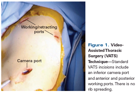
Seeking to standardize the definition of VATS lobectomy, the Cancer and Leukemia Group B (CALGB) surgery committee began a prospective, national, multicenter feasibility trial.[6] In CALGB 39802, participating surgeons underwent a rigorous credentialing protocol, and completed courses and submitted video, pathology, and operative reports to the group. The technique was defined by two 5-mm port incisions and one utility incision at a maximum length of 8 cm (generous by today’s standards). Individual dissection of veins, arteries, and airway to the affected lobe; standard nodal dissection identical to that carried out in open technique; and absolute avoidance of rib spreading were mandated (Figure 1). When the results were reported in 2007, 111 of 127 patients had undergone successful VATS lobectomy, with a favorable morbidity and mortality profile when compared with published data on open thoracotomy. Secondary endpoints were recurrence and survival. The finding of a 78% rate of failure-free survival (defined as time between surgery and death, disease progression, or relapse) at 36 months for patients with stage I non–small-cell lung cancer (NSCLC) lent support to the concept that video-assisted, minimally invasive surgery could achieve oncologic results as good as those of open surgery.
VATS Lobectomy Gains Ground
With a standardized definition of VATS lobectomy, studies could then be designed to compare the oncologic efficacy and physiologic impact of such minimally invasive approaches to lung cancer.[7,8] It quickly became clear that VATS lobectomy offered a sound oncologic operation at reduced morbidity and improved quality of life compared with open lobectomy.[9-11]
In 1995, a survey of 229 board-certified general thoracic surgeons found that only 3.5% of practicing general thoracic surgeons considered VATS the preferred approach to lung cancer surgery.[12] Fast forward to 2010, when 45% of lobectomies in the Society of Thoracic Surgeons General Thoracic Surgery Database were performed using VATS approaches.[13] Industry began to take notice, and as the instruments for retracting and dissecting improved, so did the resolution of the cameras and displays, adding unparalleled safety and efficiency. A growing community of thoracic surgeons continued to add experience and to shape the equipment and techniques applied in VATS lobectomy. Experienced surgeons began proctoring and mentoring their colleagues in the technique, as the growing body of evidence to support VATS lobectomy brought it to the forefront of national conference headlines. Courses began to appear throughout the country, allowing pioneers in the field to share troubleshooting tactics and to help others apply VATS technology to a wider population of patients and more complex tumors.
Today, the National Comprehensive Cancer Network (NCCN) guidelines recommend that minimally invasive approaches to lobectomy be strongly considered for all patients who undergo resection for lung cancer.[14] The American College of Chest Physicians (ACCP) published generally accepted criteria for cardiopulmonary reserve, which provide a valuable tool for evaluating a patient’s fitness for lung resection.[15] In experienced hands, VATS lobectomy can be achieved in patients with significant chronic obstructive pulmonary disease[16] or marginal results on pulmonary function tests,[17] with a 30-day mortality of less than 1%.
The introduction of VATS lobectomy techniques into training programs has been instrumental to dissemination of the technology. A number of opportunities for trainees to learn and perfect VATS skills are available throughout residency. These include hands-on courses offered free or at discounted rates as part of national conferences. In addition, videos designed for teaching, simulation equipment for both home and wet laboratories, and textbooks are available. Graduated responsibility is often given throughout training, such that 58% of recent graduates who perform general thoracic procedures consider themselves proficient in VATS lobectomies.[18] There is still room for improvement, however, as the implementation of VATS lobectomy training varies from program to program, and requirements for the American Board of Thoracic Surgery lag behind trends. For instance, while general thoracic surgery residents who are applying to sit for their certifying examinations are required to have completed at least 40 general VATS procedures (eg, pleural biopsy, drainage of effusion), the requirement for VATS lobectomy is 10 cases over the course of their training.[19]
VATS lobectomy has gained international acceptance as well, and reports from the European Society of Thoracic Surgeons Database corroborate such benefits of minimally invasive surgery as shorter length of stay and fewer postoperative complications. A recent analysis of more than 2,000 patients who underwent VATS lobectomy suggests that results obtainable at experienced centers in the United States are achievable in the greater global community.[20]
Minimally Invasive Lobectomy vs Sublobar Resection: Is Less More?
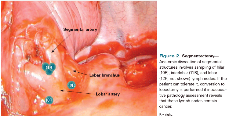
While earlier studies indicated that only a small percentage (25%) of patients with lung cancer presented with early-stage disease, the National Lung Screening Trial (NLST) confirmed that CT screening detects smaller, earlier tumors, resulting in a 20% reduction in lung cancer mortality.[21] Although national guidelines and societies support lobectomy with mediastinal lymph node dissection as the standard of care for early-stage lung cancer,[14] the fact remains that lung cancer is primarily a disease of the elderly, and many patients with lung cancer have significant comorbid conditions.[22] Patients with limited cardiopulmonary reserve may not tolerate formal lobectomy, which removes up to 20% of lung function and imposes physiologic changes that can be difficult to mitigate. For this reason, in appropriately selected patients, segmentectomy, grouped with wedge resection as a so-called sublobar resection, may be an ideal means of controlling local disease in patients who would not otherwise tolerate lobectomy. A segmentectomy, in its purest form, involves the individual dissection and division of vein, artery, and bronchus to a specific bronchopulmonary segment. As part of that dissection, lymph nodes that follow the branch points of the pulmonary artery and bronchus are encountered and can be removed distinctly as “sump” nodes for a particular tumor or en bloc with the specimen (Figure 2). Segmentectomy therefore also affords the benefit of accurate staging, in addition to preservation of functional lung parenchyma. The NLST found that the preponderance of lung cancers detected by screening CT were stage I (62.9% of the cancers detected in the study).[21] Parenchyma-preserving resection is of interest in this particular group of early-stage cancers, provided local control can be achieved. That said, anything less than lobar resection for early-stage disease is still heavily debated in the thoracic surgery community.
The Lung Cancer Study Group conducted a randomized trial that demonstrated a higher rate of recurrence and an associated trend toward decreased disease-free survival for sublobar resection as compared with lobectomy.[23] The results of this trial were published in 1995. Of note, both wedge resection (lung parenchyma stapled without dissection of the associated artery, bronchus, and vein) and segmentectomy were included in the sublobar analysis, and the majority of patients had an open thoracotomy. This established lobectomy as the standard of care for early-stage lung cancer for decades. As VATS allowed a wider application of surgical resection to patients with lung cancer, interest in VATS segmentectomy as definitive lung cancer surgery grew.
Current data that compare lobectomy with sublobar resection are mixed and mostly retrospective. At our institution, a review of 238 patients who underwent lobectomy or segmentectomy demonstrated no difference in distant recurrence, and overall and recurrence-free survival were similar for the two groups, despite the fact that the segmentectomy patients were older and had worse pulmonary function.[24] Other studies have demonstrated conflicting outcomes. Two randomized studies, one from Japan[25] and one from the United States,[26] aim to settle this matter. The results of these trials will influence how lung cancer surgery is handled not only in high-risk patients, but in an emerging subset of healthier patients as well.
Interest in the spectrum of adenocarcinomas of the lung that arise within foci of adenocarcinoma in situ (AIS) is growing in both the surgical and medical oncology communities. In general, these tumors demonstrate lepidic histologic characteristics and seem to follow an indolent course. Several studies have identified features of this group of adenocarcinomas, which represent a spectrum of AIS, minimally invasive adenocarcinoma (MIA), and lepidic-predominant adenocarcinoma.[27,28] These types of adenocarcinoma typically occur in women, nonsmokers, and Asians and are thought to result from acquired genetic field effects. Nonsmoking women are not generally part of the screening community yet; instead, these patients often present when studies done for other reasons (eg, chest pain) detect an incidental nodule or ground-glass opacity. In addition, approximately 28% of the adenocarcinomas detected in a Japanese CT screening study were classified as “bronchoalveolar carcinoma,” a term whose use is now discouraged in favor of “MIA,” “AIS,” or “lepidic-predominant adenocarcinoma.”[29] Most relevant to our discussion, these adenocarcinomas often present as multifocal disease or as dominant tumors with multifocal ground-glass opacities. Current data suggest that these multifocal indolent cancers do not behave like their aggressive counterparts. In a review published in 2014, tumor slides from 1,038 patients with stage I lung adenocarcinoma were reviewed.[30] Patients with AIS and MIA experienced no recurrences, and patients with lepidic-predominant invasive tumors had a significantly lower risk of recurrence than those with nonlepidic-predominant tumors. In fact, the American Joint Committee on Cancer staging guidelines indicate (albeit in the fine print) that these synchronous tumors should be staged separately.[31] The surgical approach to such patients requires considerable thought and deliberation over the location, number, and character of all the lesions and the feasibility of resection. Patients with such multifocal, indolent disease can require multiple resections in a lifetime, making sublobar resection a practical strategy for preserving as much lung parenchyma as possible over the course of their life.
TO PUT THAT INTO CONTEXT
[[{"type":"media","view_mode":"media_crop","fid":"54024","attributes":{"alt":"","class":"media-image","id":"media_crop_2022183319933","media_crop_h":"0","media_crop_image_style":"-1","media_crop_instance":"6761","media_crop_rotate":"0","media_crop_scale_h":"0","media_crop_scale_w":"0","media_crop_w":"0","media_crop_x":"0","media_crop_y":"0","style":"height: 144px; width: 144px;","title":" ","typeof":"foaf:Image"}}]]
David W. Johnstone, MD
Medical College of Wisconsin, Milwaukee,
WisconsinWhat Is the Current Place of Minimally Invasive Techniques in the Pulmonary Surgeon’s Armamentarium?The evolution of surgical treatment for lung cancer is well outlined in this review by White and Swanson, which nicely captures the major technical advances made over the past 80 years. We are now at a point where anatomic resection for lung cancer via smaller incisions with no rib spreading is accepted as safe, oncologically equivalent to open thoracotomy in competent hands, and less morbid in the short term. By now, any pulmonary surgeon who does not have video-assisted thoracic surgery (VATS) skills of this sort is outmoded.What Is the Role for Open Thoracotomies in an Era of VATS?A VATS resection is not suitable for all patients, and not all thoracotomies are morbid. With modern muscle- and nerve-sparing approaches, and good perioperative chest wall analgesic techniques, most standard thoracotomy exposures add little to length of stay and little long-term morbidity. As with VATS resections, success depends on attention to detail, technique, and experience. Related to this point is my strong opinion that residents must learn both open and VATS approaches, and that each richly informs the other, making both better. Training programs must provide good experience in both approaches to produce competent and safe surgeons.Will Even VATS Someday Be Eclipsed by Nonsurgical Treatment for Some Lung Cancers?Finally, as we have moved slowly towards sublobar resection for small peripheral cT1N0 lung cancers, we will have to incorporate results of three ongoing randomized trials involving radiotherapy, including the STABLE-MATES trial in the United States (ClinicalTrials.gov identifier: NCT02468024). In 10 years, it is possible that this subgroup of patients will be offered radiation or surgery as equivalent treatments.
VATS segmentectomy is an acquired skill set and can be technically challenging, particularly for less experienced VATS surgeons. At our institution, which teaches both VATS lobectomy and VATS segmentectomy as part of a cardiothoracic residency training program, we find that understanding the anatomy and dissection required for a segmentectomy often makes trainees better lobectomy surgeons-and vice versa. VATS segmentectomy is technically the same as open segmentectomy, in which the vein, artery, and bronchus to the segment of interest are individually dissected and ligated, along with the associated lymph nodes. The lung parenchyma is then divided with a stapling device or cautery. In appropriately selected segmentectomy patients, the intralobar lymph nodes are sent for immediate pathologic analysis; if the nodes are positive, a completion lobectomy is performed. In order to avoid compromising the oncologic efficacy of the procedure, patients for segmentectomy should ideally have tumors that are well centered in the segment of interest and are less than 2.0 cm in diameter.
Minimally Invasive Surgery vs Nonsurgical Therapy for Early-Stage Lung Cancer
National guidelines from the ACCP and the NCCN highlight the importance of the involvement of a board-certified thoracic surgeon when nonoperative treatments for lung cancer are considered. In particular, stereotactic ablative radiotherapy (SABR) has gained considerable momentum in the United States and in other countries. Data from prospective phase II trials of SABR for the treatment of patients with inoperable lung cancer showed promise,[32] mostly because survival rates without treatment or with conventional radiation were abysmal and these novel techniques could be applied without significant morbidity. These small (< 100 patients) studies received considerable corroboration as more and more retrospective and institutional reviews were published, with 3-year overall survival rates in the 55% range for early-stage T1a tumors. Interestingly, in some cases, the majority of the patients did not have histologic confirmation that the treated tumor was cancer, and surgeon-led evaluation of operability was rarely reported. In addition, how each study defined local and regional failure demonstrated considerable variability. Nonetheless, many of the data regarding SABR are being extrapolated to patients with operable lung cancer.
Two prospective randomized controlled trials were conducted to compare SABR with surgery for early-stage lung cancer. Both were terminated because of poor accrual, and neither was designed to compare best radiotherapy approaches with best surgical approaches with respect to morbidity, mortality, or survival. The STARS trial (ClinicalTrials.gov identifier: NCT00840749) included 28 sites in the United States, China, and France, but only 7 sites actually accrued a total of 31 patients. The ROSEL trial (ClinicalTrials.gov identifier: NCT00687986) attempted to accrue patients from 10 centers in the Netherlands, but only 4 sites contributed a total of 22 patients. A subsequent analysis combined the 53 patients from the STARS and ROSEL trials in an attempt to compare SABR and surgery.[33] The authors declared an equipoise between the two approaches, quoting overall and disease-free survival data that favored SABR; however, no histologic diagnosis was required for enrollment in the ROSEL trial, and 70% of patients in the surgical arm underwent open lobectomy. Strikingly, the 16% locoregional recurrence rate for SABR and surgical complication rates that were much higher than accepted norms were underemphasized. Thus, it is impossible to conclude that best surgical and best SABR approaches were effectively compared. Evaluating treatment efficacy when comparing surgery and SABR for early-stage lung cancer is akin to comparing clinical with pathologic staging. Pathologic data such as margin and receptor status, differentiation, tumor grade, and presence of lymphovascular invasion have a proven impact on treatment outcome, but this information is not available when the efficacy of nonoperative treatments is evaluated. For instance, NCCN guidelines promote adjuvant chemotherapy for the 15% to 20% of lung cancer patients with occult lymph node metastasis on surgical pathologic review. Does that imply that 15% to 20% of patients who undergo SABR or nonoperative therapies such as ablation are perpetually understaged or undertreated?
A study presented at the American Association for Thoracic Surgery Annual Meeting in May 2016 reviewed the overall survival of patients with cT1N0 tumors who underwent stereotactic body radiation therapy (SBRT) or wedge resection in the National Cancer Data Base from 2003 to 2011. Wedge resection was associated with improved survival when compared with SBRT in patients with operable clinical stage IA NSCLC.[34] Two additional prospective randomized trials have been designed to ascertain which patients stand to benefit the most from SABR. The US Department of Veterans Affairs’ VALOR study and the SABRTooth study in the United Kingdom will compare best surgical practice with best SABR treatment and should help shed light on this important question. Ideally, quality of life and cost analysis will be incorporated into the findings of these studies and will inform future directions.
Conclusion
In summary, lobectomy is the standard of care for early-stage lung cancer, and minimally invasive approaches should be considered for all patients with operable tumors. In centers with experienced surgeons, VATS lobectomy and sublobar resection, preferably segmentectomy, can be performed with reasonable safety in high-risk patients and in patients with multifocal disease.
Financial Disclosure: Dr. Swanson serves as an educational consultant to Covidien and Ethicon. Dr. White has no significant financial interest in or other relationship with the manufacturer of any product or provider of any service mentioned in this article.
References:
1. Graham E, Singer JJ. Successful removal of an entire lung for carcinoma of the bronchus. JAMA. 1933;101:1371-4.
2. Lewis RJ, Caccavale RJ, Sisler GE, Mackenzie JW. One hundred consecutive patients undergoing video-assisted thoracic operations. Ann Thorac Surg. 1992;54:421-6.
3. Walker WS, Codispoti M, Soon SY, et al. Long-term outcomes following VATS lobectomy for non-small cell bronchogenic carcinoma. Eur J Cardiothorac Surg. 2003;23:397-403.
4. Solaini L, Prusciano F, Bagioni P, et al. Video assisted thoracic surgery major pulmonary resection: present experience. Eur J Cardiothorac Surg. 2003;20:437-42.
5. Kaseda S, Aoki T, Hangai N, Shimizu K. Better pulmonary function and prognosis with video-assisted thoracic surgery than with thoracotomy. Ann Thorac Surg. 2000;70:1644-6.
6. Swanson SJ, Herndon JE, D’Amico TA, et al. Video-assisted thoracic surgery lobectomy: report of CALGB 39802-a prospective, multi-institution feasibility study. J Clin Oncol. 2007;25:4993-7.
7. Walker WS, Leaver HA. Immunologic and stress responses following video-assisted thoracic surgery and open pulmonary lobectomy in early stage lung cancer. Thorac Surg Clin. 2007;17:241-9.
8. Whitson BA, D’Cunha J, Andrade RS, et al. Thoracoscopic versus thoracotomy approaches to lobectomy: differential impairment of cellular immunity. Ann Thorac Surg. 2008;86:1735-44.
9. Nicastri DG, Wisnivesky JP, Little VR, et al. Thoracoscopic lobectomy: report on safety, discharge independence, pain, and chemotherapy tolerance. J Thorac Cardiovasc Surg. 2008;135:642-7.
10. Whitson BA, Groth SS, Duval SJ, et al. Surgery for early-stage non-small cell lung cancer: a systematic review of the video-assisted thoracoscopic surgery versus thoracotomy approaches to lobectomy. Ann Thorac Surg. 2008;86:2008-16.
11. Bendixen M, Jørgensen OD, Kronborg C, et al. Postoperative pain and quality of life after lobectomy via video-assisted thoracoscopic surgery or anterolateral thoracotomy for early stage lung cancer: a randomised controlled trial. Lancet Oncol. 2016;17:836-44.
12. Mack MJ, Scruggs GR, Kelly KM, et al. Video-assisted thoracic surgery: has technology found its place? Ann Thorac Surg. 1997;64:211-5.
13. Ceppa DP, Kosinski AS, Berry MF, et al. Thoracoscopic lobectomy has increasing benefit in patients with poor pulmonary function: a Society of Thoracic Surgeons Database analysis. Ann Surg. 2012;256:487-93.
14. National Comprehensive Cancer Network Guidelines. Non-small cell lung cancer. Version 4.2016. https://www.nccn.org/professionals/physician_gls/pdf/nscl.pdf. Accessed May 6, 2016.
15. Brunelli A, Kim AW, Berger KI, Addrizzo-Harris DJ. Physiologic evaluation of the patient with lung cancer being considered for resectional surgery: diagnosis and management of lung cancer, 3rd ed. American College of Chest Physicians evidence-based clinical practice guidelines. Chest. 2013;143(suppl 5):e166S-e190S.
16. Nwogu CE, D’Cunha J, Pang H, et al. VATS lobectomy has better perioperative outcomes than open lobectomy: CALGB 31001, an ancillary analysis of CALGB 140202 (Alliance). Ann Thorac Surg. 2015;99:399-405.
17. Burt BM, Kosinski AS, Shrager JB, et al. Thoracoscopic lobectomy is associated with acceptable morbidity and mortality in patients with predicted postoperative forced expiratory volume in 1 second or diffusing capacity for carbon monoxide less than 40% of normal. J Thorac Cardiovasc Surg. 2014;148:19-28.
18. Boffa DJ, Gangadharan S, Kent M, et al. Self-perceived video-assisted thoracic surgery lobectomy proficiency by recent graduates of North American thoracic residencies. Interact Cardiovasc Thorac Surg. 2012;14:797-800.
19. American Board of Thoracic Surgery case requirements. https://www.abts.org. Accessed May 5, 2016.
20. Falcoz PE, Puyraveau M, Thomas PA, et al. Video-assisted thoracoscopic surgery versus open lobectomy for primary non-small cell lung cancer: a propensity-matched analysis of outcome from the European Society of Thoracic Surgeons database. Eur J Cardiothorac Surg. 2016;49:602-9.
21. Aberle DR, Adams AM, Berg CD, et al. Reduced lung cancer mortality with low-dose computed tomographic screening. N Engl J Med. 2011;365:395-409.
22. Dela Cruz S, Tanoue LT, Matthay RA. Lung cancer: epidemiology, etiology, and prevention. Clin Chest Med. 2011;32:605-44.
23. Ginsburg RJ, Rubinstein LV. Randomized trial of lobectomy versus limited resection for T1N0 non-small cell lung cancer. Lung Cancer Study Group. Ann Thorac Surg. 1995;60:615-20.
24. Wolf AS, Richards WG, Jaklitsch MT, et al. Lobectomy versus sublobar resection for small (2 cm or less) non-small cell lung cancers. Ann Thorac Surg. 2011;92:1819-25.
25. Nakamura K, Saji H, Nakajima R, et al. A phase III randomized trial of lobectomy versus limited resection for small-sized peripheral non-small cell lung cancer (JCOG0802/WJOG4607L). Jpn J Clin Oncol. 2010;40:271-4.
26. Fox N, Bauer T. CALGB 140503: a randomized phase III trial of lobectomy versus sublobar resection for small (<2cm) peripheral non-small cell lung cancer. Oncology Issues. November/December 2008.
27. Boland JM, Froemming AT, Wampfler JA, et al. Adenocarcinoma in situ, minimally invasive adenocarcinoma, and invasive pulmonary adenocarcinoma-analysis of interobserver agreement, survival, radiographic characteristics, and gross pathology in 296 nodules. Hum Pathol. 2016;51:41-50.
28. Beasley MB, Dembitzer FR, Flores RM. Surgical pathology of early stage non-small cell lung carcinoma. Ann Transl Med. 2016;4:238.
29. Kondo RL, Yoshida K, Kawakami S, et al. Different efficacy of CT screening for lung cancer according to histological type: analysis of Japanese-smoker cases detected using a low-dose CT screen. Lung Cancer. 2011;74:433-40.
30. Kadota K, Villena-Vargas J, Yoshizawa A, et al. Prognostic significance of adenocarcinoma in situ, minimally invasive adenocarcinoma, and nonmucinous lepidic predominant invasive adenocarcinoma of the lung in patients with stage I disease. Am J Surg Pathol. 2014;38:448-60.
31. American Joint Committee on Cancer (AJCC) Cancer Staging System. https://www.cancerstaging.org. Accessed May 25, 2016.
32. Fakiris AJ, McGarry RC, Yiannoutsos CT, et al. Stereotactic body radiation therapy for early-stage non-small-cell lung carcinoma: four-year results of a prospective phase II study. Int J Radiat Oncol Biol Phys. 2009;75:677-82.
33. Chang JY, Senan S, Paul MA, et al. Stereotactic ablative radiotherapy versus lobectomy for operable stage I non-small-cell lung cancer: a pooled analysis of two randomised trials. Lancet Oncol. 2015;16:630-7.
34. Yerokun BA, Yang JC, Gulack BC, et al. A population-based analysis of surgical resection versus stereotactic body radiation therapy for stage I non-small cell lung cancer. Presented at the American Association for Thoracic Surgery Annual Meeting; May 14-18, 2016; Baltimore, MD. http://www.aats.org/annualmeeting/Program-Books/2016/3.cgi. Accessed October 19, 2016.


