In Mouse Model, Imaging Strategy Follows Prostate Cancer Bone Metastasis Response to Cabozantinib
A mouse model of bone metastasis can be used to follow real-time response to therapeutics in preclinical development, such as cabozantinib, according to results presented in the poster session of the 2013 AACR annual meeting.
A mouse model of bone metastasis can be used to follow real-time response to therapeutics in preclinical development, such as cabozantinib, according to results presented in the poster session of the 2013 American Association for Cancer Research annual meeting.

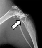
Representative x-ray image from hindlimbs of mouse, arrow indicates osteolytic lesion; source: Dunn LK et al.
PLoS ONE
2009:e6896
Prostate cancer, once metastasized, is incurable. A common site for dissemination of prostate tumors is the bone, where the microenvironment can make cancer cells difficult to target. It is thought that disrupting the interaction between the tumor and the microenvironment could be useful for combating metastasis, but drug development is challenged by the lack of preclinical models for initiating and monitoring metastasis. The work by Timothy Graham et al highlights a new model for bone metastasis in response to cabozantinib.
Cabozantinib (Cometriq) is a small molecule inhibitor of the receptor tyrosine kinases c-Met and VEGFR2 that is being developed by Exelixis. It has been reported in phase II trials to improve progression-free survival in patients with castration-resistant prostate cancer. It has also previously received FDA approval for medullary thyroid cancer.
Direct intratibial injection of the prostate cancer cell line VCaP, which was originally isolated from a prostate cancer vertebral metastasis, was used to initiate tumors that mimic metastases. Immunodeficient, castrated mice were used for the study to mimic as closely as possible the castration-resistant phenotype. Luciferase was introduced into the cells prior to the injection in order to allow for non-invasive imaging. Once the tumors were formed, mice were treated with 30 mg/kg daily cabozantinib (n = 7) or placebo (n = 7) orally for 15 days. Tumor burden could be imaged by injecting mice with luciferin and measuring the luminescence given off as a result of luciferase activity. Tumor size was also measured using T2-weighted magnetic resonance.
Photon flux measurements from luminescence detection demonstrated a significant reduction (52%, P = .016) of tumor size. Volume, as estimated by magnetic resonance, was found to be 104 ± 43 mm3 in the placebo control on average, while the treated group had 21.9 ± 5.1 mm3 tumors on average (P = .038). These findings suggested to the researchers that cabozantinib would be an effective therapeutic in prostate cancer that has metastasized to the bone.
In addition to the effect on orthotopic tumor size, the researchers analyzed the effects of cabozantinib on cellularity of the bone by determining the apparent diffusion coefficient after treatment. This feature was found to increase in treated but not control mice, suggesting that the apparent diffusion coefficient could be used as a clinical biomarker of treatment response in prostate cancer bone metastasis.