Low-grade gliomas are infiltrative primary brain tumors that most commonly occur in young adults. They are relatively slow growing compared with high-grade gliomas. The World Health Organization classification system was updated in 2016 to define low-grade gliomas using molecular markers in addition to histology. IDH mutation is an independent marker associated with better outcomes. Management is individualized based on tumor histology, molecular characterization, and patient risk factors. Given the longer course and natural history of low-grade gliomas, the goals of treatment should be to prolong overall survival and minimize neurocognitive decline. Early maximum safe resection is the first line of treatment. While low-risk patients may be followed with observation after surgery, patients with high-risk factors (subtotal resection, age > 40 years, IDH wild-type tumors) should be treated with radiation and chemotherapy. Improved understanding of the molecular characteristics of low-grade gliomas will further guide risk stratification and allow the identification of treatment approaches that are more effective and less toxic.
Introduction
Low-grade infiltrating gliomas are defined as World Health Organization (WHO) grade II primary brain neoplasms, and include astrocytomas and oligodendrogliomas.[1] Accounting for approximately 15% of all gliomas and 5% of all primary brain tumors, they present most commonly in the second to fourth decade of life, with peak incidence occurring between 35 and 44 years of age.[2] Presenting symptoms vary depending upon the neuroanatomic location of the tumor; seizures can occur in up to 80% of patients, as well as headaches, focal deficits, cognitive changes, and behavior changes. However, patients are often asymptomatic, and these brain lesions may be found incidentally.[3]
Low-grade gliomas are slow-growing tumors associated with a median survival time ranging from 4 to 13 years, depending on the subtype; in almost all cases, the tumors undergo malignant transformation, ultimately leading to death.[4,5] Treatment consists of surgery, radiation therapy (RT), and chemotherapy. Given the relatively long disease course, the optimal management approach is a matter of debate.[1] The uniform goal has been to prolong overall survival (OS), by delaying malignant transformation; and to maintain a good quality of life, by minimizing treatment-related sequelae. Treatment decisions for patients with low-grade gliomas are based on histologic and molecular characterizations of the tumors and the presence or absence of specific risk factors.
Classification
Low-grade gliomas are a heterogeneous group of tumors with differing morphologic characteristics. Recent advances in genomic analysis have led to the identification of molecular markers that play an important role in the classification, prognostication, and management of low-grade gliomas. Astrocytomas and oligodendrogliomas are the two primary histologic subtypes of low-grade glioma. The term “oligoastrocytoma,” an entity previously described based on histologic characterization, has been phased out in the 2016 WHO classification system, which uses both histologic and molecular information to define gliomas.[1]
IDH1 and IDH2 are the most commonly mutated genes in low-grade gliomas, with mutations estimated to occur in > 70% of cases.[6] IDH mutations are considered an early driver of glioma pathogenesis but are associated with better treatment outcome, since IDH-mutated tumors are more susceptible to chemotherapy.[7] Other molecular markers are used to differentiate between oligodendrogliomas and astrocytomas. A combined loss of chromosomal arms 1p and 19q (1p/19q codeletion) is seen almost exclusively in oligodendrogliomas and is now pathognomonic for this malignancy.[8] Mutations in CIC and FUBP1 also occur primarily in oligodendrogliomas, and their loss of expression is potentially associated with earlier recurrence and poorer outcome.[9,10] Low-grade astrocytomas commonly demonstrate a mutation in TP53 or inactivation of ATRX, both of which rarely occur in oligodendrogliomas.[11]
A landmark study by The Cancer Genome Atlas (TCGA) Research Network, using comprehensive genomic analysis of low-grade gliomas, revealed different molecular subtypes of low-grade glioma, with distinct prognoses based on IDH mutational status and 1p/19q codeletion status. Patients with low-grade gliomas whose tumors had IDH mutation and 1p/19q codeletion (oligodendroglioma) had the most favorable prognosis, with a median survival time of 8 years; in contrast, patients with IDH mutation without 1p/19q codeletion (astrocytoma) had a median survival time of 6.4 years. Comparatively, patients with IDH wild-type low-grade gliomas had a markedly worse median survival time of 1.7 years, similar to the 1.1-year median survival time of glioblastoma patients with wild-type IDH.[12]
Surgery
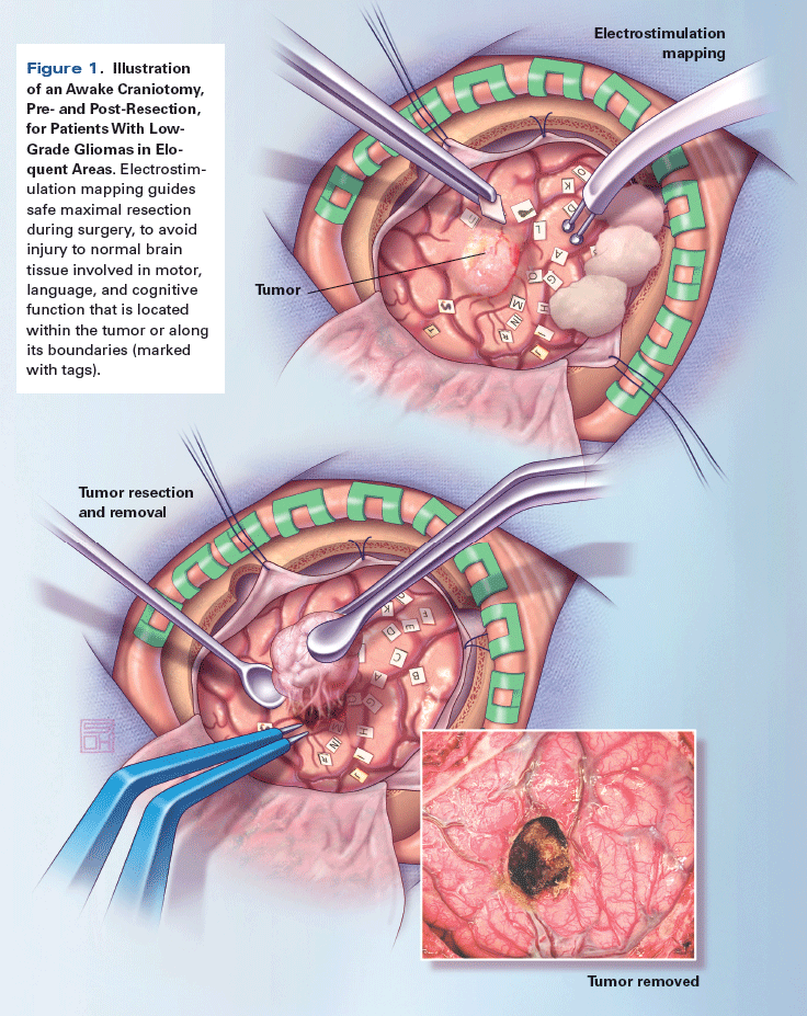
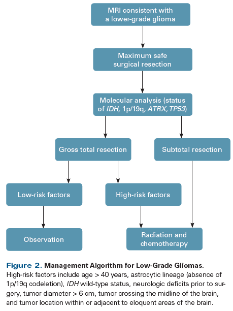
Surgical resection is the primary modality of treatment for low-grade gliomas. Historically, the timing of surgery (early vs delayed) was a matter of debate, with early surgery offered to symptomatic patients and a watch-and-wait approach employed for patients with incidentally found tumors. However, retrospective evidence has demonstrated improved outcomes in patients who undergo early resection.[13] Use of tissue samples obtained by resection enables a more accurate diagnosis than needle biopsy alone, which is associated with a relatively high rate of misdiagnosis.[14] In addition to data showing that early surgery improved patient outcomes, there is evidence that an increased extent of resection leads to better progression-free survival (PFS) and OS.[15,16] In a retrospective volumetric analysis of extent of resection in low-grade gliomas, Smith et al found that patients who underwent > 90% extent of resection had a 5-year OS rate of 97%, compared with a rate of 76% in patients who had < 90% extent of resection.[17] Intraoperative electrostimulation mapping during an awake craniotomy, for patients with tumors in eloquent areas of the brain, has demonstrated improved clinical outcomes, since it allows for more extensive resection while minimizing neurologic injury (Figure 1).[18,19] Thus, the appropriate initial management of low-grade gliomas is early maximum safe resection rather than watchful waiting. Figure 2 illustrates a management algorithm for patients with low-grade gliomas.
RT and Chemotherapy
The optimal use of RT and/or chemotherapy after surgery for low-grade gliomas is not yet fully defined. Several prognostic factors have been proposed to better identify patients at high risk for malignant transformation, who may benefit from aggressive management with adjuvant chemoradiation. Besides IDH wild-type status, other high-risk factors include age > 40 years, subtotal resection/biopsy only, astrocytic lineage (lack of 1p/19q codeletion), neurologic deficits prior to surgery, tumor diameter > 6 cm, tumor crossing the midline of the brain, and tumors located within or adjacent to eloquent areas of the brain.[20-22] Patients without these risk factors can be considered at low risk; therefore, after gross total resection, they should be observed closely with surveillance MRI every 3 months initially, and if there is no demonstrated tumor growth over a 1-year period, then the imaging interval can be increased to every 4 months for another year, and eventually every 6 months for the remainder of the patient’s life.
RT
For decades, RT has been a critical component of the management of low-grade gliomas, although the optimal timing and dose continue to be investigated. The European Organisation for Research and Treatment of Cancer (EORTC) conducted a study comparing early RT after surgery vs RT delayed until time of progression; while no significant difference in OS (7.4 years vs 7.2 years) was demonstrated, patients who received early RT had improvements in seizure control and median PFS (5.3 years vs 3.4 years with delayed RT).[23] A second EORTC study evaluated high-dose (59.4 Gy) vs low-dose (45 Gy) RT; no significant differences in PFS and OS were shown, and long-term analysis showed improved quality of life in patients treated at the lower radiation dose.[24,25] RT can cause both acute and chronic toxicities, including fatigue, neurocognitive decline, vasculopathy, endocrinopathy, and secondary malignancies; consequently, its use should be reserved for high-risk patients and carefully considered.
Proton therapy. This novel form of RT uses a heavier particle than photon RT, enabling more precise dosing and sparing of the surrounding normal brain tissue from radiation and its resultant toxicities. Proton therapy is therefore an attractive option for younger patients with low-grade gliomas. Long-term data on the efficacy and toxicity of proton therapy are not yet available; however, retrospective and prospective studies have demonstrated it to be well tolerated and associated with no early neurocognitive decline, albeit with side effects that commonly include focal alopecia, fatigue, and neuroendocrine dysfunction.[26,27] However, proton therapy is not accessible to all patients. There are currently only 25 proton radiation centers in the United States, primarily associated with university and tertiary care centers.[28] In addition, the cost of proton therapy can be up to twice as high as that of standard photon radiation, and data are limited regarding the cost-effectiveness of this technique in adult patients with brain tumors.[29,30]
KEY POINTS
- Low-grade gliomas are slowly progressive infiltrating brain tumors that preferentially affect younger people and account for approximately 5% of all brain tumors.
- Patients with IDH mutation have a significantly better prognosis. Codeletion of 1p and 19q is also associated with a more favorable outcome.
- Treatment consists of maximum safe resection and is followed by chemoradiation if high-risk factors are present.
Chemoradiation therapy
Use of chemotherapy in low-grade gliomas has historically been more controversial compared with other therapeutic modalities, although mounting evidence has demonstrated its benefit in both upfront treatment and the management of recurrent disease. Recently, results from the Radiation Therapy Oncology Group 9802 study, a large phase III trial in which patients with high-risk low-grade gliomas were randomized to receive RT or RT plus combination chemotherapy with PCV (procarbazine, lomustine, and vincristine), demonstrated an almost two-fold increase in OS for patients in the chemoradiation therapy arm, compared with patients who received RT alone (13.3 years vs 7.8 years).[31] Notably, this difference in survival only became evident on long-term follow-up, illustrating the challenges of conducting prospective trials in patients with low-grade gliomas. Considering the pivotal data, high-risk patients with low-grade gliomas should receive chemoradiation therapy rather than RT alone.
Although RTOG 9802 used PCV, in clinical practice, patients are commonly treated with temozolomide instead, due to its superior side effect profile; however, to date there has been no head-to-head comparison of temozolomide and PCV. A more direct comparison is being performed in the ongoing CODEL trial, a phase III study of patients with 1p/19q codeleted WHO grade II and III gliomas randomized to receive either RT followed by PCV or RT with concurrent and then adjuvant temozolomide. [32]
Conclusion
Low-grade gliomas are relatively slow-growing infiltrative primary brain tumors that almost invariably undergo malignant transformation. The standard of care consists of maximum safe resection; the potential use of chemoradiation for high-risk patients; and lifelong radiographic surveillance, with the goal of treatment being to delay malignant transformation and maximize quality of life. The management of low-risk patients is less well defined. Risk stratification and prognostication in low-grade gliomas will continue to be refined by improvements in our understanding of the tumor biology of this malignancy, through molecular profiling. It is hoped that these advances will lead to novel and more personalized treatments that improve patient survival and minimize treatment-related toxicities.
Financial Disclosure:The authors have no significant financial interest in or other relationship with the manufacturer of any product or provider of any service mentioned in this article.
Acknowledgment:The authors thank Matthew C. Tate, MD, PhD, for providing the clinical images that were used to create the artwork for this article.
References:
1. Louis DN, Perry A, Reifenberger G, et al. The 2016 World Health Organization classification of tumors of the central nervous system: a summary. Acta Neuropathol. 2016;131:803-20.
2. Ostrom QT, Gittleman H, Fulop J, et al. CBTRUS statistical report: primary brain and central nervous system tumors diagnosed in the United States in 2008-2012. Neuro Oncol. 2015;17(suppl 4):iv1-iv62.
3. Ruda R, Bello L, Duffau H, Soffietti R. Seizures in low-grade gliomas: natural history, pathogenesis, and outcome after treatments. Neuro Oncol. 2012;14(suppl 4):iv55-iv64.
4. Schomas DA, Laack NN, Rao RD, et al. Intracranial low-grade gliomas in adults: 30-year experience with long-term follow-up at Mayo Clinic. Neuro Oncol. 2009;11:437-45.
5. van den Bent MJ. Practice changing mature results of RTOG study 9802: another positive PCV trial makes adjuvant chemotherapy part of standard of care in low-grade glioma. Neuro Oncol. 2014;16:1570-4.
6. Yan H, Parsons DW, Jin G, et al. IDH1 and IDH2 mutations in gliomas. N Engl J Med. 2009;360:765-73.
7. Houillier C, Wang X, Kaloshi G, et al. IDH1 or IDH2 mutations predict longer survival and response to temozolomide in low-grade gliomas. Neurology. 2010;75:1560-6.
8. Wesseling P, van den Bent M, Perry A. Oligodendroglioma: pathology, molecular mechanisms and markers. Acta Neuropathol. 2015;129:809-27.
9. Bettegowda C, Agrawal N, Jiao Y, et al. Mutations in CIC and FUBP1 contribute to human oligodendroglioma. Science. 2011;333:1453-5.
10. Chan AK, Pang JC, Chung NY, et al. Loss of CIC and FUBP1 expressions are potential markers of shorter time to recurrence in oligodendroglial tumors. Mod Pathol. 2014;27:332-42.
11. Sahm F, Reuss D, Koelsche C, et al. Farewell to oligoastrocytoma: in situ molecular genetics favor classification as either oligodendroglioma or astrocytoma. Acta Neuropathol. 2014;128:551-9.
12. Cancer Genome Atlas Research Network, Brat DJ, Verhaak RG, et al. Comprehensive, integrative genomic analysis of diffuse lower-grade gliomas. N Engl J Med. 2015;372:2481-98.
13. Jakola AS, Myrmel KS, Kloster R, et al. Comparison of a strategy favoring early surgical resection vs a strategy favoring watchful waiting in low-grade gliomas. JAMA. 2012;308:1881-8.
14. Muragaki Y, Chernov M, Maruyama T, et al. Low-grade glioma on stereotactic biopsy: how often is the diagnosis accurate? Minim Invasive Neurosurg. 2008;51:275-9.
15. Sanai N, Berger MS. Glioma extent of resection and its impact on patient outcome. Neurosurgery. 2008;62:753-64.
16. Duffau H. Surgery of low-grade gliomas: towards a ‘functional neurooncology.’ Curr Opin Oncol. 2009;21:543-9.
17. Smith JS, Chang EF, Lamborn KR, et al. Role of extent of resection in the long-term outcome of low-grade hemispheric gliomas. J Clin Oncol. 2008;26:1338-45.
18. Sanai N, Mirzadeh Z, Berger MS. Functional outcome after language mapping for glioma resection. N Engl J Med. 2008;358:18-27.
19. Duffau H, Gatignol P, Mandonnet E, et al. Intraoperative subcortical stimulation mapping of language pathways in a consecutive series of 115 patients with grade II glioma in the left dominant hemisphere. J Neurosurg. 2008;109:461-71.
20. Pignatti F, van den Bent M, Curran D, et al. Prognostic factors for survival in adult patients with cerebral low-grade glioma. J Clin Oncol. 2002;20:2076-84.
21. Daniels TB, Brown PD, Felten SJ, et al. Validation of EORTC prognostic factors for adults with low-grade glioma: a report using intergroup 86-72-51. Int J Radiat Oncol Biol Phys. 2011;81:218-24.
22. Chang EF, Smith JS, Chang SM, et al. Preoperative prognostic classification system for hemispheric low-grade gliomas in adults. J Neurosurg. 2008;109:817-24.
23. van den Bent MJ, Afra D, de Witte O, et al. Long-term efficacy of early versus delayed radiotherapy for low-grade astrocytoma and oligodendroglioma in adults: the EORTC 22845 randomised trial. Lancet. 2005;366:985-90.
24. Karim AB, Maat B, Hatlevoll R, et al. A randomized trial on dose-response in radiation therapy of low-grade cerebral glioma: European Organization for Research and Treatment of Cancer (EORTC) study 22844. Int J Radiat Oncol Biol Phys. 1996;36:549-56.
25. Kiebert GM, Curran D, Aaronson NK, et al. Quality of life after radiation therapy of cerebral low-grade gliomas of the adult: results of a randomised phase III trial on dose response (EORTC trial 22844). EORTC Radiotherapy Co-operative Group. Eur J Cancer. 1998;34:1902-9.
26. Hauswald H, Rieken S, Ecker S, et al. First experiences in treatment of low-grade glioma grade I and II with proton therapy. Radiat Oncol. 2012;7:189.
27. Shih HA, Sherman JC, Nachtigall LB, et al. Proton therapy for low-grade gliomas: results from a prospective trial. Cancer. 2015;121:1712-9.
28. Particle Therapy Co-Operative Group. Particle therapy centers. https://www.ptcog.ch. Accessed August 4, 2017.
29. Verma V, Shah C, Rwigema JC, et al. Cost-comparativeness of proton versus photon therapy. Chin Clin Oncol. 2016;5:56.
30. Verma V, Mishra MV, Mehta MP. A systematic review of the cost and cost-effectiveness studies of proton radiotherapy. Cancer. 2016; 122:1483-501.â©
31. Buckner JC, Shaw EG, Pugh SL, et al. Radiation plus procarbazine, CCNU, and vincristine in low-grade glioma. N Engl J Med. 2016; 374:1344-55.
32. Jaeckle K, Vogelbaum M, Ballman K, et al. CODEL (Alliance-N0577; EORTC-26081/22086; NRG-1071; NCIC-CEC-2): phase III randomized study of RT vs. RT+TMZ vs. TMZ for newly diagnosed 1p/19q-codeleted anaplastic oligodendroglial tumors. Analysis of patients treated on the original protocol design. Neurology. 2016;86(suppl 16):abstr PL02.005.


