PEM joins fray in breast cancer diagnosis and screening
Small field of view positron imaging, optimized for breast cancer, is jockeying for position among several adjuncts to x-ray mammography. A proponent of the technology, Kathy Schilling, MD, believes it has an edge over MRI.
By Greg Freiherr
WASHINGTONSmall field of view positron imaging, optimized for breast cancer, is jockeying for position among several adjuncts to x-ray mammography. A proponent of the technology, Kathy Schilling, MD, believes it has an edge over MRI.
Preliminary work done by Dr. Schilling and her colleagues at the Center for Breast Care in Florida's 400-bed Boca Raton Community Hospital has made her "very optimistic" that positron emission mammography (PEM) will be the definitive tool not only in diagnosis but also in screening and preoperative planning of breast cancer.
Pilot study
Results from a pilot study of women with biopsy-proven breast cancer presented at the Society of Nuclear Medicine annual meeting (abstract 474) indicate that the technology is more helpful than MRI in preoperative planning. The study documented that PEMwith both sensitivity and specificity greater than 90%was as sensitive as MRI in detecting invasive and noninvasive breast cancer, and could identify atypical pathologies better than breast MRI.
"Everything is moving to molecular imaging," said Dr. Schilling, director of breast imaging and intervention at Boca Raton Community Hospital. "I think this will be breast cancer's molecular imaging tool." Boca Raton Community Hospital, which does some 55,000 breast exams annually, routinely applies PEM as part of a research protocol aimed at determining the clinical value of the fledgling tool. PEM images are interpreted by a staff of six radiologists there.
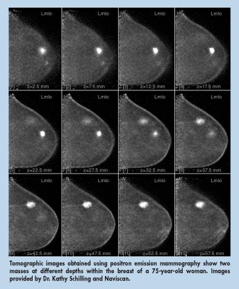
With an intrinsic resolution between 1.5 mm and 2 mm, PEM has obvious application as an adjunct in breast cancer diagnosis and possibly screening. The technology characterizes small lesions metabolically, potentially spotting malignancies at an earlier stage than any other technology, according to Dr. Schilling.
Advantages over MRI
Unlike MRI, which requires substantial training to interpret the enhancement patterns created using injected contrast media, expert interpretations using PEM come after experience with 5 to 10 cases, said Dr. Schilling, who teaches breast MRI to physicians interested in applying the modality.
"So many things light up in patients having MRIs, especially patients who are perimenopausal or on hormones," she said, noting that these problems do not affect PEM. "There are a lot fewer false positives on PEM, compared to breast MRI, and yet it is just as sensitive."
Physicians specializing in women's health are easily frustrated by breast MRI, Dr. Schilling said, not only by the difficulty in interpreting images but also in scheduling patients on a busy scanner.
"So this will be an alternative for these somewhat frustrated physicians," she said.
PEM camera
The PET cases at Boca Raton are captured using a small field of view PET camera from Naviscan PET Systems. The FDA-cleared device, called the PEM Flex Solo II (see photograph), needs no special shielding or wiring, and takes up about the same space as x-ray mammography equipment. PEM views of the breast obtained with the patient seated are comparable to those obtained using x-ray mammography, whereas MR imagescaptured when the patient is supineare difficult to match with mammograms.

Access to 18-fluorodeoxyglucose, the positron-emitting workhorse of the PET community, is a must, but if the radioisotope is there, so is the money. Reimbursement for FDG-exams of the breast is widely available, according to Dr. Schilling.
There is one big challenge, however. The PEM Flex Solo II does not image the axilla, which may harbor metastatic breast cancer. Dr. Schilling and her colleagues get around the problem by doing a whole body PET exam to look for these lesions.
"This means we are doing two exams and charging for only one," she said, "but if you want all the information, that's what you do."
Currently, physicians typically use just two modalities, mammography and ultrasound, to screen for breast cancer, referring patients with suspicious lesions for biopsy. Recent studies have shown the value of using MRI as an adjunct diagnostic aid or even as a screening tool.
Lumpectomy patients
Dr. Schilling said she believes PEM may be even better, at least under specific circumstances. She and her colleagues are conducting the research that may help determine whether this optimism is warranted.
They are focusing on presurgical planning in patients with newly diagnosed breast cancer who are considered candidates for lumpectomy after a full workup. PEM has the potential to find more and smaller malignancies, compared with breast MRI. Dr. Schilling wants to determine whether PEM, MRI, or both result in changes in surgical management, compared with conventional imaging, and to determine if the changes were appropriate, with histopathology as the gold standard.
The research protocol is also being carried out at four other medical centers: Anne Arundel Medical Center in Anne Arundel, Maryland; Scripps Cancer Center in San Diego, California; American Radiology Services/Johns Hopkins Green Spring in Lutherville, Maryland; and the University of North Carolina School of Medicine in Chapel Hill.
Dr. Schilling said she is confident that, when the results are tallied, PEM will be the clear choice in the diagnosis and screening of breast cancer.