Major technologic advances in genomics have made possible the identification of critical genetic alterations in cancer, which has led to a rapid paradigm shift in what is now considered personalized cancer treatment. This exponential growth in the understanding of cancer genomics is reshaping the drug development process, from drug discovery to novel designs of clinical trials. It is also exposing the critical importance of rigorous validation of molecular diagnostics platforms, such as the use of whole-exome sequencing and patient-derived xenografts, which may eventually be utilized to guide treatment decisions and may ultimately enable the true practice of personalized oncology. This review describes the achievements in therapeutic and molecular diagnostics, details evolving molecular platforms, and highlights the challenges for the translation of these developments to daily clinical practice.
Introduction
The central goal of personalized medicine is to tailor diagnosis and treatment to a patient’s individual biologic profile. In oncology, this concept has been reinvigorated by the substantial impact of therapeutic and diagnostic advancements. In this article, we provide a comprehensive review of personalized medicine initiatives in oncology, describing the major achievements in therapeutic and molecular diagnostics, and we highlight the challenges for the translation to daily clinical practice.
In recent years, technologic advances in genomics coupled with major multi-institutional initiatives have improved our understanding of tumor biology by revealing the major genetic alterations that drive cancer growth. For instance, the Cancer Genome Atlas Network, led by the National Institutes of Health (NIH), played a central role in the discovery of the landscape of genomic alterations that drive carcinogenesis across multiple malignancies, which provides a critical foundation for novel therapeutic approaches.[1-5] However, the results of these massive genetic, epigenetic, and transcriptomic analyses also revealed the astounding biologic heterogeneity observed among the histologic subtypes of each tumor, as well as the challenge of reconciling this information with the practice of personalized medicine. This exponential growth in the understanding of cancer genomics is not only reshaping the drug development process, leading to novel clinical trial designs, but it is also exposing the critical importance of rigorous validation of molecular diagnostics platforms that will be utilized to guide treatment decisions and ultimately make possible the practice of personalized oncology. Given the complexity, cost, and clinical implications for patients, these platforms should be required to demonstrate clear analytic validation, clinical validity (correlation of the presence of genetic alterations with clinical responses), and more importantly, clinical utility-by improving the outcomes of cancer patients. In this review, we discuss current concepts, advances, and future perspectives in personalized anticancer therapy.
Precision Medicine Victories
The growing knowledge base of tumor genomics has led to unprecedented advances in medical oncology. The evolving treatments of late-stage melanoma and non–small-cell lung cancer (NSCLC) exemplify these advances and provide the framework that is expanding into other histologies. The discovery of the importance of the driver gene mutation BRAF V600E, present in approximately half of patients with metastatic melanoma, provided the rationale for trials evaluating the efficacy of BRAF inhibitors in this disease.[6] The results of a phase III trial that included patients with metastatic BRAF V600E–mutated melanoma, randomized to treatment with the BRAF kinase inhibitor vemurafenib or dacarbazine, showed a significant increase in progression-free survival (PFS) among those who received vemurafenib.[7] The benefits of BRAF inhibition in this setting were confirmed in another phase III trial with a similar patient population treated with the BRAF inhibitor dabrafenib or dacarbazine.[8]
Advances in the molecular understanding of NSCLC have also fostered the development of several new approved therapies. Approximately 10% to 40% of lung adenocarcinomas harbor epidermal growth factor receptor (EGFR) aberrations (more than 80% of these mutations involve in-frame deletions within exon 19 or the L858R mutation within exon 21).[9] Currently, four tyrosine kinase inhibitors (erlotinib, gefitinib, afatinib, and osimertinib) are available for the treatment of EGFR-aberrant NSCLC, with overall response rates as high as 80% and reported median overall survival (OS) of more than 20 months.[10-12] In addition, rearrangements in the anaplastic lymphoma kinase (ALK) gene are present in about 7% of patients with NSCLC.[13] Patients with ALK-rearranged NSCLC who are treated with crizotinib achieve response rates estimated at 74%, with a 1-year OS rate of 84% in the first-line setting.[14] The second-generation ALK inhibitors ceritinib and alectinib have been approved for ALK-positive metastatic NSCLC in the second-line setting.
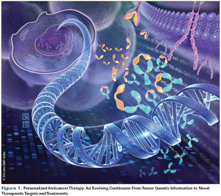
The incorporation of precision medicine into the practice of medical oncology is not only transforming treatment algorithms for several diseases, but it is also leading to a more collaborative management approach among disciplines, as demonstrated in molecular oncology tumor boards.[15,16] This interface allows for interactions between molecular pathologists, oncologists, hematologists, basic scientists, bioinformaticians, and genetic counselors, which ultimately strengthen the management and treatment recommendations for patients with advanced malignancies, who frequently have exhausted standard-of-care therapeutic options. These discussions also facilitate and expedite scientific investigation of potential novel therapeutic targets identified through genomic testing in clinical practice.[17] Furthermore, this approach serves to educate medical professionals about the nuances of complex genomic analysis and the growing number of molecular testing options, a critical development that will be further discussed below (Figure).
Tumor Genomics
Clinical application of next-generation sequencing
The widespread availability of genomic analysis of tumors in clinical practice has been primarily driven by the advent of next-generation sequencing (NGS). The term “NGS” includes several types of genomic sequencing methodologies, such as targeted sequencing or “hotspot panels,” whole-exome sequencing (WES), whole-genome sequencing (WGS), RNA sequencing (RNA-seq) (transcriptome), and bisulfite sequencing. For clinical applications, hotspot panels-as they have commonly become known-are rapidly becoming part of standard practice. These panels utilize massive multiplexed parallel sequencing to target predefined areas of the genome. The two most well-known NGS manufacturers, Ion Torrent and Illumina, offer the AmpliSeq panels and TruSeq panels, respectively, for targeted sequencing. The panels and associated library preparation kits can range in size from 20 to 400 genetic alterations (including insertions and deletions), but the most commonly used panels contain approximately 20 to 60 cancer-related genes and can utilize DNA from any source, including paraffin-embedded tissue. Although each company uses a fundamentally different technology to perform sequencing (semiconductor chips for Ion Torrent vs fluorescence detection for Illumina), analytic performance is very similar for the two platforms, with the exception of Ion Torrent’s ability to resolve homopolymers, which makes it less suitable for detecting large insertions.[18,19] The most significant differences between the platforms are associated with cost and throughput specifics that are more relevant to laboratory management than to clinical decisions.
Among a growing number of service providers in molecular pathology, Foundation Medicine performs targeted sequencing on approximately 360 cancer-related targeted genes plus introns from 28 genes often rearranged or altered in cancer.[20] These are considered laboratory-developed tests. As mentioned previously, similar-sized panels are available directly from NGS suppliers, but the bioinformatics processing required increases immensely with scaling in panel size, which makes application of such assays difficult for laboratories without significant informatics support. The challenge of how to handle the data can be prohibitive for many laboratories.
This discussion of varying panel sizes also brings up the idea of scale and specifically asks the question, what is the utility of more genomic information in treating cancer? Tumors harbor thousands of genetic (and epigenetic) alterations that are not included in the patient’s germline; however, only about 200 of the 20,000 genes in the human genome are currently considered to be “driver” genes relevant to tumorigenesis. Therefore, by focusing on “drivers,” the complexity of the cancer genome can be made intelligible. In addition, among the identified drivers, the subset that is actionable (ie, has targeting drug options) can be expected to increase.[21] This issue will grow more complicated as clinical WES, which provides information on 20,000 genes, continues to emerge as a potential diagnostic modality.[22]
The question of value added by WES was also posed in a study that compared it with targeted sequencing and array comparative genomic hybridization in a small cohort of patients. Out of the total 10 cases, the results showed only 2 targetable mutations detected that were found on all platforms; thus, no additional value for WES was demonstrated in this small cohort.[23] However, as more cancer-associated genes that can be targeted by new therapeutics are discovered, this value proposition can be expected to increase because targeted sequencing panels are inherently limited by the number of genes on the panel and the rate at which panels can be designed, since panels must account for interference between genes. Therefore, the rate at which targeted panels can be developed could be outpaced, at which point WES would become the favored platform.
Increasing the scale further to WGS adds another layer of clinical potential, as well as additional challenges. The actual physical sequencing itself and the informatics requirements of WGS are immense; however, so are the potential applications in precision medicine. The research consortium ENCODE (Encyclopedia of DNA Elements), led by the NIH, which is endeavoring to further understanding of regulatory elements of the human genome, has revealed the complexity that governs transcriptional regulation. The results from this initiative have the potential to advance knowledge of disease risk and prediction of therapeutic responses.
Two other key applications of NGS technology are RNA-seq and bisulfite sequencing for epigenomic profiling. Both applications are less commonly used than the targeted sequencing panels discussed above. Regarding bisulfite/methylation analysis: this is routinely used in the context of O6-methylguanine-DNA methyltransferase promoter methylation in glioblastoma but utilizes a pyrosequencing assay.[24] Methylation analysis by NGS is performed using a method called bisulfite amplicon sequencing and is still very much relegated to the research setting. However, preliminary clinical applications have revealed the identification of genome-wide hypomethylation from the serum of patients with various malignancies.[25,26]
RNA-seq data, often referred to as the transcriptome, have the potential for incredible clinical benefits because they can provide information on the presence of gene fusion products, as well as on gene amplification or downregulation. RNA-seq also provides a more functional understanding of the genome than does mutational analysis. The most basic example of this is through quantification of gene expression, but it can also identify and quantify specific gene isoforms that can have therapeutic ramifications. For example, vascular endothelial growth factor (VEGF)-commonly understood as a proangiogenic gene-has multiple isoforms with both proangiogenic and antiangiogenic activity.[27] Such biologic nuances add complexity to targeted treatment, given that the effects of an inhibitor could in theory vary according to the expression of these isoforms in specific tissues. Gene isoform analysis could also advance the understanding of pathogenesis and the classification of cancer into additional subtypes that better predict outcomes.[28]
The remarkable potential of this technology is balanced by numerous challenges. RNA-seq is limited by the fundamental fragility and instability of RNA derived from formalin-fixed, paraffin-embedded samples, which makes sequencing of RNA a far more challenging task than that of DNA. More important, though, is the biologic complexity. Single-cell sequencing has demonstrated differential gene expression related to therapeutic resistance within the same tumor, and similar methodologies used in tumor microenvironment studies have shown extensive variability in stromal and immune cells that can mediate processes such as immune surveillance, angiogenesis, and metastasis.[29,30] DNA sequencing by NGS cannot capture this type of information, which is critical to understanding the tumor microenvironment. However, trying to resolve this heterogeneity in a clinical setting that cannot support such time- and resource-intensive methodologies is prohibitive to the use of RNA-seq. For example, in mutational analysis, heavy inflammatory cell infiltration into a tumor decreases the mutation allele frequency detected by sequencing because there is less total tumor DNA. But in RNA-seq, such infiltration could potentially lead to inaccurate conclusions about the actual cancer cells’ transcriptome expression.
In general, as analysis methods become exceedingly more granular at the biologic level, the ability to extrapolate to the phenotypic level becomes inherently limited. As we will discuss in more depth later, integrated analysis methods that utilize multiple sources of data can potentially break through these limitations. Central to these approaches are advances in bioinformatics methods that make it possible to interpret the extensive data produced by such analyses. In the case of tumor heterogeneity, significant progress has been made in resolving mutational heterogeneity for the evaluation of clonal evolution, but these methods are limited to WES and WGS data, which are rarely used in the clinical setting.[31] Somewhat similar methods of deconvoluting gene expression profiles of individual cell types in heterogeneous tissue samples also exist, but the methods are arguably even more computationally intensive and are still in the early stages of development.[32] Nevertheless, major efforts have been directed to overcoming these challenges, and promising solutions that are in development could lead to clinical translation in the near future.[33] Widespread adoption of RNA-seq would likely lead to many new insights into treatment resistance and could play an important role in the treatment of patients.[34]
The restricted capacity of NGS platforms for the capture of alterations in epithelial-stromal interactions, angiogenesis, and immune modulation represents another major limitation. However, emerging evidence shows that the mutational profile of cancer cells influences communication with the surrounding environment. For example, patients with BRAF-mutant melanoma treated with BRAF inhibitors exhibited increased tumor infiltration by CD4+ and CD8+ lymphocytes on sequential tumor samples; the level of intratumoral CD8+ lymphocytes correlated with a reduction in tumor size and an increase in necrosis in posttreatment biopsies.[35] It has also been shown that the beta-catenin pathway in melanoma cells is associated with a lack of T-cell infiltration and resistance to anti–programmed death ligand 1 (PD-L1) and anti–cytotoxic T-lymphocyte–associated antigen 4 (CTLA-4) treatments.[36] These interactions between tumor genomic aberrations and the cancer immune microenvironment provide the impetus for clinical trials that incorporate advanced diagnostics concepts to guide therapeutic approaches, including sequencing of targeted therapies and immune checkpoint inhibitors.[37]
TO PUT THAT INTO CONTEXT
[[{"type":"media","view_mode":"media_crop","fid":"47778","attributes":{"alt":"","class":"media-image","id":"media_crop_5362711997660","media_crop_h":"0","media_crop_image_style":"-1","media_crop_instance":"5660","media_crop_rotate":"0","media_crop_scale_h":"0","media_crop_scale_w":"0","media_crop_w":"0","media_crop_x":"0","media_crop_y":"0","style":"height: 138px; width: 144px;","title":" ","typeof":"foaf:Image"}}]]
Razelle Kurzrock, MD
Center for Personalized Cancer Therapy, University of California, San Diego Moores Cancer Center, San Diego, CaliforniaWhat Are the Important ‘Take-Aways’ From This Review on Personalized Medicine?1. First, is it “personalized” medicine or “precision” medicine? In fact, it is both. We now have tools to define the precise defects present in each person’s tumor, and drugs that precisely target these defects-hence the term “precision medicine.” But it turns out that each patient with advanced disease has a complex and mostly unique molecular portfolio. Therefore, in order to be “precise,” we must “personalize” therapy.2. Assays are evolving at a breathtaking rate. Although 40- to 60-gene panels are often utilized, they may be missing substantial numbers of relevant alterations. Indeed, with a panel of ~200 genes, 90% of patients will have a potentially actionable alteration[1]-a far higher number than the ~20% often quoted for smaller panels.3. Omics may also be crucial for selecting patients for immunotherapy. Indeed, programmed death ligand 1 (PD-L1) is an excellent example of the imperfect, but still useful, dichotomy of single biomarkers: 0%–17% of PD-L1–negative patients vs 36%–100% of PD-L1–positive patients respond to PD-1 inhibitors.[2]4. While there is a need for collaboration and for exploitation of computer intelligence-indeed, computers may eventually give us completely validated answers-in the meantime, it is important to realize that the practice of medicine has never depended on perfect validation, but rather on highly trained physicians making informed decisions.REFERENCES1. Schwaederle M, Daniels GA, Piccioni DE, et al. On the road to precision cancer medicine: analysis of genomic biomarker actionability in 439 patients. Mol Cancer Ther. 2015;14:1488-94.2. Patel SP, Kurzrock R. PD-L1 expression as a predictive biomarker in cancer immunotherapy. Mol Cancer Ther. 2015;14:847-56.Financial Disclosure:Dr. Kurzrock has received research funding from Foundation Medicine, Genentech, Guardant, Merck Serono, Pfizer, and Sequenom; she has received consultant fees from Actuate Therapeutics and Sequenom; and she has an ownership interest in CureMatch, Inc, and Novena, Inc.
Evolving platforms in precision medicine
The biologic characterization of solid tumors has primarily been studied in the context of tissue biopsy samples, but this strategy is greatly limited by temporal and spatial intratumoral heterogeneity.[38] The plasticity of tumor biology, represented by clonal evolution and the accumulation of novel intratumoral genetic alterations that may mediate treatment resistance, and changes in the tumor microenvironment highlight the critical need for dynamic platforms capable of capturing this information.
Circulating tumor cells. Circulating tumor cells (CTCs) are defined as nucleated cells in the bloodstream that express epithelial cytokeratins and do not express the white blood cell surface antigen CD45.[39] Prospective results involving patients with metastatic breast cancer showed that higher CTC levels before the initiation of therapy and the failure of chemotherapy to reduce CTC levels (to < 5 CTCs per 7.5 mL of whole blood) after systemic therapy could predict shorter time to progression and shorter OS.[40] The predictive value of CTCs was also formally tested in the Southwest Oncology Group (SWOG) S0500 clinical trial in patients with metastatic breast cancer treated in the first-line setting.[41] In addition, enumeration of CTCs in patients with castration-resistant prostate cancer and metastatic colorectal cancer also showed significant correlation between high CTC levels and poor prognosis.[42-44] In addition to the quantitative enumeration of CTCs, qualitative analysis has also been performed in various disease settings to explore potential therapeutic targets, such as expression of human epidermal growth factor receptor 2 (HER2) in circulating breast cancer cells.[45]
Circulating tumorDNA. The analysis of circulating cell-free tumor DNA (cfDNA) from peripheral blood allows for serial sampling of a patient’s tumor DNA, which is essentially impossible by surgical biopsy. In addition, surgical biopsies, even if taken in several locations, are still limited because of tumor heterogeneity.[38] Liquid biopsies could potentially avoid the spatial and temporal limitations of surgical biopsies and provide frequent or dynamic molecular monitoring of cancer in the treatment and posttreatment settings. Such a practice could provide several potential clinical applications, including measuring response to therapy and disease recurrence, in addition to monitoring tumor evolution for the emergence of resistance genes. Of these applications, assessment of treatment response and early detection of disease recurrence are the closest to being incorporated into clinical practice, because of rapid developments in digital droplet polymerase chain reaction (ddPCR) technology.
The ddPCR technology can perform highly sensitive DNA mutation or structural variant quantification without calibration. This means that if a patient has a known driver mutation, such as KRAS, ddPCR can measure the amount of tumor DNA in circulation to gauge overall tumor burden or recurrence of disease.[46,47] The ddPCR technology can also be used in conjunction with NGS to increase overall assay sensitivity, using a methodology called tagged-amplicon deep sequencing (Tam-Seq).[48] This method was used in an observational study in which women with metastatic breast cancer were monitored with cfDNA, cancer antigen 125, and CTCs. cfDNA showed a correlation with tumor burden and early response.[49] Garcia-Murillas et al found that in a small cohort of patients with breast cancer who were treated with neoadjuvant chemotherapy, cfDNA detected in a single postoperative blood test was predictive of early relapse.[50] Similarly, in a prospective study, in which plasma samples were collected 4 to 10 weeks after surgery in a large cohort of patients with stage II colon cancer, those with detectable cfDNA had a shorter recurrence-free survival.[51] Taken together, these studies support the potential role of cfDNA in the posttreatment setting for evaluation of response to therapy and early detection of disease recurrence.
Genetic tumor profiling from cfDNA for the purpose of tracking tumor evolution and the emergence of resistance genes is a rapidly advancing technique that could obviate the need for additional tumor biopsies and might overcome the limitation of often inadequate archived tissue specimens. Murtaza et al used WES in a cohort of six patients with advanced cancer and were able to detect driver mutations, such as phosphatidylinositol-4,5-bisphosphate 3-kinase, catalytic subunit alpha (PIK3CA), which developed post-therapy along with the EGFR resistance mutation T790M in the serum of a patient, and which were not detectable in the tissue biopsy.[52] Clearly, more research on this topic will need to be performed; however, the utilization of cfDNA demonstrates the possibility of dynamically adjusting therapeutics in response to “real-time” molecular tumor evaluation.
“Omics.” Similar to the revolution in genomics, other “omics” fields have seen rapid advances from new profiling technologies. The disciplines of proteomics, lipidomics, and metabolomics have benefited greatly from improvements in mass spectrometry (MS) technologies, both in terms of hardware and software/informatics. Particularly for proteomics, advances in soft desorption MS, also known as “top-down proteomics,” have allowed for analysis of macromolecules in a way that was previously impossible with “bottom-up” methods that require analysis after fragmentation.[53] As an example, these advances have been applied to the screening of urine from prostate cancer patients, which has led to the discovery of novel protein markers and protein profiles with potential utility as screening or prognostic tools.[54,55] The emergence of lipidomics has been an even more recent advance than clinical proteomics because of the intrinsic difficulty of lipid analysis, which has required additional advances in MS technologies to become feasible.[56] One instance of a lipidomic application in oncology is the use of matrix-assisted laser desorption/ionization (MALDI)-MS analysis for biomarker discovery (MALDI is also used in microbiology laboratories). Kim et al showed differential lipid expression in HER2-positive breast cancer tissue that correlated with clinicopathologic outcomes.[57]
Metabolomics is an integrative systems biology approach that uses low-molecular-weight molecules; it includes certain classes of lipids and has several potential functions in precision medicine for cancer patients. As with standard lipidomics or proteomics, the most obvious application is in disease and biomarker discovery; this has been pursued in several tumor types, including colorectal, ovarian, and prostate cancer.[58-60] However, the potentially far more transformative function of metabolomics is its integration with pharmacogenomics into what has become known as pharmacometabolomics. Generally speaking, this integrative field involves defining pretreatment and posttreatment signatures that can potentially provide insight into treatment outcomes, as well as elucidating metabolomic pathways linked with phenotypic responses to certain therapeutics.[61] As an example, this application was used in patients with metastatic colon cancer in whom higher levels of low-density lipoprotein–derived lipids were predictive of toxicity with single-agent capecitabine.[62]
Ex vivo functional assays. Numerous types of chemotherapy sensitivity assays have been developed (eg, adenosine triphosphate [ATP]-based assay, ChemoFx assay, methylthiazolyldiphenyl-tetrazolium bromide assay, and extreme drug resistance assay) as part of early attempts to individualize treatment; however, the vast majority of the supportive studies have failed to show improved outcomes with their use.[63] Cree et al reported the results of a prospective trial of patients with advanced ovarian cancer in which the participants were randomized to an ATP-based tumor chemosensitivity assay or the physician’s choice of chemotherapy.[64] Among the 147 evaluable patients, 31.5% achieved a partial or complete response in the physician’s choice group compared with 40.5% in the assay-directed group. An intention-to-treat analysis showed a median PFS of 93 days in the physician’s choice group and 104 days in the assay-directed group, but without a statistically significant difference in OS. Second-generation assays that allow for the use of patient-derived tumor material, such as tissue organoids and CTCs cultivated in three-dimensional organotypic cultures, could improve the analytic validity of these assays because they may more closely mimic the original tumor environment.[65]
Ex vivo xenografts involving soft-tissue sarcoma specimens resected from patients and implanted into immunodeficient mice have been investigated in order to identify drug targets and drugs for clinical use.[66] The results of drug sensitivity testing were used to personalize cancer treatment. Of the 29 implanted tumors, 22 (76%) successfully engrafted, permitting the identification of treatment regimens for these patients. In fact, a correlation between xenograft results and clinical outcome was observed in 13 of 16 patients (81%). In another study, tumors resected from patients with refractory advanced cancers were engrafted in immunodeficient mice and treated with 63 drugs across 232 treatment regimens.[67] In the majority of cases, an effective treatment regimen was identified with the xenograft model and resulted in durable partial remission. These results are encouraging and are fostering additional investigation.
Impact of Precision Medicine on the Developmental Therapeutics Paradigm
The rapid advances in the genomic profiling of malignancies and in molecular diagnostics technologies have had a profound impact on the general infrastructure and strategy of developmental therapeutics programs. This impact can be seen from the computational modeling of compounds to the matching of specific targets identified through sequencing, and from the planning of preclinical experiments to the novel design of clinical trials. Some of the challenges of large, lengthy, phase III clinical trials that enroll thousands of patients to document a benefit in a small fraction have been resolved in part by pathway-driven clinical trials. In fact, a meta-analysis of 570 phase II oncology trials showed that the use of biomarker-based precision medicine strategies resulted in improved outcomes.[68]
A multivariable analysis of 641 single-agent treatment arms demonstrated that personalized therapy compared with nonpersonalized treatment was associated with a higher response rate (31% vs 10.5%), prolonged PFS (5.9 vs 2.7 months), and longer OS (13.7 vs 8.9 months).[68] Another meta-analysis of clinical trials of US Food and Drug Administration–approved anticancer drugs between 1998 and 2013 (57 randomized and 55 nonrandomized trials; total of 38,104 patients) also supported the benefit of a personalized strategy.[69] Specifically looking only at the experimental arms of 112 registration trials, personalized therapy led to a higher response rate (48% vs 23%), longer PFS (8.3 vs 5.5 months), and longer OS (19.3 vs 13.5 months). Nevertheless, the first prospective randomized trial that compared conventional treatment of advanced solid tumors with treatment with targeted agents selected based on the genomic profile of tumors showed disappointing results.[70]
The SHIVA trial randomized 195 patients with metastatic solid tumors (26 histologic types) and molecular alterations affecting three signaling pathways (estrogen/progesterone/androgen receptor, PI3K/AKT/mammalian target of rapamycin [mTOR], and RAF/MEK) to either matched approved targeted agents (including off-label indications of agents such as sorafenib, erlotinib, imatinib, vemurafenib, abiraterone, and letrozole) or physician’s choice.[70] Molecular analyses included assessment of mutations by NGS; gene copy number alterations; and expression of estrogen, progesterone, and androgen receptors by immunohistochemistry. This study failed to meet its primary endpoint, with a median PFS of 2.3 months in the experimental group vs 2 months in the control group. Numerous limitations might have contributed to these results, including potential lack of adequate matching of patients with multiple molecular alterations treated with single or inadequate targeted therapy, restricted number of signaling pathways, available approved drugs, and the challenge of prioritization of therapies with monotherapy. For instance, 55 patients (28%) assigned to one of the monotherapy groups had two or more potentially targetable molecular alterations. Despite the overall results of the SHIVA trial, this important proof-of-concept study highlights the critical need for further validation and refinement of the personalized treatment strategy, with close integration between clinical trial design and molecular diagnostics. Innovative clinical designs (ie, basket and umbrella trials) and major initiatives, such as the National Cancer Institute–sponsored Molecular Analysis for Therapy Choice (MATCH) and the American Society of Clinical Oncology Targeted Agent and Profiling Utilization Registry (TAPUR) trials, hold promise to further advance the field.
Basket trials are based on the premise that a molecular marker can predict tumor response to a targeted therapy independent of tumor histologic origin.[71] Two basket trials published in 2015 exemplify this strategy. Hyman et al conducted a phase II study that included patients with BRAF V600–mutated nonmelanoma cancers who were treated with the BRAF inhibitor vemurafenib.[72] The most frequent tumor responses were observed in patients with NSCLC (8 of 20 had partial responses) and those with Erdheim-Chester disease or Langerhans cell histiocytosis. Only one partial response was documented among the 27 patients with colorectal cancer treated with vemurafenib and cetuximab, although approximately half of these patients had a reduction in tumor size that did not meet the threshold for partial response. Few responses were also observed in the subgroups of patients with anaplastic thyroid cancer, cholangiocarcinoma, salivary duct cancer, soft-tissue sarcoma, and ovarian cancer.
A second and larger phase II trial included 647 patients with intrathoracic malignancies (NSCLC, small-cell lung cancer, and thymic malignancies). After tumor genomic profiling, patients received standard-of-care treatment or one of the following five treatments based on the genomic aberration identified: erlotinib (EGFR mutations), selumetinib (KRAS, NRAS, HRAS, or BRAF mutations), MK-2206 (PIK3CA, AKT, or PTEN mutations), lapatinib (ERBB2 mutations or amplifications), or sunitinib (KIT or PDGFRA mutations or amplifications).[73] Nearly half of the total number of patients had a genomic abnormality identified in one of the genes tested. However, only 45 patients were ultimately enrolled in one of the treatment arms (212 patients were considered screen failures: 68% related to previous use of erlotinib or early closure of the selumetinib arm). The study was not able to complete accrual to 13 of the 15 treatment arms because of several limitations, including the rare incidence of some genetic abnormalities in these thoracic malignancies (ie, ERBB2, PIK3CA, PTEN, AKT, KIT, PDGFRA), restricted number of genes analyzed and the number of histologies included in the design, variability of genomic tests performed, and delay in obtaining results.
These basket trials epitomize the clinical potential and challenge of departing from the paradigm of histology-based treatments to therapies tailored to specific genomic alterations across multiple histologies. The first study that focused on one genetic lesion (BRAF V600 mutation) demonstrated the feasibility of this approach and provided hypothesis-generating data of tumor responses in rare tumor subtypes that could potentially inform future trials. On the other hand, the second trial clearly shows the logistical difficulties of testing several actionable targets in a relatively small number of histologies in two cancer centers.
Umbrella trials represent another novel design in which patients with a single tumor type are screened for several aberrations at study entry and subsequently assigned to subprotocols based on the matched molecular abnormality identified.[74] This prescreening strategy allows for a lower screening-to-accrual ratio and may avoid early closure of studies involving patients whose tumors harbor low-frequency aberrations. This innovative strategy is being tested in the phase II/III LUNG-MAP (Biomarker-Driven Master Protocol for Second-Line Therapy of Squamous Cell Lung Cancer) study. The tumors of patients who have advanced squamous cell lung cancer with one previous treatment are screened for genetic alterations in more than 200 genes using targeted sequencing. As a result of this analysis, patients are then recommended for one of five subtrials within the umbrella framework. Of the five clinical trials, four are enrichment trials, with eligibility limited to those patients whose tumors harbor a specified genomic alteration in a gene that is directly targeted by a therapy in the clinical trial.[75,76]
The adaptive randomization design of phase II trials provides another strategy for accelerating the development of new cancer drugs matched to specific tumor aberrations. In these studies, regimens with higher Bayesian prediction probability of being more effective than standard therapy will be allowed to graduate from the trial with their corresponding biomarker signature. Regimens will be dropped if they show low probability of efficacy with any signature, resulting in smaller sample sizes compared with conventional clinical trials.[77] The I-SPY 2 neoadjuvant breast cancer trial exemplifies this strategy.[78] This phase II screening trial used adaptive randomization within biomarker subtypes to evaluate a series of novel agents/combinations when added to standard neoadjuvant therapy for women with high-risk stage II/III breast cancer. The primary endpoint was pathologic complete response (pCR) at surgery. Veliparib plus carboplatin met the 85% predictive probability criterion in the hormone receptor–negative cohort of only 72 patients, and there were 62 concurrently randomized controls. In triple-negative breast cancer, the pCR was 52% in the veliparib/carboplatin arm compared with 24% in the standard chemotherapy arm, with a 92% probability of success in a phase III trial. Confirmatory phase III data are pending.[79] These innovative clinical trial design strategies are aligned with advances in genomic profiling of tumors that hold the promise of transforming the developmental therapeutics landscape.
Critical Alignment of Tumor Biomarker Assay Development
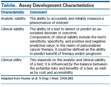
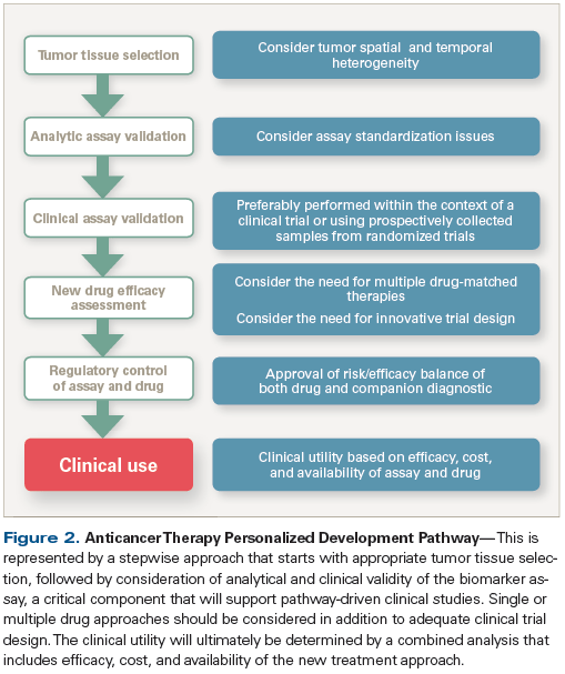
A tumor biomarker assay is any test that can measure or detect a tumor-related phenomenon. Its ultimate value to patients lies in its clinical utility. The optimal assay requires robust analytic validity and needs to impart information predictive of benefit from a given therapy. Its clinical utility also depends on easy access, low risk, and low cost of the test (Table).[80] Adequate clinical validity assessment-the process of predictive and/or prognostic information analysis of the assay-can be performed through retrospective observational studies or prospective clinical trials.[81] Methodologic constraints secondary to inadequate tumor samples, lack of standardized prospective tumor tissue and biological specimen collection, and lack of adjunct clinical data are some of the obstacles that have hampered the development of companion diagnostic assays in the past several years.
Integration of Precision Medicine With Immuno-Oncology
Immunotherapy with checkpoint inhibitors has made a significant impact on the treatment of melanoma and lung cancer. For instance, nivolumab has shown clinically meaningful activity in two phase III trials that included patients with NSCLC.[82,83] Nivolumab is a fully human immunoglobulin G4 programmed death 1 (PD-1) immune checkpoint inhibitor antibody that selectively blocks the interaction of the PD-1 receptor with its two known programmed death ligands, PD-L1 and PD-L2, disrupting the negative signal that regulates T-cell activation and proliferation.[84] In these trials patients received palliative treatment with nivolumab or docetaxel after platinum chemotherapy failure.[82,83] Nivolumab improved OS in both small-cell lung cancer patients (hazard ratio [HR], 0.73) and NSCLC patients (HR, 0.59). Intriguingly, tumor PD-L1 expression was not predictive of any efficacy endpoint in the small-cell lung cancer trial.[82] By contrast, regression modeling showed significant interaction between nivolumab treatment and all efficacy endpoints for patients with NSCLC expressing PD-L1.[83]
PD-L1 expression was also assessed in the phase III trial that compared nivolumab with dacarbazine in patients with metastatic melanoma without BRAF mutation.[85] Nivolumab improved outcomes in both PD-L1–positive and PD-L1–negative patients compared with dacarbazine, although higher tumor response rates were observed among those with positive PD-L1 status (52% vs 33%). Improved outcomes in renal cell carcinoma were also observed regardless of PD-L1 status in a large phase III study.[86] Accumulating evidence indicates that a subset of patients sustain prolonged responses and experience clinical benefit from immune checkpoint inhibitors across other tumor types, such as bladder cancer and ovarian carcinoma.[87,88] These results have fostered the search for predictive biomarkers of response to checkpoint inhibitors.
It is likely that future biomarkers will have to integrate both tumor and immune cell profiling in order to address the need for adequate patient selection. In support of this hypothesis, Le et al recently reported an improved benefit from pembrolizumab therapy in patients with metastatic solid tumors and increased genomic instability secondary to mismatch repair deficiency.[89] The underlying mechanistic hypothesis involved potential increased stimulation of the immune system against epitopes generated in the setting of increased genomic instability.[90] This study showed that mismatch repair status predicted clinical benefit of immune checkpoint blockade with pembrolizumab. Furthermore, Nagalla et al analyzed breast cancer tissue samples by hierarchical clustering of gene expression profiles, which revealed a large cluster of distant metastasis-free survival–associated genes with known immunologic functions.[91] Those genes were further subdivided into three distinct immune metagenes that likely reflected (1) B cells and/or plasma cells, (2) T cells and natural killer cells, and (3) monocytes and/or dendritic cells. Another area of intense investigation relates to the possible correlation between tumor neoantigen burden and response to immunotherapy. The ability to identify new epitopes linked to genetic aberrations is enabling researchers to explore neoantigen-specific T-cell therapy, with encouraging preliminary study results.[92,93] Two important studies have also recently shown a correlation between mutational burden in melanoma and NSCLC and improved outcomes from treatment with checkpoint inhibitors (ie, anti–CTLA-4 and anti–PD-1), paving the way for personalized immuno-oncology.[94,95]
Future Perspectives
Personalized therapy has been the cornerstone of cancer medicine in recent years, with numerous examples of unprecedented improvements in diagnostics and therapeutics. The widespread adoption of clinical NGS for solid tumor sequencing continues to generate a plethora of data regarding tumor biology, which subsequently allows for the potential development of more effective targeted therapies. Clearly, we are entering the age of true personalized medicine. However, if this review is to serve as a snapshot of the current state of precision medicine, a key take-away point is the challenge and potential of integrating the various topics discussed into coherent and synchronized clinical applications. Integration is a multifaceted issue that involves meticulous clinical validation along with continued advancements in informatics and computational technologies. The latter of these two topics is particularly interesting to consider. Medicine for the most part does not readily identify itself with computer science or artificial intelligence (predictive analytics). However, going forward, if oncologists want to optimize treatment for a patient based on integrating disparate sets of data from genomics, proteomics, metabolomics, and pharmacogenomics, they may need to incorporate artificial intelligence systems in their daily practice. Physicians will also have to guide the development of personalized medicine from its current state, in which most of the fields exist in individual silos, to clinical research settings designed to validate the new technologies, and incorporating the perspective of application beyond cancer therapeutics, with expansion to cancer prevention and early diagnosis.[96]
The 21st century of cancer medicine is clearly a time of optimism, but oncologists cannot avoid facing the challenges ahead, including the issues of tumor assay predictive validation, the resolution of tumor biologic heterogeneity, clinical trial design improvement, and regulatory points of contention (Figure 2). Personalized cancer therapies also carry the potential to forge and transform global scientific collaborations through sharing of therapeutic and diagnostic strategies, data, and standards of care that ultimately could expedite drug development efforts, facilitate progress in health education and health information technology, and inform public health policies.
Financial Disclosure:The authors have no significant financial interest in or other relationship with the manufacturer of any product or provider of any service mentioned in this article.
References:
1. Cancer Genome Atlas Research Network. Comprehensive molecular characterization of gastric adenocarcinoma. Nature. 2014;513:202-9.
2. Cancer Genome Atlas Network. Comprehensive molecular portraits of human breast tumours. Nature. 2012;490:61-70.
3. Cancer Genome Atlas Research Network. Comprehensive molecular characterization of clear cell renal cell carcinoma. Nature. 2013;499:43-9.
4. Cancer Genome Atlas Research Network. Comprehensive molecular profiling of lung adenocarcinoma. Nature. 2014;511:543-50.
5. Cancer Genome Atlas Research Network. Comprehensive molecular characterization of urothelial bladder carcinoma. Nature. 2014;507:315-22.
6. Jakob JA, Bassett RL Jr, Ng CS, et al. NRAS mutation status is an independent prognostic factor in metastatic melanoma. Cancer. 2012;118:4014-23.
7. Chapman PB, Hauschild A, Robert C, et al. Improved survival with vemurafenib in melanoma with BRAF V600E mutation. N Engl J Med. 2011;364:2507-16.
8. Hauschild A, Grob JJ, Demidov LV, et al. Dabrafenib in BRAF-mutated metastatic melanoma: a multicentre, open-label, phase 3 randomised controlled trial. Lancet. 2012;380:358-65.
9. Herbst RS, Heymach JV, Lippman SM. Lung cancer. N Engl J Med. 2008;359:1367-80.
10. Zhou C, Wu YL, Chen G, et al. Final overall survival results from a randomised, phase III study of erlotinib versus chemotherapy as first-line treatment of EGFR mutation-positive advanced non-small-cell lung cancer (OPTIMAL, CTONG-0802). Ann Oncol. 2015;26:1877-83.
11. Fukuoka M, Wu YL, Thongprasert S, et al. Biomarker analyses and final overall survival results from a phase III, randomized, open-label, first-line study of gefitinib versus carboplatin/paclitaxel in clinically selected patients with advanced non-small-cell lung cancer in Asia (IPASS). J Clin Oncol. 2011;29:2866-74.
12. Sequist LV, Yang JC, Yamamoto N, et al. Phase III study of afatinib or cisplatin plus pemetrexed in patients with metastatic lung adenocarcinoma with EGFR mutations. J Clin Oncol. 2013;31:3327-34.
13. Soda M, Choi YL, Enomoto M, et al. Identification of the transforming EML4-ALK fusion gene in non-small-cell lung cancer. Nature. 2007;448:561-6.
14. Solomon BJ, Mok T, Kim DW, et al. First-line crizotinib versus chemotherapy in ALK-positive lung cancer. N Engl J Med. 2014;371:2167-77.
15. Schwaederle M, Parker BA, Schwab RB, et al. Molecular tumor board: the University of California-San Diego Moores Cancer Center experience. Oncologist. 2014;19:631-6.
16. Tafe LJ, Gorlov IP, de Abreu FB, et al. Implementation of a molecular tumor board: the impact on treatment decisions for 35 patients evaluated at Dartmouth-Hitchcock Medical Center. Oncologist. 2015;20:1011-8.
17. Carneiro BA, Elvin JA, Kamath SD, et al. FGFR3-TACC3: a novel gene fusion in cervical cancer. Gynecol Oncol Rep. 2015;13:53-6.
18. Chen S, Li S, Xie W, et al. Performance comparison between rapid sequencing platforms for ultra-low coverage sequencing strategy. PLoS One. 2014;9:e92192.
19. Loman NJ, Misra RV, Dallman TJ, et al. Performance comparison of benchtop high-throughput sequencing platforms. Nat Biotechnol. 2012;30:434-9.
20. Frampton GM, Fichtenholtz A, Otto GA, et al. Development and validation of a clinical cancer genomic profiling test based on massively parallel DNA sequencing. Nat Biotechnol. 2013;31:1023-31.
21. Vogelstein B, Kinzler KW. The path to cancer-three strikes and you’re out. N Engl J Med. 2015;373:1895-8.
22. Van Allen EM, Wagle N, Stojanov P, et al. Whole-exome sequencing and clinical interpretation of formalin-fixed, paraffin-embedded tumor samples to guide precision cancer medicine. Nat Med. 2014;20:682-8.
23. Postel-Vinay S, Boursin Y, Massard C, et al. Seeking the driver in tumours with apparent normal molecular profile on comparative genomic hybridization and targeted gene panel sequencing: What is the added value of whole exome sequencing? Ann Oncol. 2016;27:344-52.
24. Thon N, Kreth S, Kreth FW. Personalized treatment strategies in glioblastoma: MGMT promoter methylation status. Onco Targets Ther. 2013;6:1363-72.
25. Masser DR, Stanford DR, Freeman WM. Targeted DNA methylation analysis by next-generation sequencing. J Vis Exp. 2015;96:52488.
26. Chan KC, Jiang P, Chan CW, et al. Noninvasive detection of cancer-associated genome-wide hypomethylation and copy number aberrations by plasma DNA bisulfite sequencing. Proc Natl Acad Sci USA. 2013;110:18761-8.
27. Biselli-Chicote PM, Oliveira AR, Pavarino EC, Goloni-Bertollo EM. VEGF gene alternative splicing: pro- and anti-angiogenic isoforms in cancer. J Cancer Res Clin Oncol. 2012;138:363-70.
28. Pal S, Gupta R, Davuluri RV. Alternative transcription and alternative splicing in cancer. Pharmacol Ther. 2012;136:283-94.
29. Kim KT, Lee HW, Lee HO, et al. Single-cell mRNA sequencing identifies subclonal heterogeneity in anti-cancer drug responses of lung adenocarcinoma cells. Genome Biol. 2015;16:127.
30. Brosseau JP, Lucier JF, Nwilati H, et al. Tumor microenvironment-associated modifications of alternative splicing. RNA. 2014;20:189-201.
31. Oesper L, Satas G, Raphael BJ. Quantifying tumor heterogeneity in whole-genome and whole-exome sequencing data. Bioinformatics. 2014;30:3532-40.
32. Li Y, Xie X. A mixture model for expression deconvolution from RNA-seq in heterogeneous tissues. BMC Bioinformatics. 2013;14(suppl 5):S11.
33. Cieslik M, Chugh R, Wu YM, et al. The use of exome capture RNA-seq for highly degraded RNA with application to clinical cancer sequencing. Genome Res. 2015;25:1372-81.
34. Bardelli A, Corso S, Bertotti A, et al. Amplification of the MET receptor drives resistance to anti-EGFR therapies in colorectal cancer. Cancer Discov. 2013;3:658-73.
35. Wilmott JS, Long GV, Howle JR, et al. Selective BRAF inhibitors induce marked T-cell infiltration into human metastatic melanoma. Clin Cancer Res. 2012;18:1386-94.
36. Spranger S, Bao R, Gajewski TF. Melanoma-intrinsic beta-catenin signalling prevents anti-tumour immunity. Nature. 2015;523:231-5.
37. Atkins MB, Larkin J. Immunotherapy combined or sequenced with targeted therapy in the treatment of solid tumors: current perspectives. J Natl Cancer Inst. 2016 Feb 2. [Epub ahead of print]
38. Gerlinger M, Rowan AJ, Horswell S, et al. Intratumor heterogeneity and branched evolution revealed by multiregion sequencing. N Engl J Med. 2012;366:883-92.
39. Ignatiadis M, Lee M, Jeffrey SS. Circulating tumor cells and circulating tumor DNA: challenges and opportunities on the path to clinical utility. Clin Cancer Res. 2015;21:4786-800.
40. Cristofanilli M, Budd GT, Ellis MJ, et al. Circulating tumor cells, disease progression, and survival in metastatic breast cancer. N Engl J Med. 2004;351:781-91.
41. Smerage JB, Barlow WE, Hortobagyi GN, et al. Circulating tumor cells and response to chemotherapy in metastatic breast cancer: SWOG S0500. J Clin Oncol. 2014;32:3483-9.
42. Scher HI, Heller G, Molina A, et al. Circulating tumor cell biomarker panel as an individual-level surrogate for survival in metastatic castration-resistant prostate cancer. J Clin Oncol. 2015;33:1348-55.
43. de Bono JS, Scher HI, Montgomery RB, et al. Circulating tumor cells predict survival benefit from treatment in metastatic castration-resistant prostate cancer. Clin Cancer Res. 2008;14:6302-9.
44. Cohen SJ, Punt CJ, Iannotti N, et al. Relationship of circulating tumor cells to tumor response, progression-free survival, and overall survival in patients with metastatic colorectal cancer. J Clin Oncol. 2008;26:3213-21.
45. Pestrin M, Bessi S, Puglisi F, et al. Final results of a multicenter phase II clinical trial evaluating the activity of single-agent lapatinib in patients with HER2-negative metastatic breast cancer and HER2-positive circulating tumor cells. A proof-of-concept study. Breast Cancer Res Treat. 2012;134:283-9.
46. Diehl F, Diaz LA Jr. Digital quantification of mutant DNA in cancer patients. Curr Opin Oncol. 2007;19:36-42.
47. Nygaard AD, Holdgaard PC, Spindler KL, et al. The correlation between cell-free DNA and tumour burden was estimated by PET/CT in patients with advanced NSCLC. Br J Cancer. 2014;110:363-8.
48. Forshew T, Murtaza M, Parkinson C, et al. Noninvasive identification and monitoring of cancer mutations by targeted deep sequencing of plasma DNA. Sci Transl Med. 2012;4:136ra68.
49. Dawson SJ, Tsui DW, Murtaza M, et al. Analysis of circulating tumor DNA to monitor metastatic breast cancer. N Engl J Med. 2013;368:1199-209.
50. Garcia-Murillas I, Schiavon G, Weigelt B, et al. Mutation tracking in circulating tumor DNA predicts relapse in early breast cancer. Sci Transl Med. 2015;7:302ra133.
51. Tie J, Kinde I, Wang Y, et al. Circulating tumor DNA (ctDNA) as a marker of recurrence risk in stage II colon cancer (CC). J Clin Oncol. 2014;32(suppl 5s):abstr 11015.
52. Murtaza M, Dawson SJ, Tsui DW, et al. Non-invasive analysis of acquired resistance to cancer therapy by sequencing of plasma DNA. Nature. 2013;497:108-12.
53. Cravatt BF, Simon GM, Yates JR 3rd. The biological impact of mass-spectrometry-based proteomics. Nature. 2007;450:991-1000.
54. Kim Y, Ignatchenko V, Yao CQ, et al. Identification of differentially expressed proteins in direct expressed prostatic secretions of men with organ-confined versus extracapsular prostate cancer. Mol Cell Proteomics. 2012;11:1870-84.
55. Principe S, Jones EE, Kim Y, et al. In-depth proteomic analyses of exosomes isolated from expressed prostatic secretions in urine. Proteomics. 2013;13:1667-71.
56. Stock J. The emerging role of lipidomics. Atherosclerosis. 2012;221:38-40.
57. Kim IC, Lee JH, Bang G, et al. Lipid profiles for HER2-positive breast cancer. Anticancer Res. 2013;33:2467-72.
58. Zhang A, Sun H, Yan G, et al. Metabolomics in diagnosis and biomarker discovery of colorectal cancer. Cancer Lett. 2014;345:17-20.
59. Buas MF, Gu H, Djukovic D, et al. Identification of novel candidate plasma metabolite biomarkers for distinguishing serous ovarian carcinoma and benign serous ovarian tumors. Gynecol Oncol. 2016;140:138-44.
60. Giskeodegard GF, Hansen AF, Bertilsson H, et al. Metabolic markers in blood can separate prostate cancer from benign prostatic hyperplasia. Br J Cancer. 2015;113:1712-9.
61. Kaddurah-Daouk R, Weinshilboum R. Metabolomic signatures for drug response phenotypes: pharmacometabolomics enables precision medicine. Clin Pharmacol Ther. 2015;98:71-5.
62. Backshall A, Sharma R, Clarke SJ, Keun HC. Pharmacometabonomic profiling as a predictor of toxicity in patients with inoperable colorectal cancer treated with capecitabine. Clin Cancer Res. 2011;17:3019-28.
63. Burstein HJ, Mangu PB, Somerfield MR, et al. American Society of Clinical Oncology clinical practice guideline update on the use of chemotherapy sensitivity and resistance assays. J Clin Oncol. 2011;29:3328-30.
64. Cree IA, Kurbacher CM, Lamont A, et al; TCA Ovarian Cancer Trial Group. A prospective randomized controlled trial of tumour chemosensitivity assay directed chemotherapy versus physician's choice in patients with recurrent platinum-resistant ovarian cancer. Anticancer Drugs. 2007;18:1093-101.
65. Friedman AA, Letai A, Fisher DE, Flaherty KT. Precision medicine for cancer with next-generation functional diagnostics. Nat Rev Cancer. 2015;15:747-56.
66. Stebbing J, Paz K, Schwartz GK, et al. Patient-derived xenografts for individualized care in advanced sarcoma. Cancer. 2014;120:2006-15.
67. Hidalgo M, Bruckheimer E, Rajeshkumar NV, et al. A pilot clinical study of treatment guided by personalized tumorgrafts in patients with advanced cancer. Mol Cancer Ther. 2011;10:1311-6.
68. Schwaederle M, Zhao M, Lee JJ, et al. Impact of precision medicine in diverse cancers: a meta-analysis of phase II clinical trials. J Clin Oncol. 2015;33:3817-25.
69. Jardim DL, Schwaederle M, Wei C, et al. Impact of a biomarker-based strategy on oncology drug development: a meta-analysis of clinical trials leading to FDA approval. J Natl Cancer Inst. 2015 Sep 15. [Epub ahead of print]
70. Le Tourneau C, Delord JP, Goncalves A, et al. Molecularly targeted therapy based on tumour molecular profiling versus conventional therapy for advanced cancer (SHIVA): a multicentre, open-label, proof-of-concept, randomised, controlled phase 2 trial. Lancet Oncol. 2015;16:1324-34.
71. Redig AJ, Janne PA. Basket trials and the evolution of clinical trial design in an era of genomic medicine. J Clin Oncol. 2015;33:975-7.
72. Hyman DM, Puzanov I, Subbiah V, et al. Vemurafenib in multiple nonmelanoma cancers with BRAF V600 mutations. N Engl J Med. 2015;373:726-36.
73. Lopez-Chavez A, Thomas A, Rajan A, et al. Molecular profiling and targeted therapy for advanced thoracic malignancies: a biomarker-derived, multiarm, multihistology phase II basket trial. J Clin Oncol. 2015;33:1000-7.
74. Rodon J, Saura C, Dienstmann R, et al. Molecular prescreening to select patient population in early clinical trials. Nat Rev Clin Oncol. 2012;9:359-66.
75. Herbst RS, Gandara DR, Hirsch FR, et al. Lung Master Protocol (Lung-MAP)-a biomarker-driven protocol for accelerating development of therapies for squamous cell lung cancer: SWOG S1400. Clin Cancer Res. 2015;21:1514-24.
76. Steuer CE, Papadimitrakopoulou V, Herbst RS, et al. Innovative clinical trials: the LUNG-MAP study. Clin Pharmacol Ther. 2015;97:488-91.
77. Berry DA. Bayesian clinical trials. Nat Rev Drug Discov. 2006;5:27-36.
78. Rugo HS, Olopade O, DeMichele A, et al. Veliparib/carboplatin plus standard neoadjuvant therapy for high-risk breast cancer: first efficacy results from the I-SPY 2 trial. Cancer Res. 2013;73(suppl 24):abstr S5-02.
79. Glimelius B, Lahn M. Window-of-opportunity trials to evaluate clinical activity of new molecular entities in oncology. Ann Oncol. 2011;22:1717-25.
80. Hunter DJ, Khoury MJ, Drazen JM. Letting the genome out of the bottle-will we get our wish? N Engl J Med. 2008;358:105-7.
81. Simon RM, Paik S, Hayes DF. Use of archived specimens in evaluation of prognostic and predictive biomarkers. J Natl Cancer Inst. 2009;101:1446-52.
82. Brahmer J, Reckamp KL, Baas P, et al. Nivolumab versus docetaxel in advanced squamous-cell non-small-cell lung cancer. N Engl J Med. 2015;373:123-35.
83. Borghaei H, Paz-Ares L, Horn L, et al. Nivolumab versus docetaxel in advanced nonsquamous non-small-cell lung cancer. N Engl J Med. 2015;373:1627-39.
84. Wang C, Thudium KB, Han M, et al. In vitro characterization of the anti-PD-1 antibody nivolumab, BMS-936558, and in vivo toxicology in non-human primates. Cancer Immunol Res. 2014;2:846-56.
85. Robert C, Long GV, Brady B, et al. Nivolumab in previously untreated melanoma without BRAF mutation. N Engl J Med. 2015;372:320-30.
86. Motzer RJ, Escudier B, McDermott DF, et al. Nivolumab versus everolimus in advanced renal cell carcinoma. N Engl J Med. 2015;373:1803-13.
87. Powles T, Eder JP, Fine GD, et al. MPDL3280A (anti-PD-L1) treatment leads to clinical activity in metastatic bladder cancer. Nature. 2014;515:558-62.
88. Hamanishi J, Mandai M, Ikeda T, et al. Safety and antitumor activity of anti-PD-1 antibody, nivolumab, in patients with platinum-resistant ovarian cancer. J Clin Oncol. 2015;33:4015-22.
89. Le DT, Uram JN, Wang H, et al. PD-1 blockade in tumors with mismatch-repair deficiency. N Engl J Med. 2015;372:2509-20.
90. Segal NH, Parsons DW, Peggs KS, et al. Epitope landscape in breast and colorectal cancer. Cancer Res. 2008;68:889-92.
91. Nagalla S, Chou JW, Willingham MC, et al. Interactions between immunity, proliferation and molecular subtype in breast cancer prognosis. Genome Biol. 2013;14:R34.
92. Tran E, Turcotte S, Gros A, et al. Cancer immunotherapy based on mutation-specific CD4+ T cells in a patient with epithelial cancer. Science. 2014;344:641-5.
93. Carreno BM, Magrini V, Becker-Hapak M, et al. Cancer immunotherapy. A dendritic cell vaccine increases the breadth and diversity of melanoma neoantigen-specific T cells. Science. 2015;348:803-8.
94. Snyder A, Makarov V, Merghoub T, et al. Genetic basis for clinical response to CTLA-4 blockade in melanoma. N Engl J Med. 2014;371:2189-99.
95. Rizvi NA, Hellmann MD, Snyder A, et al. Cancer immunology. Mutational landscape determines sensitivity to PD-1 blockade in non-small cell lung cancer. Science. 2015;348:124-8.
96. Kensler TW, Spira A, Garber JE, et al. Transforming cancer prevention through precision medicine and immune-oncology. Cancer Prev Res (Phila). 2016;9:2-10.


