PET Imaging: Update on Sarcomas
Sarcomas are a group of tumors with highly variable character istics and clinical outcomes. Their locations in almost all body locations present unique challenges for diagnosis and management. These challenges have presented opportunities for evaluation and validation of new imaging techniques. Positron-emission tomography (PET) has been evaluated for use in cancer over the years, and in particular, it has been evaluated in sarcoma diagnosis and treatment evaluation.
Sarcomas are a group of tumors with highly variable character istics and clinical outcomes. Their locations in almost all body locations present unique challenges for diagnosis and management. These challenges have presented opportunities for evaluation and validation of new imaging techniques. Positron-emission tomography (PET) has been evaluated for use in cancer over the years, and in particular, it has been evaluated in sarcoma diagnosis and treatment evaluation.
PET is unique in its capabilities for cancer imaging. It generates high-resolution three-dimensional images and utilizes radiotracers specific for biologic processes. Fluorine-18 fluorodeoxyglucose (FDG) is the most commonly used imaging agent in cancer clinical imaging practice. FDG is used to report regional tissue metabolism. Because the positron radiolabel has such high imaging energies, direct measurements of radiotracer concentration in tissue can be made. This important characteristic is the basis for determinations of tumor grade, quantitation of changes in response to treatment, and patient outcome prediction that are made with FDG imaging.
Optimum Parameters for FDG Imaging
FIGURE 1

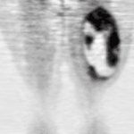
Thigh Sarcoma
The most commonly used measurement of tissue-uptake quantitation for a PET radiotracer is the standardized uptake variable (SUV). This the uptake in a tissue region such as a tumor mass, which is normalized for the amount of radioactivity injected, patient weight, and tissue background activity. SUV is a robust value for comparison of one imaging study to another, the same patient at a later time, and patients in different groups. However, it can be affected by imaging and patient parameters such as patient serum glucose, and imaging time after FDG administration.
Generally patients are asked to fast for at least 5 hours prior to the study, and serum glucose levels should be 150 mg/mL or less for the most reliable imaging results. This optimization is for uptake suppression of metabolically active tissues such as the myocardium and other muscles, reduced suppression of tumor uptake, and reliability of the SUV measurement. The absolute upper limit for serum glucose that allows for reliable imaging is 200 mg/dL. The serum glucose level is assessed prior to administration of FDG, as is a brief history of when the most recent meal was consumed.
These optimum parameters for FDG imaging in cancer were recently described in a report of a National Cancer Institute (NCI) consensus committee, where standard techniques for the use of FDG as a biomarker for cancer treatment are presented.[1,2] The use of FDG-PET as a biomarker or surrogate endpoint for patient outcome is the basis for clinical research studies aimed at determining the sensitivity and specificity of the method for tumor staging and grade establishment, following response to treatment, and assessing normal tissue damage as a result of treatment.
SUV and Tumor Characteristics
FIGURE 2

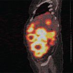
Midabdominal GIST
One of the first comparisions of FDG-PET quantitative imaging measurements with the now clinically used SUV was performed in sarcomas. A group of about 70 patients underwent full 1-hour dynamic imaging with rapid blood sampling after FDG infusion to establish the tumor FDG metabolic rate with Patlak analysis methods (a comparison of the relative rates of blood clearance and tumor uptake). SUV measurements of tumor and normal tissues were made from the same image sets. The investigators found that the FDG metabolic rate and SUV had a linear relationship throughout most of the uptake ranges present in a mixed group of sarcomas. This work formed the basis for the use of FDG SUV to explore sarcoma biology and patient outcome prediction.[3]
Later work focused on the use of FDG-PET to determine malignancy in bone and soft-tissue tumors. Several investigators found that tumor FDG uptake correlates with tumor grade in patient groups with diverse sarcomas.[4-7] The most significant discrimination ability for the use of the tumor FDG SUV was in distinguishing low- from high-grade tumors. Using this imaging agent, there was less ability to distinguish low-grade from benign tumors, as in the case of lipomatous tumors.[8] Folpe et al examined a mixed group of sarcomas and found that the FDG SUV was correlated with mitosis rate and cellularity.[9]
These relationships of FDG-PET SUV to tumor characteristics can be used clinically to assist in accurate diagnosis. Sarcomas are often large, with heterogenous tissue characteristics. Random or convenient choice of biopsy site may not always report the most aggressive tumor areas. Tumor heterogeneity is a common cause of error in sarcoma tumor grade and histologic type for treatment planning. The FDG-PET image provides a complete assessment of the tumor and can indicate the most aggressive areas for biopsy, seen as areas of more intense FDG uptake.
Figure 1 shows an example of a tumor with spatial heterogeneity in FDG uptake. Newer PET imaging devices utilize a computed tomography (CT) scan to generate the attenuation correction portion of the PET emission scan. This image can also be used for further anatomic localization of sites of abnormal tissue FDG uptake and has become quite popular for clinical imaging in all cancer types.
Treatment Response
The ability of FDG-PET to identify treatment response is an important goal for the sarcoma patient population. These tumors often do not change size in response to neoadjuvant chemotherapy because they can be made up of tissue elements that are static to the tumor response, or very slow in size reduction, such as bone, cartilage, scar, and myxoid areas. Consequently, the Response Evaluation Criteria in Solid Tumors (RECIST criteria) for treatment response do not apply well to this group.[10,11] Newer therapies directed at specific molecular targets may be cytostatic and result in tumor growth arrest, which may indicate effective therapy for a patient, as opposed to direct cell-killing mechanisms and tumor shrinkage. In fact, early detection of treatment response that indicates improved patient outcome for newer molecular-targeted therapies is an area of active research.
The most dramatic example of using FDG-PET as a biomarker for treatment response assessment is in gastrointestinal stromal tumors (GIST) treated with imatinib mesylate (Gleevec). GIST are FDG-avid and can yield impressive PET images (Figure 2). Goerres et al found that the median survival of patients who demonstrated an FDG-PET response was 100% at 2 years compared to a group with residual tumor uptake after treatment. The study also demonstrated the ability to separate patients by time to tumor progression based on tumor FDG-uptake levels.[12]
Early response to imatinib in the GIST population detected by FDG-PET has also been shown. As much as a 65% decrease in tumor FDG uptake was demonstrated at the end of 1 week of effective therapy, and other groups have found as high as a 95% response detected by 1 month after treatment initiation. Response detection using CT criteria was less accurate, with no significant CT responses noted in FDG-responsive patients.[13-15] These investigators recommend FDG-PET imaging for GIST patients at baseline to observe for maximum tumor activity levels and for accurate staging. Repeat imaging is suggested in the first month after therapy initiation to observe for response and to predict treatment effect. Another image may be helpful if treatment resistance is suspected, and a new baseline tumor uptake and location for treatment observation needs to be established.
Treatment response in other sarcomas using FDG-PET has also been demonstrated. In an extremity soft-tissue sarcoma group treated with doxorubicin-based neoadjuvant chemotherapy, Scheutze et al showed that separating patients by their FDG response (> 40%) showed a significant difference in survival between the two groups.[16] Patients in the FDG-PET nonresponse group had a 90% risk of disease recurrence at 4 years compared to FDG responders.
These data, and those of others, have provided an argument for the effectiveness and survival increase in soft-tissue sarcoma patients treated with neoadjuvant chemotherapy prior to tumor resection. This imaging technique can also be used to identify patients with tumor resistance during the course of therapy, who might benefit from treatment intensification, or early resection. A similar finding has been shown in the Ewing's sarcoma population reported by Hawkins et al.[17] Patients whose tumors showed increased SUV ratios between scans at baseline and preresection (after neoadjuvant chemotherapy) had significantly improved survival. In the future, good responders in this patient group may be identified for less toxic treatment protocols.
Patient Outcome
Since the sarcoma FDG uptake determined by PET has been shown to be correlated with tumor grade, mitotic rate, and cellularity, several investigators have evaluated the ability of this technique to predict patient outcome. Eary and her group evaluated the outcome of a large group of sarcoma patients with different histologic tumor types and grades, and examined the relationship of tumor FDG SUV and patient disease recurrence and survival.[18] The FDG SUV in this patient group was found to be an independent predictor of overall and event-free survival in a multivariate analysis that included standard patient and tumor clinical variables. This value also showed a dose-response relationship. For every integer increase above the group SUV median, there was a 20% increase in risk of decreased survival. Schwarzbach et al showed a similar result in a group of patients with respectable soft-tissue sarcomas.[19]
Future Directions
The use of PET can be extended with the employment of other PET radiopharmaceuticals that are more specific than FDG for biologically relevant tumor aspects. Today, agents that report on aspects of tumors that contribute to tumor resistance and biologic aggressiveness are under study in the sarcoma patient population. For example, there are imaging agents that can be used to determine levels of tumor hypoxia.[20,21] This tumor condition is known to confer resistance to both radio- and chemotherapy. Newer therapy strategies that target hypoxic tumor tissues after identification of these regions may improve patient outcome.
Given the tumor heterogeneity (and poor survival for high-grade histologic types), sarcoma patients are likely to benefit from these more specific imaging techniques. Molecular imaging research efforts in PET are being directed toward identifying tumor characteristics that can be used to define patient risk for aggressive disease behavior and for noninvasive treatment monitoring. With PET radiopharmaceuticals, most aspects of tumor cell biology, specific receptors, and therapy targets can be imaged and quantitated noninvasively, and repeatedly. This type of imaging will likely play a large role in individualizing therapy for sarcoma patients in the near future.
Financial Disclosure:The authors have no significant financial interest or other relationship with the manufacturers of any products or providers of any service mentioned in this article.
These articles are part of a series based on the proceedings of a special symposium of the Intergroup Coalition Against Sarcomas, "Progress in Translational and Clinical Research for Sarcomas of Adults," which was held October 8, 2006, in Seattle. The series is guest edited by Margaret von Mehren, MD, Director of Sarcoma Oncology at Fox Chase Cancer Center, Philadelphia.
References:
References
1. Shankar LK, Hoffman JM, Bacharach S, et al, for the National Cancer Institute: Consensus recommendations for the use of 18F-FDG PET as an indicator of therapeutic response in patients in National Cancer Institute Trials. J Nucl Med 47:1059-1066, 2006.
2. Kelloff GJ, Hoffman JM, Johnson B, et al: Progress and promise of FDG-PET imaging for cancer patient management and oncologic drug development. Clin Cancer Res 11:2785-2808, 2005.
3. Eary JF, Mankoff DA: Tumor metabolic rates in sarcoma using FDG PET. J Nucl Med 39:250-254, 1998.
4. Eary JF, Conrad EU, Bruckner JD, et al: Quantitative [F-18] fluorodeoxyglucose positron emission tomography in pretreatment and grading of sarcoma. Clin Can Res 4:1215-1220, 1998.
5. Adler LP, Blair HF, Makley JT, et al: Non-invasive grading of musculoskeletal tumors using PET. J Nucl Med 32:1508-1512, 1990.
6. Schwarzbach M, Willeke F, Dimitrakopoulou Strauss A, et al: Functional imaging and detection of local recurrence in soft tissue sarcomas by positron emission tomography. Anticancer Res 19:1343-1349, 1999.
7. Griffeth LK, Dedashti F, McGuire AH, et al: PET evaluation of soft tissue masses with fluorine-18 fluoro-2-deoxy-glucose. Radiology 182:185-194, 1992.
8. Nieweg OE, Pruim J, van Ginkel RJ, et al: Fluorine-18-fluorodeoxyglucose PET imaging of soft-tissue sarcoma. J Nucl Med 37:257-261, 1996.
9. Folpe AL, Lyles RH, Sprouse JT, et al: (F-18) fluorodeoxyglucose positron emission tomography as a predictor of pathologic grade and other prognostic variables in bone and soft tissue sarcoma. Clin Cancer Res 6:1279-1287, 2000.
10. Therasse P, Arbuck SG, Eisenhauer EA, et al: New guidelines to evaluate the response to treatment in solid tumors. J Natl Cancer Inst 92:205-216, 2000.
11. Ratain MJ, Eckhardt SG: Phase II studies of modern drugs directed against new target: If you are fazed, too, then resist RECIST. J Clin Oncol 22:4442-4445, 2004.
12. Goerres GW, Stupp R, Barghouth G, et al: The value of PET, CT and in-line PET/CT in patients with gastrointestinal stromal tumors: Long-term outcome of treatment with imatinib mesylate. Eur J Med Mol Imaging 32:153-162, 2005.
13. Antoch G, Kanja J, Bauer S, et al: Comparison of PET, CT, and dual-modality PET/CT imaging for monitoring of imatinib (STI571) therapy in patients with gastrointestinal stromal tumors. J Nucl Med 45:357-365, 2004.
14. Gayed I, Vu T, Johnson M, et al: Comparison of bone and 2-deoxy-2-[18F]fluoro-D-glucose positron emission tomography in the evaluation of bony metastases in lung cancer. Mol Imaging Biol 5:26-31, 2003.
15. Stroobants S, Goeminne J, Seegers M, et al: 18-FDG-positron emission tomography for the early prediction of response in advanced soft tissue sarcoma treated with imatinib mesylate (Glivec). Eur J Cancer 39:2012-2020, 2003.
16. Schuetze SM, Rubin BP, Vernon C, et al: Use of positron emission tomography in localized extremity soft tissue sarcoma treated with neoadjuvant chemotherapy. Cancer 103:339-348, 2005.
17. Hawkins DS, Schuetze SM, Butrynski JE, et al: [18F]Fluorodeoxyglucose positron emission tomography predicts outcome for Ewing sarcoma family of tumors. J Clin Oncol 23:8828-8834, 2005.
18. Eary JF, O'Sullivan F, Powitan Y, et al: Sarcoma tumor FDG uptake measured by PET and patient outcome: A retrospective analysis. Eur J Nucl Med 29:1149-1154, 2002.
19. Schwarzbach MHM, Hinz U, Dimitrakopoulou-Strauss A, et al: Prognostic significance of preoperative [18-F] fluorodeoxyglucose (FDG) positron emission tomography (PET) imaging in patients with resectable soft tissue sarcomas. Ann Surg 241:286-294, 2005.
20. Rasey JS, Koh WJ, Evans ML, et al: Quantifying regional hypoxia in human tumors with positron emission tomography of [F-18]fluoromisonidazole: a pre-therapy study of 37 patients. Int J Radiat Oncol Biol Phys 36:417-428, 1996.
21. Rajendran JG, Krohn KA: Imaging hypoxia and angiogenesis in tumors. Radiol Clin North Am 43:169-187, 2005.
Sarcoma Awareness Month 2023 with Brian Van Tine, MD, PhD
August 1st 2023Brian Van Tine, MD, PhD, speaks about several agents and combination regimens that are currently under investigation in the sarcoma space, and potential next steps in research including immunotherapies and vaccine-based treatments.