Sentinel Lymph Node Dissection in Vulvar Carcinoma: What Is the Acceptable False-Negative Rate?
Although vulvar cancer is relatively rare, accounting for less than 5% of all cancers of the female genital organs, lymph node metastasis associated with vulvar carcinoma is a common event and occurs in about 25% of cases.[1] The presence and number of lymph node metastases is the single most important prognostic factor in vulvar cancer and a critical component to the International Federation of Gynecology and Obstetrics (FIGO) staging system, as well as a major determinant in the need for adjuvant therapy
Although vulvar cancer is relatively rare, accounting for less than 5% of all cancers of the female genital organs, lymph node metastasis associated with vulvar carcinoma is a common event and occurs in about 25% of cases.[1] The presence and number of lymph node metastases is the single most important prognostic factor in vulvar cancer and a critical component to the International Federation of Gynecology and Obstetrics (FIGO) staging system, as well as a major determinant in the need for adjuvant therapy.[2]
Radiologic imaging and histologic features of the primary tumor have been unreliable in accurately predicting groin spread, making surgical staging of the regional lymph nodes mandatory in the staging of vulvar cancer since 1988.[2] Clearly, traditional inguinofemoral lymphadenectomy is associated with significant morbidity, especially related to local wound breakdown, lymphocysts, and lower-extremity lymphedema, making the reduction of these complications the number 1 scientific priority in vulvar cancer research for most large cooperative groups, including the Gynecologic Oncology Group (GOG).
Previous strategies to limit morbidity following groin dissection have been met with mixed results. Although the adoption of the triple-incision technique pioneered by DiSaia[3] has resulted in decreased wound breakdown and infection and improved cosmesis, the problems of lymphedema, cellulitis, and lymphocyst formation remain problematic. Superficial inguinofemoral lymphadenectomy was developed to address some of these complications, but the technique fell out of favor once the groin relapse rate of 7.3% was reported by the Gynecologic Oncology Group (GOG) as part of their analysis of protocol 74.[4] Finally, preservation of the greater saphenous vein was first championed by Zhang et al[5] in 2000 as a method for reducing lymphedema. Although three additional studies have reported similar results,[6-8] this technique has not been validated prospectively.
Sentinel Lymph Node Dissection
Over the past decade, traditional axillary lymph node dissection in treating breast cancer has been replaced by axillary sentinel lymph node (SLN) dissection when the SLN is negative. Similar advancements have also been made in other solid tumors, particularly melanoma. Notwithstanding the successful adoption of SLN dissection among surgeons caring for patients with breast cancer and melanoma, the gynecologic oncology community in the United States must recognize salient differences before SLN biopsies can become the community standard in vulvar cancer.
Most patients with breast cancer receive some form of adjuvant therapy even when the SLN is negative. Conversely, clinical trials have yet to identify an effective adjuvant for melanoma. For vulvar cancer, however, the decision to administer adjuvant therapy (typically radiotherapy or chemoradiation) is often predicated on margin status of the primary lesion and/or, most importantly, the presence of metastases in the inguinofemoral lymph nodes. For these reasons, a false-negative rate of 5% to 10%, which has been described for SLN dissection in the axilla, may not be acceptable for SLN biopsies in the groin.
Clinical Trials
Many lessons and key points can now be made about SLN procedures in treating vulvar vancer and are nicely reviewed by Drs. Frumovitz and Levanback in their article entitled, "Lymphatic Mapping and Sentinel Node Biopsy in Vulvar, Vaginal, and Cervical Cancers."[9] These insights should allow most centers to perform SLN biopsies routinely, if the data from the soon-to-be-competed GOG trial (protocol 173, described below) confirm the results recently published by Dr. Van der Zee and his European colleagues from the GROningen INternational Study on Sentinel nodes in Vulvar cancer (GROINSS-V) study.[10]
GROINSS-V was a prospective nonblinded observational study designed to evaluate the safety of SLN biopsies and compare the adverse events following complete lymphadenectomy compared to SLN procedures. A total of 259 patients with unifocal vulvar lesions and a negative SLN were followed for a median of 35 months. Six groin recurrences were diagnosed (2.3%; 95% confidence interval [CI] = 0.6%–5%), with a 3-year survival rate of 97% (95% CI = 91%–99%). Importantly, the short-term morbidity was decreased in patients after SLN dissection only when compared to patients with a positive sentinel node who underwent inguinofemoral lymphadenectomy (wound breakdown in groin: 11.7% vs 34.0%, respectively; P < .0001; cellulitis: 4.5% vs 21.3%, respectively; P < .0001). Long-term morbidity was also less frequently observed after removal of only the SLN compared with sentinel node removal and inguinofemoral lymphadenectomy (recurrent erysipelas: 0.4% vs 16.2%, respectively; P < .0001; lymphedema of the legs: 1.9% vs 25.2%, respectively; P < .0001).

GOG 173 is a prospective evaluation of the negative (NPV) and positive (PPV) predictive value of a negative or positive SLN in patients with squamous cell carcinoma of the vulva and a lesion ≥ 2 cm in size with a depth of invasion greater than > 1 mm (TNM stage > T1b). Lesions with a lower risk of nodal metastases (TNM stage T1b) are not eligible for this clinical trial, in order to enrich the study population with women at higher risk of groin spread. This important point seems to indicate that SLN procedures already have an acceptable NPV and PPV among those with TNM stage T1b lesions (where risk of positive nodes is approximately 10%). GOG 173 is designed to declare SLN biopsies "minimally effective" if the lower limit of the 90% confidence interval for detecting true-positive SLNs is greater than 81%. In other words, this trial is designed to test whether SLN biopsies can detect positive nodes in the groin at least 81% of the time when positive nodes are present.
Before embarking upon a discussion of an acceptable false-negative rate, we must first ask whether the correct endpoint was selected for the GROINSS-V and GOG 173 studies. Because local extension of the primary tumor and lymphatic embolization occur more commonly than hematogenous dissemination for epithelial vulvar tumors, it follows that among those with FIGO surgical stage IB/II disease destined to relapse, local and/or groin recurrence typically precedes distant failure. With vulvar cancer being relatively uncommon, however, data describing the natural history of the disease following primary therapy are limited.
False-Negative Rate
During March 2008 at the 39th Annual Meeting of the Society of Gynecologic Oncologists, Kunos et al updated the GOG's experience with pelvic irradiation vs pelvic lymphadenectomy for patients with positive groin nodes (protocol 37) and reported distant failures with an extended median survivor follow-up of 74 months (range = 63–92 months).[11] In another presentation at this meeting, Butler et al reported on 144 women with recurrent vulvar cancer, of whom 15.4% comprised distant failures at a median follow-up of 47 months (range = 1–415 months).[12] Therefore, while groin failure appears to be an appropriate surrogate for a false-negative SLN dissection, caution must be exercised when interpreting the GROINSS-V study, as the consequences of a false-negative SLN may also be reflected ultimately in the distant failure rate.

An acceptable false-negative rate must be carefully selected. Taking into consideration the results from GOG protocol 74, an acceptable false-negative rate for SLN dissections in vulvar cancer will need to be below 7.3%, and most certainly should be under 5%, since adjuvant therapy decisions will continue to weigh heavily on the status of the inguinofemoral lymph nodes. It is interesting to note that the failure rate of 2.3% reported by Van der Zee in the European study is nearly identical to that discovered by Frumovitz and Levenback in their current literature review (ie, 2.4%).
The soon-to-be-completed GOG trial will define the true positive predictive value of a positive sentinel node and finally answer the question as to whether SLN procedures should become standard at centers where sufficient clinical volume allows expertise in this unique morbidity-sparing procedure. SLN surgery is a multidisciplinary effort involving gynecologic oncology, nuclear medicine, and pathology. Each member of the team must rely on each other's contribution for the accurate and efficient performance of the procedure. A close correlation has been observed between the number of procedures performed by the team and the positive predictive value of the technique.
Future Directions
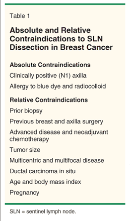
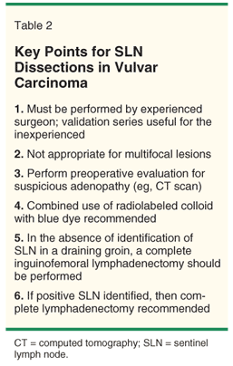
Many issues remain unaddressed. In addition to recognition of the learning curve, if GOG protocol 173 declares SLN biopsies safe, it will also be important for gynecologic oncologists to carefully identify which patients are appropriate candidates for this procedure, and more importantly, which are not. Table 1 lists the absolute and relative contraindications to the performance of SLN dissection for breast cancer,[13] and we may envision the need for a comparable table for vulvar cancer. Table 2 collects many key points as they relate to SLN procedures in vulvar cancer.
Additional research into this promising field should further refine eligibility through an evaluation of patient-related factors (eg, body mass index, prior vulvar/groin surgery, etc) as well as tumor-related factors including lymph-vascular space invasion, cell type, depth of invasion, tumor size, clinically palpable groin nodes, and preoperative therapy. Finally, important issues relating directly to SLN dissections such as the need for pretherapeutic lymphoscintigraphy, frozen section on the sentinel, the role of immunohistochemical staining, and discovery of multiple sentinels, must also be studied.
-Bradley J. Monk, MD
-Krishnansu S. Tewari, MD
Disclosures:
Financial Disclosure: Dr. Monk is a member of the speakers bureau and has received honoraria from GlaxoSmithKline.
References:
References
1. Kosary CL: Cancer of the vulva, in Ries LAG, Young JL, Keel GE, et al (eds): SEER Survival Monograph: Cancer Survival Among Adults: US SEER Program, 1988-2001-Patient and Tumor Characteristics, chapter 18, pp 147-154. Bethesda, MD; National Cancer Institute; SEER Program, NIH Pub No. 07-6215, 2007.
2. Homesley HD, Bundy BN, Sedlis A, et al: Prognostic factors for groin node metastasis in squamous cell carcinoma of the vulva: A Gynecologic Oncology Group study. Gynecol Oncol 49:279-283, 1993.
3. DiSaia PJ, Creasman WT, Rich WM: An alternate approach to early cancer of the vulva. Am J Obstet Gynecol 133:825-832, 1979.
4. Stehman RB, Bundy BN, Dvoretsky PM, et al: Early stage I carcinoma of the vulva treated with ipsilateral superficial inguinal lymphadenectomy and modified radical hemivulvectomy: A prospective study of the Gynecologic Oncology Group. Obstet Gynecol 79:490-497, 1992.
5. Zhang SH, Sood AK, Sorosky JL, et al: Preservation of the saphenous vein during inguinal lymphadenectomy decreases morbidity in patients with carcinoma of the vulva. Cancer 105:722-726, 2000.
6. Rouzier R, Haddad B, Dubernard G, et al: Inguinofemoral dissection for carcinoima of the vulva: Effect of modifications of extent and technique on morbidity and survival. J Am Coll Surg 196:442-450, 2003.
7. Dardarian TS, Gray HJ, Morgan MA, et al: Saphenous vein sparing during inguinal lymphadenectomy to reduce morbidity in patients with vulvar carcinoma. Gynecol Oncol 101:140-142, 2006.
8. Zhang X, Sheng X, Niu J, et al: Sparing of saphenous vein during inguinal lymphadenectomy for vulval malignancies. Gynecol Oncol 105:722-726, 2007.
9. Frumovitz M, Levenback CF: Lymphatic mapping and sentinel node biopsy in vulvar, vaginal, and cervical cancers. Oncology (Williston Park) 22:529-536, 2008.
10. Van der Zee AGJ, Oonk MH, De Hullu JA, et al: Sentinel node dissection is safe in the treatment of early-stage vulvar cancer. J Clin Oncol 26:884-889, 2008.
11. Kunos C, Simpkins F, Gibbons H, et al: Radiation therapy versus pelvic node resection for carcinoma of the vulva with positive groin nodes: An update of a Gynecologic Oncology Group study (abstract 5). Presented at the 39th Annual Meeting of the Society of Gynecologic Oncologists, Tampa, Fla, 2008.
12. Butler JS, Blake P, Bridges JE, et al: Outcomes of patients with recurrent vulval squamous cell cancer (abstract 317). Presented at the 39th Annual Meeting of the Society of Gynecologic Oncologists, Tampa, Fla, 2008.
13. Filippakis GM, Zografos G: Contraindications of sentinel lymph node biopsy: Are there any really? World J Surg Oncol 5:10, 2007.