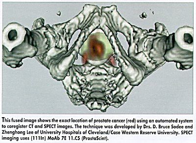
Automated Method Fuses MRI and SPECT Prostate Images
ST. LOUIS-An automated technique for coregistering MRI or CT images with SPECT (single photo emission computed tomography) images has the potential to improve the accuracy of prostate cancer staging, according to research presented at the Society of Nuclear Medicine 47th Annual Meeting.
ST. LOUISAn automated technique for coregistering MRI or CT images with SPECT (single photo emission computed tomography) images has the potential to improve the accuracy of prostate cancer staging, according to research presented at the Society of Nuclear Medicine 47th Annual Meeting.
Sophisticated Software
Zhenghong Lee, PhD, D. Bruce Sodee, MD, and their colleagues at University Hospitals of Cleveland/Case Western Reserve University used sophisticated new software to automatically coregister results with SPECT imaging of ProstaScint (a monoclonal antibody labeled with indium-111) and MRI in prostate cancer patients.
The resulting image shows the site of SPECT metabolic activity superimposed on the same MRI-defined anatomic slice. The researchers have also used the technique to coregister SPECT and CT prostate cancer images.

The software maximizes Mutual Information, or relative entropy between the two volumes, to calculate the optimal alignment of the SPECT/ProstaScint and MRI images.
Virtual 3D Presentation
Data from this fused image is presented in virtually three dimensions, or virtual 3D. The result, Dr. Sodee said, is a significantly clearer image of the tumors size and exact location.
The 3D virtual software will also have applications in coregistering PET (positron emission tomography) images of metabolic cancer activity with anatomic CT or MRI correlation, Dr. Lee said.
Prolaris in Practice: Guiding ADT Benefits, Clinical Application, and Expert Insights From ACRO 2025
April 15th 2025Steven E. Finkelstein, MD, DABR, FACRO discuses how Prolaris distinguishes itself from other genomic biomarker platforms by providing uniquely actionable clinical information that quantifies the absolute benefit of androgen deprivation therapy when added to radiation therapy, offering clinicians a more precise tool for personalizing prostate cancer treatment strategies.