A color atlas of skin lesions
Prepared by The Skin Cancer Foundation, www.skincancer.org,and the American Academy of Dermatology


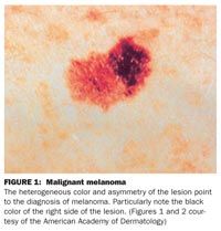
FIGURE 1: Malignant melanoma
The heterogeneous color and asymmetry of the lesion point to the diagnosis of melanoma. Particularly note the black color of the right side of the lesion. (Figures 1 and 2 courtesy of the American Academy of Dermatology)


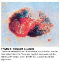
FIGURE 2: Malignant melanoma
There are several colors clearly visible in this lesion, consistent with melanoma. There are mottled black areas of the lesion, with central tumor growth that is eroded and less pigmented.


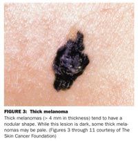
FIGURE 3: Thick melanoma
Thick melanomas (> 4 mm in thickness) tend to have a nodular shape. While this lesion is dark, some thick melanomas may be pale. (Figures 3 through 11 courtesy of The Skin Cancer Foundation)


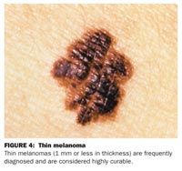
FIGURE 4: Thin melanoma
Thin melanomas (1 mm or less in thickness) are frequently diagnosed and are considered highly curable.


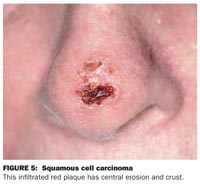
FIGURE 5: Squamous cell carcinoma
This infiltrated red plaque has central erosion and crust.


FIGURE 6: Squamous cell carcinoma
Erythematous and infiltrated lesion in a maximally sun-exposed area, with an erosive center.


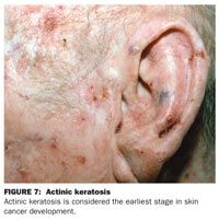
FIGURE 7: Actinic keratosis
Actinic keratosis is considered the earliest stage in skin cancer development.


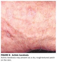
FIGURE 8: Actinic keratosis
Actinic keratosis may present as a dry, rough-textured patch on the skin.


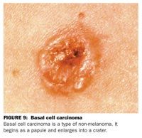
FIGURE 9: Basal cell carcinoma
Basal cell carcinoma is a type of non-melanoma. It begins as a papule and enlarges into a crater.


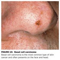
FIGURE 10: Basal cell carcinoma
Basal cell carcinoma is the most common type of skin cancer and often presents on the face and head.


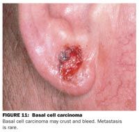
FIGURE 11: Basal cell carcinoma
Basal cell carcinoma may crust and bleed. Metastasis is rare.


![A third of patients had a response [to lifileucel], and of the patients who have a response, half of them were alive at the 4-year follow-up.](/_next/image?url=https%3A%2F%2Fcdn.sanity.io%2Fimages%2F0vv8moc6%2Fcancernetwork%2F6b7c9a3270c71a70749ba86000cfc78a29d74309-2988x1702.png%3Ffit%3Dcrop%26auto%3Dformat&w=3840&q=30 "A third of patients had a response [to lifileucel], and of the patients who have a response, half of them were alive at the 4-year follow-up.")


