High-Dose Interleukin-2: Is It Still Indicated for Melanoma and RCC in an Era of Targeted Therapies?
In this review, we examine the currently approved options available for these disease processes, including the newer agents and selected combinatorial approaches under investigation, and we attempt to identify the role of high-dose IL-2 in the context of current clinical practice.
Immunotherapy with interleukin-2 (IL-2) has been the mainstay of systemic therapy for advanced kidney cancer and melanoma. Although IL-2 treatment is limited to healthy patients, a select group of these patients have derived substantial, durable benefit from it-in some translating into cures with no ongoing therapy or chronic toxicity. Over the past 10 years, insights into the biology of renal cell carcinoma and into key signaling mechanisms in melanoma, and growth in our understanding of immune checkpoints, have led to the development and approval of targeted and immune-modulatory therapeutic options with clinically relevant benefit. Our improved understanding of the relationship between the host environment, immune system, and malignancy has helped identify compounds and therapies that are changing the way we think about cancer and our approach to cancer therapeutics. While the newer options may be applicable to most patients, durable responses measured in years are rare. In this review, we examine the currently approved options available for these disease processes, including the newer agents and selected combinatorial approaches under investigation, and we attempt to identify the role of high-dose IL-2 in the context of current clinical practice.
Immune Response
FIGURE 1
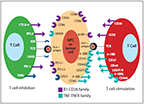
Co-Stimulatory (on Red T Cell) and Co-Inhibitory Proteins (on Green T Cell) and Associated Ligands That Are Expressed on APCs (or Tumor Cells) and T Cells and That Further Fine-Tune the Immune Response
The generation of antitumor immune response by T lymphocytes is a complex multi-step process that is modulated by several signals. Primary signal requires that antigen be presented by antigen-presenting cells (APCs) in the context of self–human leukocyte antigen (HLA) molecules-for recognition by T cells. HLA class I molecules present antigens to CD8+ T lymphocytes, and class II molecules present antigens to CD4+ T lymphocytes. CD4+ T helper cells subsequently produce a number of cytokines, including interleukin-2 (IL-2), that can propagate the immune response (Figure A). IL-2, a nonspecific T-cell growth factor, can result in the expansion of all T-cell subsets, including T-cytotoxic, helper, and regulatory cells. After initial recognition of the antigen, the T-cell function is modified by complex interactions between co-stimulatory or co-inhibitory proteins and their ligands expressed on the APCs (or in some cases tumor cells) and on the T cells; these interactions constitute the secondary regulatory signal that fine-tunes the immune response. A number of immune-modulatory proteins, termed checkpoints, have been identified; the list continues to expand as we get better insights into the anatomy of the immune response; some of these checkpoints are illustrated in Figure 1. Most of these proteins belong to the immunoglobulin superfamily or to the tumor necrosis factor (TNF)/TNF receptor (TNFR) family. While not inherently active, these regulatory molecules exert their effect via the recruitment of TNFR-associated factor (TRAF) adapter proteins. Cytotoxic T lymphocyte antigen 4 (CTLA-4) and programmed death protein 1 (PD-1) have been targeted successfully in cancer immunotherapy; several other checkpoints are under active investigation.
FIGURE A
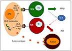
Generation of Anti-Tumor Immune Response by T Lymphocytes
Immunotherapy With IL-2–Based Approaches
Malignant melanoma and renal cell carcinoma are immunologically modulated malignancies, as demonstrated by observations of spontaneous regression without systemic intervention.[1,2] These malignancies show relative resistance to cytotoxic chemotherapy, and therefore, until recently, patients have had limited treatment options.
IL-2 was initially described as a growth factor that was necessary and sufficient for T-cell growth and activation.[3] Identification of IL-2 as a critical T-cell growth factor and its subsequent availability for clinical use facilitated the development of immunotherapeutic approaches against diseases amenable to immune modulation.[4-6] The Surgery Branch of the National Cancer Institute (NCI) demonstrated in 1985 that administration of exogenous IL-2 could result in modulation of the immune response with generation of durable regression of established tumors in a murine model.[7]
Based on the animal model, IL-2, with or without the addition of lymphokine-activated killer (LAK) cells, was administered to patients with advanced renal cell carcinoma and metastatic melanoma, eliciting responses,[8] with a small proportion of these measured in years.[9]
FIGURE 2
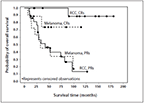
Survival in Patients With Advanced Melanoma (n = 117) and RCC (n = 104) Who Were Treated With High-Dose IL-2 at the Levine Cancer Institute (unpublished data)
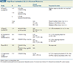
Seven phase II studies, including 255 patients with metastatic renal cell carcinoma who were treated with high-dose (HD) IL-2 (600,000–720,000 IU/kg) yielded an overall response rate of 15%, with 7% complete responders. Median duration of complete responses could not be calculated at that time because > 50% of the patients had not experienced recurrence of their disease.[10] Based on the duration of response, HD IL-2 was approved by the US Food and Drug Administration (FDA) in 1992 for the treatment of advanced renal cell carcinoma. These findings have been confirmed by the Cytokine Working Group,[11] as well as by our group in 104 patients (Figure 2). Updated response data from patients with advanced renal cell carcinoma who were treated at the Surgery Branch of the NCI between 1986 and 2006 include some responses that have lasted over 20 years.[12] The above studies are summarized in Table 1.
TABLE 1
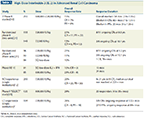
High-Dose Interleukin-2 (IL-2) in Advanced Renal Cell Carcinoma
The data set that led to FDA approval of HD IL-2 for melanoma consisted of a series of eight phase II studies that included a total of 270 patients who were treated with HD IL-2 at doses of 600,000–720,000 IU/kg. The overall response rate was 16%; 6% had a complete response. The median duration of response had not been reached at the time of reporting.[13] Thus, HD IL-2 was approved for advanced melanoma in 1998. The Cytokine Working Group conducted three phase II studies of HD IL-2 (at a dose of 600,000 IU/kg) along with gp100 peptide that showed an overall response in 16%; complete response was observed in 9%.[14] In 684 consecutive patients with metastatic melanoma treated at the NCI with HD IL-2 alone or in combination with a vaccine, the overall response rate was 13% for patients who received IL-2 alone and 16% for patients who received IL-2 with the vaccine.[15] A phase III study comparing HD IL-2 alone to HD IL-2 with gp100 vaccine showed the overall response rate to be 6% for IL-2 alone and 16% for the combination.[16] Our own experience with HD IL-2 in 117 patients has been consistent with the above outcomes (see Figure 2). These data are summarized in Table 2.
TABLE 2
High-Dose Interleukin-2 (IL-2) in Advanced Melanoma
Despite these responses and the likely cures for some patients, adoption of HD IL-2 treatment has been hampered by its toxicity profile-primarily capillary leak syndrome, which manifests as oliguria, mild hypoxemia, generalized edema, tachyarrhythmias, and hypotension. Toxicities also include fever, nausea, diarrhea, catheter-related sepsis, and death.[17] These toxicities require clinical expertise and hospitalization for monitoring but can be managed with limited mortality.[18,19] In our 17-year experience treating 221 patients with 744 cycles of HD IL-2, only one treatment-related death has occurred.
In order to mitigate the toxicities of HD IL-2, several studies have been performed with low doses of IL-2 in both malignant melanoma and renal cell carcinoma. However, these have consistently shown the apparent superiority of HD IL-2 as the optimal regimen for appropriately selected patients.[11,20,21]
FIGURE B
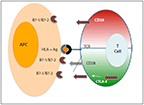
Secondary Regulatory Interaction Between CD28 (Co-stimulatory Molecule) on the T Cell and Molecules in the B7 Family on the APC, Resulting in Activation of the T Cell (Red); Subsequent Interaction Involving CTLA-4 (Co-stimulatory Molecule), Which Displaces CD28 and Leads to Inhibition of the T Cell (Green)
CTLA-4 Checkpoint Inhibition
Cytotoxic T lymphocyte antigen 4 (CTLA-4) is an inhibitory checkpoint that is expressed on activated T cells. T-cell activation requires presentation of antigen in the appropriate context by APCs; this constitutes the initial signal in the activation process. Once the antigen is recognized as non-self, a secondary regulatory interaction occurs between CD28 expressed on the T cell and molecules in the B7 family (CD80, CD86) on the APC. This constitutes a stimulatory signal that results in the activation of the T cell. Subsequent down-regulation of the T-cell activation ensues when the co-inhibitory regulator CTLA-4 is expressed on activated T cells. CTLA-4 binds to CD80 and CD86 with much higher affinity than does CD28, thus displacing CD28 and leading to inhibition of the T cell (Figure B). Blocking the CTLA-4 checkpoint results in unrestrained activation of the T cell, which, when appropriately harnessed, translates into enhanced antitumor activity.[22]
FIGURE 3

Blocking of the CTLA-4 Checkpoint, Which Results in Unrestrained Activation of the T Cell and Enhanced Antitumor Activity
Ipilimumab (Yervoy) is a fully human immunoglobulin G1 (IgG1) antibody that binds to the CTLA-4 molecule and blocks it (Figure 3). Demonstration that CTLA-4 blockade could result in clinically meaningful tumor regression in patients with advanced melanoma led to its clinical development in this setting.[23] Monotherapy with ipilimumab in the phase II setting has shown overall responses in the range of 6% to 16%, and the disease control rate (defined as the percentage of complete responses, partial responses, and stable disease) has ranged between 27% and 32% in patients with advanced melanoma, including initially surprising response rates in visceral disease, including in the liver. The overall 5-year survival rate for treatment-naive patients has reached as high as 49%.[24,25] In a phase III study, ipilimumab alone was compared with the glycoprotein 100 peptide (gp100) vaccine and with a combination of both.[26] The median overall survival in the ipilimumab-containing arms of the trial was 10 months, compared with 6.4 months for the vaccine-alone arm. The overall survival rates for ipilimumab alone vs gp100 alone were 45.6% vs 25.2% at 12 months, and 23.5% vs 13.7% at 24 months. The disease control rate was 28.5% in the ipilimumab-alone arm and 11% in the gp100 vaccine−alone arm. This was the first phase III study to show a survival benefit for immunotherapy in the setting of advanced melanoma, and it led to FDA approval of ipilimumab for this indication in 2011. This study allowed individuals to receive reinduction with the same regimen if they demonstrated clinical benefit and then subsequent progression. Thirty-one individuals met the criteria and were reinduced. Those reinduced patients who had previously received ipilimumab alone showed a response rate of 37.5% and a disease control rate of 75%.[27]
In a second phase III study, the combination of ipilimumab and dacarbazine was compared to dacarbazine alone.[28] The median overall survival in the combination arm was 11.2 months compared with 9.1 months in the dacarbazine-alone arm. The estimated survival rate for the combination was 47.3% at 1 year, compared with 36.3% for dacarbazine alone; the rates were 28.5% and 17.9% at 2 years, and 20.8% and 12.2% at 3 years, respectively (hazard ratio [HR] = 0.72; P < .001). While no difference was noted in the disease control rate (32.2% for the combination vs 30.2% for dacarbazine alone), the median duration of response in complete and partial responders was 19.3 months in the combination arm vs 8.1 months in the dacarbazine-alone arm.
Ipilimumab is now an established therapeutic option for patients with advanced melanoma, yet it comes with attendant expected toxicity on account of the associated breakdown in self-tolerance. The most common autoimmune toxicities include dermatologic reactions; gastrointestinal complications, such as colitis; endocrinopathies; and hepatitis. Administration of ipilimumab therefore requires substantial care, with an emphasis on patient education, ongoing close clinical vigilance, and timely appropriate response to control severe autoimmune manifestations.
FIGURE 4
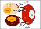
Blocking the Interaction Between PD-1 and PDL-1 Prevents Down-Regulation of T Cells
PD-1 Checkpoint Inhibition
PD-1 is an inhibitory molecule that is expressed on T cells and that interacts with molecules in the B7 family-B7-H1 (also called PD ligand 1 [PD-L1]) and B7-DC (or PD ligand 2 [PD-L2])-to down-regulate the peripheral T-cell response to infection and to limit autoimmunity.[29] PD-1 is also involved in controlling T-cell exhaustion that may result with continued antigenic stimulation.[30] PD-L1 can be expressed on hematopoietic cells, in normal tissues (pancreatic islets, heart, endothelium, small intestine, and placenta) and on several tumor cell types, thereby providing the tumor cells with a protective mechanism against tumor cell–specific T-cell responses, a concept known as adaptive immune resistance (Figure C).[31,32] Blocking the interaction of the PD-1 receptor or its ligand PD-L1 using an anti–PD-1 or an anti–PD-L1 antibody prevents down-regulation of T cells (Figure 4). Several antibodies exploiting this approach are in clinical testing (MDX-1106, MK-3475, Amp224, CT-011, MDX-1105).[33]
FIGURE C

PD-1, Expressed on T Cells, Interacts With Molecules in the B7 Family of Molecules to Down-Regulate the Peripheral T-Cell Response; B7 Family Molecules Are Also Found on Tumor Cells, Protecting Them Against Tumor Cell–Specific T-Cell Activity
Initial evidence of clinical activity with PD-1 blockade using the anti–PD-1 antibody BMS-936558 (MDX-1106 [nivolumab]) was noted in a phase I study that included 39 patients with various malignancies. One durable complete response was noted in a patient with colorectal cancer, and two partial responses were seen in patients with melanoma and renal cell carcinoma; two additional patients with diagnoses of melanoma and non–small-cell lung cancer had significant tumor regression.[34] A subsequent phase I study included 296 patients with advanced solid malignancies (melanoma, non–small-cell lung cancer, renal cell carcinoma, castration-resistant prostate cancer, and colorectal cancer) who were treated with escalating doses of the anti–PD-1 antibody BMS-936558. The incidence of grade 3 or 4 adverse events attributed to the drug was 14%; there were 3 deaths due to pulmonary toxicity. The autoimmune breakthrough toxicities were different from those observed with the anti–CTLA-4 antibody: the incidence of diarrhea was lower, but the incidence of pneumonitis was higher. Responses occurred in 28% of the patients with melanoma, in 18% of those with non–small-cell lung cancer, and in 27% of those with renal cell carcinoma. The responses were durable: 20 out of 31 lasted more than 1 year. In 42 patients, pretreatment tumor samples were obtained and tested for expression of PD-L1. None of the 17 patients with PD-L1–negative tumors had a response; 9 of the 25 patients with PD-L1–positive tumors had an objective response, suggesting PD-L1 expression as a potential tumor marker for response prediction, although this needs validation in larger studies.[35] Building on the concept of preventing interaction between PD-1 and PD-L1, 207 patients with advanced solid malignancies (non–small-cell lung cancer, melanoma, colorectal cancer, renal cell carcinoma, ovarian cancer, pancreatic cancer, gastric cancer, and breast cancer) were treated with the anti–PD-L1 antibody BMS-936559 (MDX-1105) in a phase I study. Objective responses occurred in 17% of patients with melanoma, in 10% of those with non–small-cell lung cancer, in 12% of those with renal cell carcinoma, and in 6% of those with ovarian cancer. Responses were durable and lasted more than 1 year in 50% of patients.[36] Very impressive early results in melanoma have been reported by Hamid et al with another anti–PD-1 antibody, lambrolizumab (previously known as MK-3475). In a phase I study that included 135 patients with advanced melanoma, the overall response rate was 38%. At the highest dose level (10 mg/kg every 2 weeks), the overall response was 52%; responses were observed both in patients who had previously received ipilimumab monotherapy and in those who were ipilimumab-naive.[37] Both anti–PD-1 and anti–PD-L1 antibodies are in advanced stages of clinical development and are expected to gain FDA approval.
Currently Approved Advanced Melanoma Treatment Options
Chemotherapy
Dacarbazine is the only FDA-approved chemotherapy for malignant melanoma. The approval was based on response rate, not survival. Temozolomide (Temodar) has shown equivalence to dacarbazine and is widely used in community practice for advanced melanoma, although it is not approved by the FDA for this indication. The combination of carboplatin and paclitaxel has been used in multiple randomized studies as a control arm. While occasional complete responses have been reported with chemotherapy, no reliably durable responses have occurred. More recently, a phase III study compared nanoparticle albumin-bound paclitaxel (nab-paclitaxel [Abraxane]) with dacarbazine. The overall response rate for nab-paclitaxel was 15%, compared with 11% for dacarbazine. The median overall survival for nab-paclitaxel was 12.8 months, which was not statistically different from the 10.7-month overall survival seen with dacarbazine. The median progression-free survival for nab-paclitaxel was 4.8 months, compared with 2.5 months for dacarbazine-an increase that was statistically significant.[38] Table A presents selected, control-arm data for chemotherapy from recent phase III studies.
BRAF pathway inhibition
BRAF is a serine-threonine kinase. It is a component of the mitogen-activated protein kinase (MAPK) signaling pathway that promotes cell proliferation. Mutations in the BRAF genes are identified in almost 50% of cutaneous melanomas and result in constitutive activation of the pathway. Most of these mutations involve a substitution of glutamic acid for valine at amino acid 600 (V600E). Identification of mutated BRAF as a pertinent target for therapy led to development of the BRAF inhibitors. In the phase I setting, patients treated with vemurafenib (Zelboraf) showed an overall response rate of 81%.[39] A subsequent phase II study with 132 previously treated patients with advanced melanoma showed an overall response rate of 53% (complete responses, 6%; partial responses, 47%). The median duration of response was 6.7 months, and the median overall survival was 15.9 months.[40] A phase III study comparing vemurafenib with dacarbazine showed the median overall survival to be 13.6 months for vemurafenib vs 9.7 months for dacarbazine. The overall response rate was 48% for vemurafenib and 5% for dacarbazine.[41,42] Based on these data, vemurafenib was approved by the FDA for treatment of BRAF V600E–positive advanced melanoma in 2011.
BRAF inhibition with vemurafenib for patients with melanomas that harbor a V600 mutation induces rapid and substantial responses, often in visceral disease, and is therefore an excellent choice of therapy, especially for large, rapidly progressing tumors. While the ease of administration makes it an attractive option, care has to be taken to ensure that a close dermatologic survey is conducted for new squamous cell carcinomas, that the QT interval and liver enzyme levels are monitored, that there is adequate control of arthralgias, and that the patient is educated regarding significant potential for photosensitivity.
BRAF inhibition plus MEK inhibition
Responses observed with BRAF inhibitors are rapid and substantial, but generally not durable. Mechanisms of resistance to BRAF inhibition are currently an area of intense investigation. One of the strategies employed in this context is vertical blockade of the MAPK pathway using a combination of a BRAF inhibitor plus a MEK inhibitor. Although not approved by the FDA, this approach is briefly discussed here given the strong evidence of its anticancer activity, which may result in approval. In a phase I/II study, 162 previously treated patients with advanced melanoma were treated with dabrafenib (Tafinlar; a selective BRAF inhibitor) or a combination of dabrafenib and trametinib (Mekinist; a selective MEK inhibitor). The median progression-free survival was 9.2–9.4 months for the combination (P = .006) vs 5.8 months for monotherapy (P < .001). The response rate was 60%–76% for the combination compared with 54% for monotherapy; median duration of response was 9.5–10.5 months for the combination and 5.6 months for the monotherapy.[43] A phase III study comparing monotherapy with BRAF inhibition vs the combination of BRAF and MEK inhibition has just completed accrual, but results are not yet available.
TABLE 3
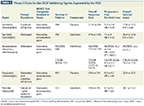
Phase III Data for the VEGF-Inhibiting Agents Approved by the FDA
Currently Approved Advanced Renal Cell Carcinoma Treatment Options
VEGF pathway inhibition
Identification of the Von Hippel Lindau (VHL) gene and elucidation of a critical pathway in renal cell carcinoma mediated via the hypoxia-inducing factors facilitated development of small-molecule inhibitors of the vascular endothelial growth factor (VEGF) axis.[44,45] Phase III data that led to approval of the VEGF tyrosine kinase inhibitors (TKIs) and the combination of bevacizumab (Avastin) plus interferon are summarized in Table 3.
Development of the VEGF TKIs has clearly had a significant impact on the natural history of metastatic renal cell carcinoma. The oral route of administration has made systemic treatment for advanced renal cell carcinoma accessible to a large number of patients. However, these agents do come with attendant toxicities that are ongoing and that occur on a daily basis and need to be controlled-including hypertension, dermatologic reactions, diarrhea, cardiac toxicity, embolic phenomena, fatigue, and hematologic toxicity.[46]
TABLE 4

Phase III Data for mTOR-Inhibiting Agents Approved by the FDA
Inhibition of the mTOR pathway
Mammalian target of rapamycin (mTOR) is an intracellular serine/threonine kinase that integrates signals from several upstream growth factor signal transduction pathways (notably the phosphatidylinositol-3-kinase[PI3K]/Akt pathway) and acts as a central sensor for optimizing both cell growth and the monitoring and regulation of the cell’s nutrient/energy requirements. Activation of mTOR results in (1) increased angiogenesis and (2) increased nutrient uptake, with an improvement in metabolism that results in cell growth and a proliferation advantage. The downstream effects of mTOR activation are carried out by phosphorylation of 4E-binding protein 1 and S6 kinase 1, which impact the translational efficiency of messenger RNA (mRNA). In addition to several proteins that are involved in improvement in the nutritional status of the cell, mTOR also increases hypoxia-inducible factors and impacts the VEGF axis, deemed critical in the setting of renal cell carcinoma. Discovery of rapamycin and the demonstration of its inhibition of mTOR by binding to the FK506 binding protein led to development of its analogs, temsirolimus (Torisel) and everolimus (Afinitor), which have shown clinically relevant activity in the setting of advanced renal cell carcinoma.[47] Phase III data that led to the approval of these agents are summarized in Table 4.
The availability of the mTOR inhibitors for advanced renal cell carcinoma has added to the growing list of therapeutic options. However, these agents have associated toxicities that can impact quality of life for patients on active treatment. These may include fatigue, stomatitis, hypersensitivity reactions, increased risk of infections, elevated triglyceride levels, and hyperglycemia, as well as risk of noninfectious pneumonitis.
Selected New Combinatorial Approaches
BRAF inhibition + CTLA-4 inhibition in advanced melanoma
Inhibition of the CTLA-4 immune checkpoint has induced durable responses; however, 16 to 24 weeks are usually required to identify non-responders. Patients with rapidly progressive disease or large tumor burden require prompt cytoreduction and may not have 16 to 24 weeks for elucidation of response. Rapid and substantial responses have been reproducibly elicited with BRAF inhibition in melanomas that harbor a BRAF V600 mutation. Combining BRAF inhibition with CTLA-4 inhibition is therefore a clinically logical approach for this patient group.
Combinatorial therapy should ideally be supported by proof of mechanistic synergy and absence of overlapping toxicity, as well as by sound clinical logic. Several studies have shown that BRAF inhibition does not compromise immune competence,[48,49] and in fact may enhance the immune response, with improved antigen presentation and increased CD8+ T-cell infiltration into the tumor.[50,51] Exposure to a BRAF inhibitor or a BRAF inhibitor plus a MEK inhibitor has been shown to increase both expression of melanoma antigens by the tumor and infiltration by CD8+ T cells, thus making the immune environment more conducive to antitumor immune response. BRAF inhibition has also been shown to increase PD-L1 expression, suggesting a combinatorial role for PD-1 blockade.[52]
A phase I study combining BRAF inhibition with vemurafenib and CTLA-4 blockade with ipilimumab, administered concurrently, showed excessive hepatotoxicity and has been closed to further accrual.[53] Sequential dosing with vemurafenib followed by ipilimumab is now being studied.
VEGF TKIs + PD-1 inhibition in advanced renal cell carcinoma
Several VEGF TKIs have been approved for advanced renal cell carcinoma (see Table 3) and have clearly had a significant impact on the natural history of the disease. The median survival for advanced disease has almost doubled-from 12 to 22 months-since the introduction of VEGF TKIs.[54] However, durable long-term responses have not been consistently reported. In a phase I study, PD-1 checkpoint blockade produced objective responses in 27% of patients, some of them durable. It is therefore clinically logical to assess the combination of VEGF TKIs plus PD-1 inhibition. As discussed previously, however, the combinatorial approach should have a basis in mechanistic synergy and ideally should not give rise to overlapping toxicities.
Sunitinib (Sutent) has been shown to inhibit T-regulatory cells and myeloid-derived suppressor cells in patients with renal cell carcinoma, and it may thus enhance the immune response.[55,56] Theoretically, combining an agent that inhibits immunosuppressive influences (sunitinib) with an agent that adds to immune activation (anti–PD-1) may allow for enhanced antitumor effect. This approach is being studied in the phase I setting.
PD-1 inhibition + CTLA-4 inhibition in advanced melanoma and renal cell carcinoma
Synergistic antitumor activity has been shown with blockade of PD-1 and CTLA-4 in the animal model.[57] While both CTLA-4 and PD-1 are inhibitory checkpoints, they operate at different levels and different times during the immune response. The CTLA-4 checkpoint is more important in the initial phases of an immune response, while the PD-1 checkpoint may have a greater role in the later phases of the immune response, including the regulation of T-cell exhaustion. CTLA-4 expression has been shown to increase after PD-1 blockade in tumor-infiltrating lymphocytes and peripheral blood lymphocytes, and PD-1 expression is increased on T cells after CTLA-4 blockade.[58] Combining blockades of these two checkpoints is therefore rational and is currently being tested in the phase I setting. The initial results of this approach in melanoma, reported at the 2013 meeting of the American Society of Clinical Oncology, showed an overall response rate of 40%. The cohort in which the maximum tolerated dose was used (nivolumab at a dose of 1 mg/kg and ipilimumab at a dose of 3 mg/kg), showed an overall response in 53% of patients, and 41% had tumor reduction of 80% or greater at 12 weeks. Grade 3/4 adverse events were observed in 53% of patients but were generally reversible.[59]
TKIs + autologous dendritic cells in advanced renal cell carcinoma
AGS-003 is a unique immunotherapy platform that allows autologous dendritic cells (DC) loaded with autologous tumor mRNA to present tumor antigens to T cells. The final product (autologous DC) is also electroporated with CD40. CD40 is a member of the TNFR superfamily expressed on dendritic cells as well as other cells. The ligand for CD40 (CD40L) is expressed on activated T cells. Interaction between CD40 and CD40L plays a critical role in antigen presentation.[60] Sunitinib is a VEGF TKI that is considered standard-of-care for first-line treatment of patients with advanced renal cell carcinoma. Sunitinib elicits positive immunomodulatory effects by suppression of myeloid-derived suppressor cells and T-regulatory cells. A combinatorial approach employing DCs loaded with autologous tumor mRNA plus CD40 and sunitinib was tested in a phase II study in 21 intermediate-risk or poor-risk patients with advanced renal cell carcinoma; the approach showed an overall response of 43%. Stratifying by risk level, the median overall survival was 9.1 months in the poor-risk cohort and 39.5 months in the intermediate-risk cohort. A statistically significant correlation was noted between overall survival and increase in the absolute number of memory T cells measured prospectively.[61] This approach is being evaluated in a phase III study (ADAPT).
Discussion
Insights into the biology of renal cell carcinoma and melanoma have facilitated the development of targeted agents that have revolutionized oncology therapeutics in these areas. The overall survival of 6 to 12 months for patients with advanced renal cell carcinoma has almost doubled-to 22 months in the era of VEGF axis inhibitors. Similarly, the overall survival of 6 to 9 months for advanced melanoma has clearly been affected by the introduction of BRAF inhibitors, which have yielded a median survival of up to 16 months in the second-line setting. However, these responses are rarely complete or durable. In a retrospective analysis, Albiges et al[62] reported outcomes for patients with renal cell carcinoma treated with TKIs who had achieved complete remission. Of the 28 patients who achieved a complete response with TKIs alone, 17 maintained the response with a median follow-up of 255 days. Ahmann et al[63] reported retrospective data from 503 patients with advanced melanoma treated with chemotherapy on study. Only 3 patients achieved a complete response and survived more than 5 years. Kim et al[64] reported retrospective data from 397 patients with advanced melanoma treated with dacarbazine- or temozolomide-based regimens. Only 7 patients achieved a durable, complete response. In contrast, HD IL-2 has consistently elicited durable responses in 10% to 15% of patients with advanced melanoma, and in 15% to 25% of patients with advanced renal cell carcinoma.
Standard HD IL-2 is administered in the inpatient setting and requires hospitalization for about 2 weeks during each treatment course. Close clinical monitoring by a dedicated team is required during this period because of the physiologic changes that result from capillary leak syndrome. Patients subjectively experience a flulike syndrome and severe fatigue. They rapidly recover to their baseline performance status within 72 hours of discontinuing IL-2 dosing, usually with no sequelae or ongoing toxicities that chronically impact quality of life. Changes in hemodynamic and physiologic parameters that result from capillary leak syndrome can be high-grade, yet these occur in a carefully monitored and controlled environment. This is in contrast to toxicities produced by TKIs or mTOR inhibitors for renal cell carcinoma-or by chemotherapy, BRAF inhibitor therapy, or CTLA-4 inhibition for advanced melanoma. While the toxicities from these agents are usually lower grade, subjectively they are experienced by patients on an ongoing basis, with a chronic impact on quality of life. Furthermore, some of the toxicities from the above agents may have an innocuous initial presentation and may go unrecognized in an unmonitored environment-but have potential for progression to clinically ominous outcomes if not diagnosed and treated early (eg, colitis secondary to CTLA-4 inhibition or pneumonitis secondary to mTOR inhibitor treatment).
Identification of biologic markers that may predict for response to therapeutic oncologic interventions has been an area of intense study, yet for melanoma and renal cell carcinoma, such markers remain elusive targets. Given the response rates seen with HD IL-2 (10% to 25%), with some responses being durable and meeting the definition of cure, the ideal situation would be to treat only those patients most likely to achieve that endpoint. Efforts have been made to identify patients most likely to respond to HD IL-2. The renal cell SELECT trial in patients with advanced renal cell carcinoma attempted to validate a potential biomarker-carbonic anhydrase 9-but it was unable to confirm the value of this biomarker in predicting responders. However, this trial did show an overall response in 29% of the patients. The overall response in those with clear-cell carcinoma was 30%.[65] The SELECT trial in patients with melanoma, currently in progress, is evaluating tumor gene expression profiles for patients being treated with HD IL-2, in order to identify predictive markers.
Combinatorial approaches with targeted therapies have opened an exciting new area of drug development. Ideally, the toxicity from a single-agent intervention or combination needs to balance favorably against the potential therapeutic outcome. Logic would require one to accept a higher grade of toxicity from an intervention if the potential outcomes include cure. Analogously, temporary disease control/stability should come at an acceptable (ie, reduced) “price” in terms of toxicity, with preservation of quality of life. Exciting data are now emerging with some of the newer immunotherapeutic modalities, showing both substantial response rates and promise for durability. Given the established durability of responses to HD IL-2 compared with the available data on other therapeutic modalities, and given the ability of centers with expertise to administer the therapy safely, without long-term impact on patient quality of life, our institutional approach (supported by the Society for the Immunotherapy of Cancer consensus statement [manuscript in preparation]) calls for HD IL-2 to remain a viable first-line therapy for appropriately selected patients with advanced melanoma and renal cell carcinoma. Future trials will address the feasibility and utility of synchronously or metachronously combining HD IL-2 with the other modalities reviewed in this paper.
Acknowledgements:The authors thank Cissy Swartz for manuscript preparation assistance, Kendall Carpenter for data management assistance, and James T. Symanowski, PhD, for preparation of survival graphs.
Financial Disclosure:Dr. Amin has served on speakers bureaus and advisory boards of Genentech, Prometheus, and Bristol-Myers Squibb. Dr. White serves on the speakers bureaus of Merck and Prometheus.
References:
REFERENCES
1. Vogelzang NJ, Priest ER, Borden L. Spontaneous regression of histologically proved pulmonary metastases from renal cell carcinoma: a case with 5-year follow-up. J Urol. 1992;148:1247-8.
2. Bulkley GB, Cohen MH, Banks PM, et al. Long-term spontaneous regression of malignant melanoma with visceral metastases. Report of a case with immunologic profile. Cancer. 1975;36:485-94.
3. Morgan DA, Ruscetti FW, Gallo R. Selective in vitro growth of T lymphocytes from normal human bone marrows. Science. 1976;193:1007-8.
4. Gillis S, Smith KA. Long-term culture of tumour-specific cytotoxic T cells. Nature. 1977;268:154-6.
5. Taniguchi T, Matsui H, Fujita T, et al. Structure and expression of a cloned cDNA for human interleukin-2. Nature. 1983;302:305-10.
6. Smith KA. Interleukin-2: inception, impact, and implications. Science. 1988;240:1169-76.
7. Rosenberg SA, Mule JJ, Spiess PJ, et al. Regression of established pulmonary metastases and subcutaneous tumor mediated by the systemic administration of high-dose recombinant interleukin 2. J Exp Med. 1985;161:1169-88.
8. Rosenberg SA, Lotze MT, Muul LM, et al. Observations on the systemic administration of autologous lymphokine-activated killer cells and recombinant interleukin-2 to patients with metastatic cancer. N Engl J Med. 1985;313:1485-92.
9. Rosenberg SA, Yang JC, Topalian SL, et al. Treatment of 283 consecutive patients with metastatic melanoma or renal cell cancer using high-dose bolus interleukin 2. JAMA. 1994;271:907-913.
10. Fyfe G, Fisher RI, Rosenberg SA, et al. Results of treatment of 255 patients with metastatic renal cell carcinoma who received high-dose recombinant interleukin-2 therapy. J Clin Oncol. 1995;13:688-96.
11. McDermott DF, Regan MM, Clark JI, et al. Randomized phase III trial of high-dose interleukin-2 versus subcutaneous interleukin-2 and interferon in patients with metastatic renal cell carcinoma. J Clin Oncol. 2005;23:133-41.
12. Klapper JA, Downey SG, Smith FO, et al. High-dose interleukin-2 for the treatment of metastatic renal cell carcinoma: a retrospective analysis of response and survival in patients treated in the surgery branch at the National Cancer Institute between 1986 and 2006. Cancer. 2008;113:293-301.
13. Atkins MB, Lotze MT, Dutcher JP, et al. High-dose recombinant interleukin 2 therapy for patients with metastatic melanoma: analysis of 270 patients treated between 1985 and 1993. J Clin Oncol. 1999;17:2105-16.
14. Sosman JA, Carillo C, Urba WJ, et al. 3 phase II cytokine working group trials with gp 100 peptide plus high-dose interleukin-2 in patients with HLA A2 positive advanced melanoma. J Clin Oncol. 2008;26:2292-8.
15. Smith FO, Downey SG, Klapper JA, et al. Treatment of metastatic melanoma using interleukin-2 alone or in conjunction with vaccines. Clin Cancer Res. 2008;14:5610-8.
16. Schwartzentruber DJ, Lawson DH, Richards JM, et al. gp100 peptide vaccine and interleukin-2 in patients with advanced melanoma. N Engl J Med. 2011;364:2119-27.
17. Margolin K. The clinical toxicities of high-dose interleukin-2. In: Atkins MB, Mier JW, editors. Therapeutic applications of interleukin-2. New York, NY: Marcel Dekker; 1993. p. 331-62.
18. Kammula US, White DE, Rosenberg SA. Trends in the safety of high dose bolus interleukin-2 administration in patients with metastatic cancer. Cancer. 1998;83:797-805.
19. Schwartzentruber DJ. Guidelines for the safe administration of high-dose interleukin. J Immunother. 2001;24:287-93.
20. Atkins MB. Interleukin-2: clinical applications. Sem Oncol. 2002;29:12-17.
21. Yang JC, Sherry RM, Steinberg SM, et al. Randomized study of high-dose and low-dose interleukin-2 in patients with metastatic renal cancer. J Clin Oncol. 2003;21:3127-32.
22. Leach DR, Krummel MF, Allison JP. Enhancement of antitumor immunity by CTLA 4 blockade. Science. 1996;271:1734-6.
23. Phan GQ, Yang JC, Sherry RM, et al. Cancer regression and autoimmunity induced by cytotoxic T lymphocyte-associated antigen 4 blockade in patients with metastatic melanoma. Proc Natl Acad Sci USA. 2003;100:8372.
24. O’Day SJ, Weber JS, Hamid O, et al. Completed phase II clinical trials: experience with 10 mg/kg ipilimumab for the treatment of advanced melanoma. Presented at: World Meeting of Interdisciplinary Melanoma/Skin Cancer Centers; November 19–21, 2009; Berlin, Germany. Poster 41.
25. Lebbé C, Weber JS, Maio M, et al. Long-term survival in patients with metastatic melanoma who received ipilimumab in four phase II trials. J Clin Oncol. 2013;31(suppl):abstr 9053.
26. Hodi FS, O'Day SJ, McDermott DF, et al. Improved survival with ipilimumab in patients with metastatic melanoma. N Engl J Med. 2010;363:711-23.
27. Robert C, Schadendorf D, Messina M, et al. Efficacy and safety of retreatment with ipilimumab in patients with pretreated advanced melanoma who progressed after initially achieving disease control. Clin Cancer Res. 2103;19:2232.
28. Robert C, Thomas L, Bondarenko I, et al. Ipilimumab plus dacarbazine for previously untreated metastatic melanoma. N Engl J Med. 2011;364:2517-26.
29. Keir ME, Butte MJ, Freeman GJ, Sharpe AH. PD-1 and its ligands in tolerance and immunity. Ann Rev Immunol. 2008;26:677-704.
30. Barber DL, Wherry EJ, Masopust D, et al. Restoring function in exhausted CD8 T cells during chronic viral infection. Nature. 2006;439:682-687
31. Dong H, Strome SE, Salomao DR, et al. Tumor-associated B7-H1 promotes T cell apoptosis: a potential mechanism of immune evasion. Nature Med. 2002;8:793-800.
32. Pardoll DM. The blockade of immune checkpoints in cancer immunotherapy. Nat Rev Cancer. 2012;12:252-64.
33. Topalian SL, Weiner GJ, Pardoll DM. Cancer immunotherapy comes of age. J Clin Oncol 2011;29:4828-36.
34. Brahmer JR, Drake CG, Wollner I, et al. Phase I study of single agent anti-programmed death-1 (MDX-1106) in refractory solid tumors: Safety clinical activity, pharmacodynamics and immunologic correlates. J Clin Oncol. 2010;28:3167-75.
35. Topalian SL, Hodi FS, Brahmer JR, et al. Safety, activity, and immune correlates of anti-PD-1 antibody in cancer. N Engl J Med. 2012;366:2443-54.
36. Brahmer JR, Tykodi SS, Chow LQ, et al. Safety and activity of anti-PD-L1 antibody in patients with advanced cancer. N Engl J Med. 2012;366:2455-65.
37. Hamid O, Robert C, Daud A, et al. Safety and tumor responses with lambrolizumab (anti-PD1) in melanoma. N Engl J Med. 2013 June 2. [Epub ahead of print]
38. Hersh E, Del Vecchio M, Brown M, et al. Phase 3, randomized, open-label, multicenter trial of nab-paclitaxel (nab-P) vs dacarbazine (DTIC) in previously untreated patients with metastatic malignant melanoma (MMM). Pigment Cell Melanoma Res. 2012;25:863.
39. Flaherty KT, Puzanov I, Kim KB, et al. Inhibition of mutated, activated BRAF in metastatic melanoma. N Engl J Med. 2010;363:809-19.
40. Sosman JA, Kim KB, Schuchter L, et al. Survival in BRAF V600-mutant advanced melanoma treated with vemurafenib. N Engl J Med. 2012;366:707-14.
41. Chapman PB, Hauschild A, Robert C, et al; Group B-S. Improved survival with vemurafenib in melanoma with BRAF V600E mutation. N Engl J Med. 2011;364:2507-16.
42. Chapman PB, Hauschild A, Robert C, et al. Updated overall survival (OS) results for BRIM-3, a phase III randomized, open-label, multicenter trial comparing BRAF inhibitor vemurafenib (vem) with dacarbazine (DTIC) in previously untreated patients with BRAF V600E-mutated melanoma. J Clin Oncol. 2012;30(suppl): 8502.
43. Flaherty KT, Infante JR, Daud A, et al. Combined BRAF and MEK inhibition in melanoma with BRAF V600 mutations. N Engl J Med. 2012;367:1694-703.
44. Latif F, Tory K, Gnarra J, et al. Identification of the von Hippel-Lindau disease tumor suppressor gene. Science. 1993;260:1317-20.
45. Patel PH, Chadalavada RS, Chaganti RS, Motzer RJ. Targeting von Hippel-Lindau pathway in renal cell carcinoma. Clin Cancer Res. 2006;12:7215-20.
46. Najjar YG, Rini BI. Novel agents in renal carcinoma: a reality check. Ther Adv Med Oncol. 2012;4:183-94.
47. Meric-Bernstam F, Gonzalez-Angulo AM. Targeting the mTOR signaling network for cancer therapy. J Clin Oncol. 2009;27:2278-87.
48. Comin-Anduix B, Chodon T, Sazegar H, et al. The oncogenic BRAF kinase inhibitor PLX4032/RG7204 does not affect the viability or function of human lymphocytes across a wide range of concentrations. Clin Can Res. 2010;16:6040-8.
49. Hong DS, Vence L, Falchook G, et al. BRAF (V600) inhibitor GSK2118436 targeted inhibition of mutant BRAF in cancer patients does not impair overall immune competency. Clin Can Res. 2102;18:2326-35.
50. Boni A, Cogdill AP, Dang P, et al. Selective BRAF V600E inhibition enhances T cell recognition of melanoma without affecting lymphocyte function. Cancer Res. 2010;70:5213-9.
51. Wilmott JS, Long GV, Howle JR, et al. Selective BRAF inhibitors induce marked T cell infiltration into human metastatic melanoma. Clin Can Res. 2012;18:1386-94.
52. Frederick DT, Piris A, Cogdill AP, et al. BRAF inhibition is associated with enhanced melanoma antigen expression and a more favorable tumor microenvironment in patients with metastatic melanoma. Clin Can Res. 2013;19;1225-31.
53. Ribas A, Hodi FS, Callahan M, et al. Hepatotoxicity with combination of vemurafenib and ipilimumab. N Engl J Med. 2013;368:1365-8.
54. Heng DY, Xie W, Regan MM, et al. Prognostic factors for overall survival in patients with metastatic renal cell carcinoma treated with vascular endothelial growth factor-targeted agents: results from a large, multicenter study. J Clin Oncol. 2009;27:5794-9.
55. Finke JH, Rini B, Ireland J, et al. Sunitinib reverses type-1 immune suppression and decreases T-regulatory cells in renal cell carcinoma patients. Clin Can Res. 2008;14:6674-82.
56. Ko J, Zea AH, Rini BI, et al. Sunitinib mediates reversal of myeloid derived suppressor cell accumulation in renal cell carcinoma patients. Clin Can Res. 2009;15:2148-57.
57. Curran MA, Montalvo W, Yagita H, Allison JP. PD1 and CTLA 4 combination blockade expands infiltrating T cells and reduces regulatory T and myeloid cells within the 16 melanoma tumors. Proc Natl Acad Sci USA. 2010;107:4275-80,
58. Sznol M, Chen L. Antagonist antibodies to PD1 and PDL1 in the treatment of advanced human cancer. Clin Can Res. 2013;19:1021-34.
59. Wolchok JD, Kluger H, Callahan M, et al. Nivolumab plus ipilimumab in advanced melanoma. N Engl J Med. 2013. June 2. [Epub ahead of print]
60. Vonderheide RH, Glennie MJ. Agonistic CD40 antibodies and cancer therapy. Clin Can Res. 2013;19:1035-43.
61. Amin A, Dudek A, Logan T, et al. Prolonged survival with personalized immunotherapy (AGS-003) in combination with sunitinib in unfavorable risk mRCC. (Abstract 357) Poster presented at: Genitourinary Cancers Symposium, February 14-16, 2013, Orlando, Fla.
62. Albiges L, Oudard S, Negrier S, et al. Complete remission with tyrosine kinase inhibitors in renal cell carcinoma. J Clin Oncol. 2012;30:482-7.
63. Ahmann DL, Creagan ET, Hahn RG, et al. Complete responses and long-term survivals after systemic chemotherapy for patients with advanced malignant melanoma. Cancer. 1989;63:224-7.
64. Kim C, Lee CW, Kovacic L, et al. Long-term survival in patients with metastatic melanoma treated with DTIC or temozolomide. Oncologist. 2010;15:765-71.
65. McDermott DF, Ghebremichael MS, Signoretti S, et al. The high-dose aldesleukin (HD IL-2) "SELECT" trial in patients with metastatic renal cell carcinoma (mRCC). J Clin Oncol. 2010;28:345s.
67. Wolchok JD, Thomas L, Bondarenko I, et al. Phase III randomized study of ipilimumab (IPI) plus dacarbazine (DTIC) versus DTIC alone as first-line treatment in patients with unresectable stage III or IV melanoma. J Clin Oncol. 2011;29(suppl): LBA5.
68. Middleton MR, Grob JJ, Aaronson N, et al. Randomized phase III study of temozolomide versus dacarbazine in the treatment of patients with advanced metastatic malignant melanoma. J Clin Oncol. 2000;18:158-66.
69. Flaherty KT, Lee SJ, Schuchter LM, et al. Final results of E2603: A double-blind, randomized phase III trial comparing carboplatin (C)/paclitaxel (P) with or without sorafenib (S) in metastatic melanoma. J Clin Oncol. 2010;28:(suppl):8511.
70. Hauschild A, Agarwala SS, Trefzer U, et al. Results of a phase III, randomized, placebo-controlled study of sorafenib in combination with carboplatin and paclitaxel as second-line treatment in patients with unresectable stage III or stage IV melanoma. J Clin Oncol. 2009;27:2823-30.