Iron and the Anemia of Chronic Disease
The anemia of chronic disease traditionally is defined as a hypoproliferative anemia of no apparent cause that occurs in association with an inflammatory, infectious, or neoplastic disorder, and resolves when the underlying disorder is corrected. Disordered iron metabolism as manifested by a low serum iron, decreased serum transferrin, decreased transferrin saturation, increased serum ferritin, increased reticuloendothelial iron stores, increased erythrocyte-free protoporphyrin, and reduced iron absorption, is a characteristic feature of the anemia of chronic disease and has been thought to be a major factor contributing to the syndrome.
ABSTRACT: The anemia of chronic disease traditionally is defined as a hypoproliferative anemia of no apparent cause that occurs in association with an inflammatory, infectious, or neoplastic disorder, and resolves when the underlying disorder is corrected. Disordered iron metabolism as manifested by a low serum iron, decreased serum transferrin, decreased transferrin saturation, increased serum ferritin, increased reticuloendothelial iron stores, increased erythrocyte-free protoporphyrin, and reduced iron absorption, is a characteristic feature of the anemia of chronic disease and has been thought to be a major factor contributing to the syndrome. A mild shortening of red cell life span also occurs. However, we now know that impaired erythropoietin production and impaired responsiveness of erythroid progenitor cells to this hormone are also important abnormalities contributing to the anemia of chronic disease, and appear to be due to the effects of inflammatory cytokines. Increased intracellular iron may also have a role in the inhibition of erythropoietin production, since the oxygen sensor is a hemoprotein. While the role of inflammatory cytokines in the pathogenesis of anemia of chronic disease appears unequivocal, it has become apparent that disordered iron metabolism, while characteristic of this form of anemia, may not be central to its pathogenesis. It is undisputed that iron absorption is reduced, and that iron administered intravenously is rapidly sequestered in the reticuloendothelial system; however, iron delivery to the bone marrow is not impaired, and erythroid iron utilization is not markedly depressed in anemia of chronic disease. Importantly, recombinant erythropoietin therapy can correct the anemia of chronic disease, but it cannot correct the anemia due to iron deficiency. This refutes the concept that the lack of available iron is central to the pathogenesis of the syndrome. Indeed, it is highly likely that abnormalities such as reduced iron absorption and decreased erythroblast transferrin-receptor expression largely result from decreased erythropoietin production and inhibition of its activity by inflammatory cytokines. [ONCOLOGY 16(Suppl 10):25-33, 2002]
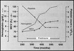
FIGURE 1

Anemia of Chronic Disease
Introduction
The anemia of chronic disease has been traditionally defined as a hypoproliferative anemia of no apparent cause associated with distinct abnormalities in iron metabolism. The syndrome occurs in the setting of an infectious, inflammatory, or neoplastic disorder and resolves when the underlying disorder is corrected (Figure 1).[1] This form of anemia is thought to be a nonspecific consequence of the elaboration of inflammatory cytokines in response to the underlying systemic disorder.[2]
The anemia of chronic disease can be normochromic and normocytic or hypochromic and microcytic. While the degree of hemoglobin reduction is usually modest, in some patients it can be severe (Figure 1). In keeping with its hypoproliferative nature, the reticulocyte count is low and the serum erythropoietin level, while elevated, is not increased to the extent seen in uncomplicated iron deficiency anemia or in hemolytic anemia.[3] The leukocyte and platelet counts are either normal or increased depending on the nature of the underlying illness and the presence of associated complications. Bone marrow histology is usually normal except for increased iron stores. Typically, the amount of nonheme storage iron is increased at the expense of red cell iron.[4]
Essential Factors for Erythropoiesis
TABLE 1

Essential Factors Involved in Erythropoiesis and Anemia of Chronic Disease
The mechanisms for impaired erythropoiesis in the anemia of chronic disease are multifaceted and can be considered most easily in the context of the essential factors involved in erythropoiesis (Table 1). Since the major function of the red cell is to transport oxygen from the lungs to the other tissues, the process of red cell production is inextricably linked to tissue oxygenation-a linkage that has both positive and negative connotations. From the positive perspective, red cell production is coupled with long-term tissue oxygen needs by the glycoprotein hormone erythropoietin. In adults, erythropoietin is produced primarily in the kidneys[5] and to only a small extent in the liver,[6] and acts in the bone marrow to trigger dormant primitive red cells into entering the cell cycle[3] and maintaining their viability[7] as they differentiate into mature erythrocytes.
Because erythropoietin is an erythroid cell viability factor, its production must be constitutive, and because it is a mitogen, its production must be inducible, since it takes more erythropoietin to trigger a dormant erythroid cell into cycle than to maintain its viability. This contention is based on the difference between the numbers of erythropoietin receptors on primitive erythroid progenitors as compared with their more mature counterparts[8] and corresponding erythropoietin dose-response curves for both types of progenitor cells.[9] Because primitive erythroid progenitor cells have fewer erythropoietin receptors, a higher concentration of erythropoietin is required to ensure that ligand-receptor interactions take place.
Mechanisms of Erythropoietin Production
Under normal circumstances, the plasma erythropoietin level is constant in a given individual, just as that individual’s red cell mass is constant.[3] With tissue hypoxia, there is recruitment of additional cells in the kidney to produce erythropoietin[10] and to a smaller extent, up-regulation of erythropoietin production in hepatocytes.[11]
Several feedback mechanisms ensure that red cell production matches tissue oxygen requirements. First, erythropoietin is metabolized by its target cells,[12,13] ensuring that erythropoiesis cannot exceed its stimulus. Second, as expansion of the red cell mass enhances tissue oxygenation, erythropoietin production is down-regulated. Third, as the red cell mass increases, plasma viscosity increases by an unknown mechanism, and this also down-regulates erythropoietin production.[14] There also appears to be a threshold hemoglobin level (10.5 g/dL) above which plasma erythropoietin does not increase outside the range of normal.[3] However, since the normal range for plasma erythropoietin is wide, 4 to 26 mU/mL, there is substantial latitude for a significant increase in the plasma erythropoietin level without exceeding the upper limit of normal.
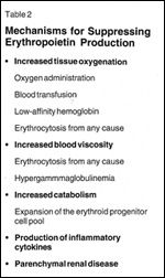
TABLE 2

Mechanisms for Suppressing Erythropoietin Production
Although tissue hypoxia is the only physiologic mechanism for increasing erythropoietin production, the plasma erythropoietin level is not always an accurate surrogate for this situation. This is because other factors such as intrinsic renal disease, liver disease,[15] inflammatory cytokines,[16,17] or chemotherapy[18] can influence erythropoietin production or its metabolism (Table 2). In the anemia of chronic disease, erythropoietin production (as reflected by its plasma level) is usually inappropriately low for the degree of anemia due to the effects of inflammatory cytokines.[16,17]
An adequate response to erythropoietin requires a normal complement of responsive erythroid progenitor cells. In the anemia of chronic disease, however, erythropoietin progenitor cell proliferation is suppressed both directly by inflammatory cytokines[2] and indirectly through the inhibition of erythropoietin production by these cytokines.[16,17] An adequate supply of nutrients is also vital for effective erythropoiesis. These include amino acids, vitamins such as folic acid, B6, B12, and iron.
Patients with the anemia of chronic disease are often in a net catabolic state as evidenced by a reduction in the plasma level of albumin and transferrin,[70] but the most important nutritional deficit involves iron-the absorption and distribution of which are profoundly altered in this situation. Finally, a mild shortening of red cell life span that appears to be extrinsic to the red cell has been documented in patients with anemia of chronic disease,[1,18] a defect for which a normally functioning marrow should easily compensate but does not in this circumstance. The situation is usually aggravated, particularly if the identity of the underlying disorder is in doubt, by the large volumes of blood removed for diagnostic testing.
Abnormalities of Iron Metabolism
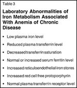
TABLE 3

Laboratory Abnormalities of Iron Metabolism Associated With Anemia of Chronic Disease
Abnormalities in iron metabolism are a striking clinical feature of the anemia of chronic disease (Table 3), and have been considered not only diagnostic for its presence, but also central to the pathogenesis of the anemia.[19-21] However, as discussed below, neither of these contentions are supported by experimental and clinical observations. The abnormalities in iron metabolism, like the anemia, are partly a consequence of the inflammatory cytokine response and partly a consequence of the anemia.[1,22,23]
FIGURE 2

Iron Distribution in AdultsFIGURE 3

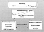
Iron Absorption
Iron is a vital mineral in cellular homeostasis, being essential for oxygen transport, DNA synthesis, and energy metabolism. Iron also represents a potential threat to cell function and tissue integrity, however, since oxidation of hemoglobin iron prevents oxygen release and leads to hemoglobin denaturation, while free iron promotes the synthesis of toxic oxygen radicals that can damage protein, lipids, and DNA. Furthermore, iron is vital for the proliferation of microbial organisms.[24] Thus, iron metabolism is carefully controlled from its absorption in the intestine through its transport in the circulation and its uptake, utilization, and storage within various organs. Figure 2 provides an overview of iron transport pathways and distribution in adults under normal circumstances,[25] and Figure 3 illustrates the salient features of iron absorption.[26]
Central to the absorption, transport, and distribution of iron within the body are a group of proteins that have only recently been identified and whose functions are incompletely characterized. These proteins include the iron transport protein transferrin and the two forms of its cellular receptor, the iron storage protein ferritin, and the hemochromatosis protein (Hfe) that appears to regulate the affinity of the transferrin receptor for transferrin and the iron regulatory proteins (IRP) IRP1 and IRP2 that control the synthesis of ferritin and the transferrin receptor, divalent metal transporter DMT1 and metal transporter MTP1 that transport iron into and out of duodenal epithelial cells, respectively. Hephaestin, a membrane-bound ferroxidase, and hepcidin, an acute-phase reactant, stimulate cellular iron uptake.[27]
The iron required to replace obligate losses due to desquamation of epithelial cells from the skin, gastrointestinal tract, and genitourinary tract; from bile, urine, and sweat; and from menstrual blood loss in women, is obtained from dietary intake in the form of heme and nonheme iron. Heme iron is easily absorbed without modification, but nonheme ferric iron requires chelation to amino acids, ascorbic acid, or sugars in the acid environment of the stomach for its absorption in the alkaline environment of the duodenum, where in the absence of chelation it would be converted to insoluble ferric hydroxides.[28] Chelated ferric3+ iron is reduced to ferrous2+ by a ferrireductase in the brush border of duodenal epithelial cells before transport into these cells by the apical (lumenal) transporter DMT1.[25]
The uptake and processing of dietary nonheme iron by duodenal epithelial cells represent the essential regulatory mechanisms for body iron balance: Body iron stores can only be adjusted by iron absorption because there is no normal mechanism for iron excretion. Therefore, duodenal enterocytes are programmed during their development in the duodenal crypts to facilitate or retard dietary iron absorption.[26,27] Although the details of this programming mechanism are not completely defined, multiple humoral regulatory pathways appear to be involved. These include a "stores" regulator that senses when body iron stores fall below a specific level, an "erythropoietic" regulator that senses an imbalance between the rate of erythropoiesis and the supply of iron, and an independent hypoxia regulator.[25,69] In addition to these humoral regulators, iron absorption appears to be subject to regulation by the relative concentrations of the specific proteins involved in iron transport and storage. These include the Hfe protein, the transferrin receptor, DMT1, MTP1, ferritin, and the acute-phase reactant hepcidin.
Regulation of Intracellular Iron
Classically, the control of intracellular iron transport and storage proteins involves both transcriptional and posttranscriptional controls.[29] Both ferritin and transferrin receptor gene transcription can be regulated by iron-dependent and iron-independent mechanisms, and these appear to be tissue-specific.[29,30] For example, in erythroid cells, transferrin receptor gene expression is controlled at the level of transcription, while in monocytes, its regulation appears to occur largely at the translational level.[29,31,32] Posttranscriptional controls influence either mRNA translation or its stability. Thus, ferritin mRNA has a single iron regulatory element (IRE) in its 5¢UTR and transferrin mRNA has five IREs in its 3'UTR that bind specific iron regulatory proteins (IRP) IRP1 and IRP2.[30] Binding of ferritin mRNA IRE by IRP represses its translation, reducing ferritin synthesis, while binding of transferrin receptor mRNA IRE by IRP increases its stability, resulting in an increase in transferrin receptor synthesis and iron uptake by the cell (Table 4).
TABLE 4

Regulation of Intracellular Iron
Under conditions of high intracellular iron, IRP1 is converted to a form (cytosolic aconitase) unable to bind IRE while IRP2 is oxidized and then degraded, providing within the cell a mechanism for adjusting its iron uptake and iron storage pool according to the intracellular free iron concentration.[30] Iron regulatory elements are present in the mRNA of other proteins involved in iron transport and utilization, such as erythrocyte 5-aminolevulinate synthesis (ALAS) and also DMT1 and MTP1.[30] However, for the latter two proteins, the role of the IRE in regulating their expression in an iron-dependent manner has not been established and other regulatory mechanisms involving hypoxia and changes in localization between membrane and intracellular sites may be important.[26]
The so-called "stores" regulator and the "erythropoietin" regulator are probably closely linked given the influence of intracellular iron concentration on erythropoietin production and the influence of erythropoietin on cellular iron uptake and iron utilization. The intracellular oxygen sensor that regulates erythropoietin production is thought to be a hemoprotein since in vitro chelation of intracellular iron up-regulated erythropoietin production, while an increase in intracellular iron suppressed it.[32,34,35] Similar results have been obtained in vivo following exposure to desferrioxamine or to a transferrin receptor antibody that inhibited transferrin binding.[36]
Administration of desferrioxamine (Desferal) to patients with rheumatoid arthritis was followed by an increase in erythropoietin production and, in some patients, an increase in hemoglobin production.[37] There was no evidence that these changes were due to a change in the activity of the underlying inflammatory process associated with iron chelation and excretion.[38] Importantly, erythropoietin also increased transferrin receptor synthesis by activating IRP1[39] and by enhancing transferrin receptor gene transcription as well.[29] Activation of IRP1 by erythropoietin might be the consequence of its effect on heme synthesis and the resulting decrease in intracellular iron concentration.[40,41]
Characteristic Laboratory Abnormalities of Iron Metabolism
FIGURE 4

Iron Distribution in the Anemia of Chronic Disease
The laboratory abnormalities characteristic of the anemia of chronic disease (Table 3) reflect the significant functional abnormalities of iron metabolism that occur in this situation (Figure 4). The reduction of gastrointestinal iron absorption reflects both the increase in body iron stores (as signaled by the "stores" regulator) and the diminished state of erythropoiesis. The influence of acute-phase reactants, such as hepcidin, is also seen in the increase in iron uptake by developing enterocytes in the duodenal crypts leading to their inability to absorb dietary iron from the intestinal lumen when they mature.[27,42,43] Increased plasma iron clearance is probably due to uptake by hepatocytes since erythropoiesis is suppressed. At the same time, the increase in hemosiderin iron prevents its rapid mobilization.[44]
Effects of Inflammatory Cytokines on Iron Metabolism
TABLE 5

Effects of Inflammatory Cytokines on Iron Metabolism
These well-documented changes appear to be the consequence of the inflammatory reaction to the underlying disorder responsible for the anemia of chronic disease and are common to all of the diseases causing it.[45] As tabulated in Table 5, hypoferremia can be caused by tumor necrosis factor (TNF)-alpha, interleukin (IL)-1, and IL-6,[46] which are T helper (TH)1 cytokines.[27,35] In a cell-specific fashion, TNF-alpha and IL-1 enhance iron uptake and ferritin synthesis.[24). In addition, inflammatory cytokines such as interferon-gamma can stimulate nitric oxide production.[1,63]
Nitric oxide oxidizes IRP2, leading to its degradation while causing an increase in IRP1 activity.[47] However, IRP2 appears to play a more dominant role in regulating ferritin synthesis than IRP1, at least in macrophages, and its degradation permits an increase in ferritin synthesis.[31] At the same time, both macrophage ferritin synthesis and transferrin receptor synthesis are enhanced by the TH2 cytokines IL-4 and IL-13.[32] Furthermore, other acute phase reactants such as alpha-1 antitrypsin may block erythroid cell iron uptake,[48] while the newly recognized transferrin receptor 2 that has no IRE,[49] and thus is not subject to down-regulation by intracellular iron, can facilitate increased iron-loading in tissues such as the liver.[50] Finally, there is little evidence that lactoferrin[51] has a major role in iron sequestration in the anemia of chronic disease.[52]
Erythropoietin Production and Erythropoiesis
TABLE 6

Effects of Inflammatory Cytokines on Erythropoietin ProductionTABLE 7

Effects of Inflammatory Cytokines on Erythropoiesis
In addition to their effects on iron metabolism, inflammatory cytokines also influence erythropoietin production (Table 6) and erythroid progenitor cell proliferation (Table 7). Tumor necrosis factor-alpha and IL-1 inhibit erythropoietin production by mechanisms independent of the hypoxia-inducible factor (HIF)-1 pathway,[53] although a role for an increase in intracellular iron cannot be excluded. Interestingly, IL-6 increased erythropoietin production in hepatocytes but appeared to inhibit it in the kidney.[54] The overall net effect is a limited increase in erythropoietin production that is inappropriately low for the degree of anemia, a phenomenon that is common to infectious, inflammatory, and neoplastic disorders.[3]
Tumor necrosis factor-alpha suppressed erythroid progenitor cell proliferation indirectly through interferon-beta (Betaseron),[55] while IL-1 suppressed it through interferon-gamma (Actimmune),[56] which has been demonstrated to inhibit erythropoietin receptor expression.[57] Nitric oxide, which can be generated by interferon-gamma, directly inhibited erythroid colony formation.[58] Since pharmacologic doses of recombinant erythropoietin could reverse the inhibitory effects of the inflammatory cytokines[54,60-63] while physiologic concentrations cannot, it is clear that both the lack of sufficient erythropoietin production and the impaired responsiveness of its target cells are involved in the syndrome.
TABLE 8

Role of Iron Sequestration in Anemia of Chronic Disease
Although it has been generally accepted that the iron metabolism abnormalities have a central role in the pathogenesis of the anemia of chronic disease, a variety of observations make this conclusion untenable (Table 8). First, although iron absorption is reduced in the anemia of chronic disease, there is no impairment of utilization of the iron absorbed from the gastrointestinal tract by marrow erythroid cells.[43,64] To the extent that iron uptake by erythroid cells is impaired in this situation, this impairment can be explained by reduced transferrin iron saturation and, accordingly, a reduction in diferric transferrin, and more importantly, a reduced stimulus for erythropoiesis, since erythropoietin production is reduced and its effect antagonized by inflammatory cytokines.
Reduced transferrin receptor expression, which is also a feature of the anemia of chronic disease,[65] probably also reflects the reduction in erythropoietin production.[39] Furthermore, while iron therapy can replenish depleted iron stores in patients with the anemia of chronic disease, it usually does not correct the anemia unless the underlying disease is corrected.[64] Juvenile rheumatoid arthritis, however, appears to be an exception because there is no impairment of erythropoietin production in this disorder. Rather, impaired iron absorption appears to be the principal cause for the anemia.[66]
Recombinant erythropoietin therapy cannot correct iron deficiency anemia, but it can usually correct or ameliorate the anemia of chronic disease, regardless of the associated underlying disease.[2,67] Indeed, such a correction can occur in the absence of any change in the laboratory iron abnormalities associated with this form of anemia.[43,61] Previous studies, demonstrating decreased red cell iron utilization as compared to normal, failed to take into consideration that erythropoiesis in the subjects under scrutiny was depressed because of insufficient erythropoietin and the presence of inflammatory cytokines.
In addition to not having a principal role in the pathogenesis of the anemia of chronic disease, the abnormalities in iron metabolism do not appear to define the limits of this form of anemia. When anemia patients were segregated based on hypoferremia and a normal or elevated serum ferritin level, 22% had an underlying disorder other than infection, malignancy, or inflammation.[68] These included congestive heart failure and alcohol abuse. Furthermore, 31% of the patients with the classic anemia of chronic disease had an elevated serum creatinine, and 14% had overt renal failure.
Amongst the group of anemic patients without the classic iron metabolism abnormalities, 54% had an infectious, inflammatory, or neoplastic disease; 36% had other diseases such as alcoholic liver disease, congestive heart failure, deep venous thrombosis, chronic obstructive lung disease, coronary artery disease, hypothyroidism, or diabetes mellitus.[68] Thus, it is clear that abnormalities of iron metabolism do not define the limits of the anemia of chronic disease, that renal insufficiency is also not uncommon in this situation, and that erythropoietin deficiency is the principal abnormality responsible for this form of anemia.
References:
1. Cartwright GE: The anemia of chronic disorders. SeminHematol 3:351-375, 1966.
2. Means RT, Krantz SB: Progress in understanding thepathogenesis of the anemia of chronic disease. Blood 80:1639-1647, 1992.
3. Spivak JL: The clinical physiology of erythropoietin. SeminHematol 30(4 suppl 6):2-11, 1993.
4. Baynes RD, Flax H, Bothwell TH, et al: Haematological andiron-related measurements in active pulmonary tuberculosis. Scand J Haematol36:280-287, 1986.
5. Jacobson LO, Goldwasser E, Fried W, et al: Role of thekidney in erythropoiesis. 1957. J Am Soc Nephrol 11:589-590, 2000.
6. Tan CC, Eckardt KU, Firth JD, et al: Feedback modulationof renal and hepatic erythropoietin mRNA in response to graded anemia andhypoxia. Am J Physiol 263(3 pt 2):F474-F481, 1992.
7. Koury MJ, Bondurant MC: A survival model of erythropoietinaction. Science 248:378-381, 1992.
8. Sawada K, Krantz SB, Dai CH, et al: Purification of humanblood burst-forming units-erythroid and demonstration of the evolution oferythropoietin receptors. J Cell Physiol 142:219-230, 1990.
9. Eaves CJ, Eaves AC: Erythropoietin (Ep) dose-responsecurves for three classes of erythroid progenitors in normal human marrow and inpatients with polycythemia vera. Blood 52:1196-1210, 1978.
10. Koury ST, Koury MJ, Bondurant MC, et al: Quantitation oferythropoietin-producing cells in kidneys of mice by in situ hybridization:Correlation with hematocrit, renal erythropoietin mRNA, and serum erythropoietinconcentration. Blood 74:645-651, 1989.
11. Koury ST, Bondurant MC, Koury MJ, et al: Localization ofcells producing erythropoietin in murine liver by in situ hybridization. Blood77:2497-2503, 1991.
12. Sawyer ST, Krantz SB, Goldwasser E: Binding andreceptor-mediated endocytosis of erythropoietin in Friend virus-infectederythroid cells. J Biol Chem 262:5554-5562, 1987.
13. Cazzola M, Guarnone R, Cerani P, et al: Red blood cellprecursor mass as an independent determinant of serum erythropoietin level. Blood91:2139-2145, 1987.
14. Singh A, Eckardt KU, Zimmermann A, et al: Increasedplasma viscosity as a reason for inappropriate erythropoietin formation. JClin Invest 91:251-256, 1993.
15. Miller CB, Jones RJ, Zahurak ML, et al: Impairederythropoietin response to anemia after bone marrow transplantation. Blood80:2677-2682, 1992.
16. Faquin WC, Schneider TJ, Goldberg MA: Effect ofinflammatory cytokines on hypoxia-induced erythropoietin production. Blood79:1987-1994, 1992.
17. Jelkmann W, Pagel H, Wolff M, et al: Monokines inhibitingerythropoietin production in human hepatoma cultures and in isolated perfusedrat kidneys. Life Sci 50:301-308, 1992.
18. Dinant HJ, de Maat CE: Erythropoiesis and mean red-celllifespan in normal subjects and in patients with the anaemia of activerheumatoid arthritis. Br J Haematol; 39:437-444, 1978.
19. Weiss G: Iron and anemia of chronic disease. KidneyInt Suppl; 69:S12-S17, 1978.
20. Jurado RL: Iron, infections, and anemia of inflammation. ClinInfect Dis 25:888-895, 1997.
21. Freireich E, Miller A, Emerson CP, et al: The effect ofinflammation on the utilization of erythrocyte and transferrin bound radioironfor red cell production. J Hemat 12:11, 1957.
22. Lukens JN: Control of erythropoiesis in rats withadjuvant-induced chronic inflammation. Blood 41:37-44, 1973.
23. Zarrabi MH, Lysik R, DiStefano J, et al: The anaemia ofchronic disorders: Studies of iron reutilization in the anaemia of experimentalmalignancy and chronic inflammation. Br J Haematol 35:647-658, 1977.
24. Kluger MJ, Rothenburg BA: Fever and reduced iron: Theirinteraction as a host defense response to bacterial infection. Science203:374-376, 1979.
25. Andrews NC: Disorders of iron metabolism. N Engl J Med341:1986-1995, 1999.
26. Roy CN, Enns CA: Iron homeostasis: New tales from thecrypt. Blood 96(4020-4027, 1999.
27. Fleming RE, Sly WS: Hepcidin: A putative iron-regulatoryhormone relevant
to hereditary hemochromatosis and the
anemia of chronic disease. Proc Natl Acad Sci U S A 98:8160-8162, 2001.
28. Schade SC: Effect of hydrochloric acid on ironabsorption. N Engl J Med 279:672-674, 1968.
29. Ponka P: Cellular iron metabolism. Kidney Int Suppl69:S2-11, 1999.
30. Eisenstein RS: Iron regulatory proteins and the molecularcontrol of mammalian iron metabolism. Annu Rev Nutr 20:627-662, 2000.
31. Recalcati S, Taramelli D, Conte D, et al: Nitricoxide-mediated induction of ferritin synthesis in J774 macrophages byinflammatory cytokines: Role of selective iron regulatory protein-2downregulation. Blood 91:1059-1066, 1998.
32. Weiss G, Bogdan C, Hentze MW: Pathways for the regulationof macrophage iron metabolism by the anti-inflammatory cytokines IL-4 and IL-13.J Immunol 158:420-425, 1997.
33. Wang GL, Semenza GL: Desferrioxamine induceserythropoietin gene expression and hypoxia-inducible factor 1 DNA-bindingactivity: Implications for models of hypoxia signal transduction. Blood82:3610-3615, 1993.
34. Ho VT, Bunn HF: Effects of transition metals on theexpression of the erythropoietin gene: Further evidence that the oxygen sensoris a heme protein. Biochem Biophys Res Commun 223:175-180, 1996.
35. Daghman NA, McHale CM, Savage GM, et al: Regulation oferythropoietin gene expression depends on two different oxygen-sensingmechanisms. Mol Genet Metab 67:113-117, 1999.
36. Kling PJ, Dragsten PR, Roberts RA, et al: Irondeprivation increases erythropoietin production in vitro, in normal subjects andpatients with malignancy. Br J Haematol 95:241-248, 1996.
37. Salvarani C, Baricchi R, Lasagni D, et al: Effects ofdesferrioxamine therapy on chronic disease anemia associated with rheumatoidarthritis. Rheumatol Int 16:45-48, 1996.
38. Polson RJ, Jawad AS, Bomford A, et al: Treatment ofrheumatoid arthritis with desferrioxamine. QJM 61:1153-1158, 1986.
39. Weiss G, Houston T, Kastner S, et al: Regulation ofcellular iron metabolism by erythropoietin: Activation of iron-regulatoryprotein and upregulation of transferrin receptor expression in erythroid cells. Blood89:680-687, 1997.
40. Tong X, Kawabata H, Koeffler HP: Iron deficiency canupregulate expression of transferrin receptor at both the mRNA and proteinlevel. Br J Haematol 116:458-464, 2002.
41. Nordstrom D, Lindroth Y, Marsal L, et al: Availability ofiron and degree of inflammation modifies the response to recombinant humanerythropoietin when treating anemia of chronic disease in patients withrheumatoid arthritis. Rheumatol Int 17:67-73, 1997.
42. O’Toole PA, Sykes H, Phelan M, et al: Duodenal mucosalferritin in rheumatoid arthritis: Implications for anaemia of chronic disease. QJM89:509-514, 1996.
43. Raymond FD, Bowie MA, Dugan A: Iron metabolism inrheumatoid arthritis. Arthr Rheum 8:233-243, 1965.
44. Savage D, Lindenbaum J: Anemia in alcoholics. Medicine(Baltimore) 65:322-338, 1986.
45. Douglas SW, Adamson JW: The anemia of chronic disorders:Studies of marrow regulation and iron metabolism. Blood 45:55-65, 1975.
46. Moldawer LL, Marano MA, Wei H, et al: Cachectin/tumornecrosis factor-alpha alters red blood cell kinetics and induces anemia in vivo.FASEB J 3:1637-1643, 1975.
47. Lappin MB, Campbell JD: The Th1-Th2 classification ofcellular immune responses: Concepts, current thinking and applications inhaematological malignancy. Blood Rev 14:228-239, 2000.
48. Graziadei I, Gaggl S, Kaserbacher R, et al: Theacute-phase protein alpha 1-antitrypsin inhibits growth and proliferation ofhuman early erythroid progenitor cells (burst-forming units-erythroid) and ofhuman erythroleukemic cells (K562) in vitro by interfering with transferrin ironuptake. Blood 83:260-268, 1994.
49. Tong X, Kawabata H, Koeffler HP: Iron deficiency canupregulate expression of transferrin receptor at both the mRNA and proteinlevel. Br J Haematol 116:458-464, 2002.
50. Fleming RE, Migas MC, Holden CC, et al: Transferrinreceptor 2: Continued expression in mouse liver in the face of iron overload andin hereditary hemochromatosis. Proc Natl Acad Sci U S A 97:2214-2219,2000.
51. Van Snick JL, Masson PL: The binding of human lactoferrinto mouse peritoneal cells. J Exp Med 144:1568-1580, 1976.
52. Baynes R, Bezwoda W, Bothwell T, et al: The non-immuneinflammatory response: Serial changes in plasma iron, iron-binding capacity,lactoferrin, ferritin and C-reactive protein. Scand J Clin Lab Invest46:695-704, 1986.
53. Hellwig-Burgel T, Rutkowski K, Metzen E, et al:Interleukin-1 beta and tumor necrosis factor-alpha stimulate DNA binding ofhypoxia-inducible factor-1. Blood 94:1561-1567, 1999.
54. Jelkmann W: Proinflammatory cytokines loweringerythropoietin production. J Interferon Cytokine Res 18:555-559,1998.
55. Means RT, Krantz SB: Inhibition of human erythroidcolony-forming units by tumor necrosis factor requires beta interferon. JClin Invest 91: 416-419, 1993.
56. Means RT, Dessypris EN, Krantz SB: Inhibition of humanerythroid colony-forming units by interleukin-1 is mediated by gamma interferon.J Cell Physiol 150:59-64, 1992.
57. Taniguchi S, Dai CH, Price JO, et al: Interferon gammadownregulates stem cell factor and erythropoietin receptors but not insulin-likegrowth factor-I receptors in human erythroid colony-forming cells. Blood90:2244-2252, 1997.
58. Maciejewski JP, Selleri C, Sato T, et al: Nitric oxidesuppression of human hematopoiesis in vitro. Contribution to inhibitory actionof interferon-gamma and tumor necrosis factor-alpha. J Clin Invest96:1085-1092, 1995.
59. Johnson CS, Cook CA, Furmanski P: In vivo suppression oferythropoiesis by tumor necrosis factor-alpha (TNF-alpha): Reversal withexogenous erythropoietin (EPO). Exp Hematol 18:109-113, 1990.
60. Means RT, Krantz SB: Inhibition of human erythroidcolony-forming units by gamma interferon can be corrected by recombinant humanerythropoietin. Blood 78:2564-2567, 1991.
61. Pincus T, Olsen NJ, Russell IJ, et al: Multicenter studyof recombinant human erythropoietin in correction of anemia in rheumatoidarthritis. Am J Med 89:161-168, 1990.
62. Pettersson T, Rosenlof K, Laitinen E, et al: Effect ofexogenous erythropoietin on haem synthesis in anaemic patients with rheumatoidarthritis. Br J Rheumatol 33:526-529, 1994.
63. Kaltwasser JP, Kessler U, Gottschalk R, et al: Effect ofrecombinant human erythropoietin and intravenous iron on anemia and diseaseactivity in rheumatoid arthritis. J Rheumatol 28:2430-2436, 2001.
64. Vreugdenhil G, Baltus JA, van Eijk HG, et al: Predictionand evaluation of the effect of iron treatment in anaemic RA patients. ClinRheumatol 8:352-362, 1989.
65. Feelders RA, Vreugdenhil G, van Dijk JP, et al: Decreasedaffinity and number of transferrin receptors on erythroblasts in the anemia ofrheumatoid arthritis. Am J Hematol 43:200-204, 1993.
66. Cazzola M, Ponchio L, de Benedetti F, et al: Defectiveiron supply for erythropoiesis and adequate endogenous erythropoietin productionin the anemia associated with systemic-onset juvenile chronic arthritis. Blood87:4824-4830, 1996.
67. Spivak JL: The blood in systemic disorders. Lancet355:1707-1712, 2000.
68. Cash JM, Sears DA: The anemia of chronic disease:Spectrum of associated diseases in a series of unselected hospitalized patients.Am J Med 87:638-644, 1989.
69. Finch C: Regulators of iron balance in humans. Blood84:1697-1702, 1994.
70. Kurnick JE, Ward HP, Pickett JC: Mechanism of the anemiaof chronic disorders: Correlation of hematocrit value with albumin, vitamin B12, transferrin, and iron stores. Arch Intern Med 130:323-326, 1972.
71. Nordstrom D, Lindroth Y, Marsal L, et al: Availability of iron and degreeof inflammation modifies the response to recombinant human erythropoietin whentreating anemia of chronic disease in patients with rheumatoid arthritis. RheumatolInt 17:67-73, 1997.
Â
How Supportive Care Methods Can Improve Oncology Outcomes
Experts discussed supportive care and why it should be integrated into standard oncology care.