Management of Leptomeningeal Disease From Solid Tumors
Metastasis of solid tumors to the cerebrospinal fluid is a serious complication of cancer. Although it can occur with any type of cancer, tumors with a high propensity for CNS involvement (brain metastases) are the most likely to spread to the cerebrospinal fluid.
Oncology (Williston Park). 30(8):724–730.

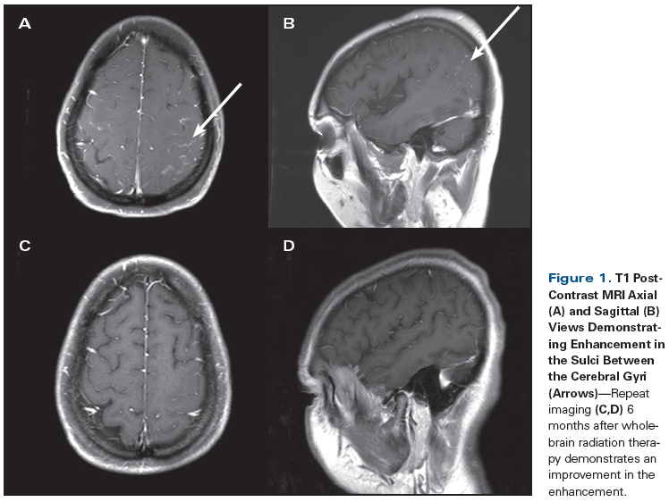
Figure 1. T1 Post-Contrast MRI Axial (A) and Sagittal (B) Views Demonstrating Enhancement in the Sulci Between the Cerebral Gyri (Arrows)

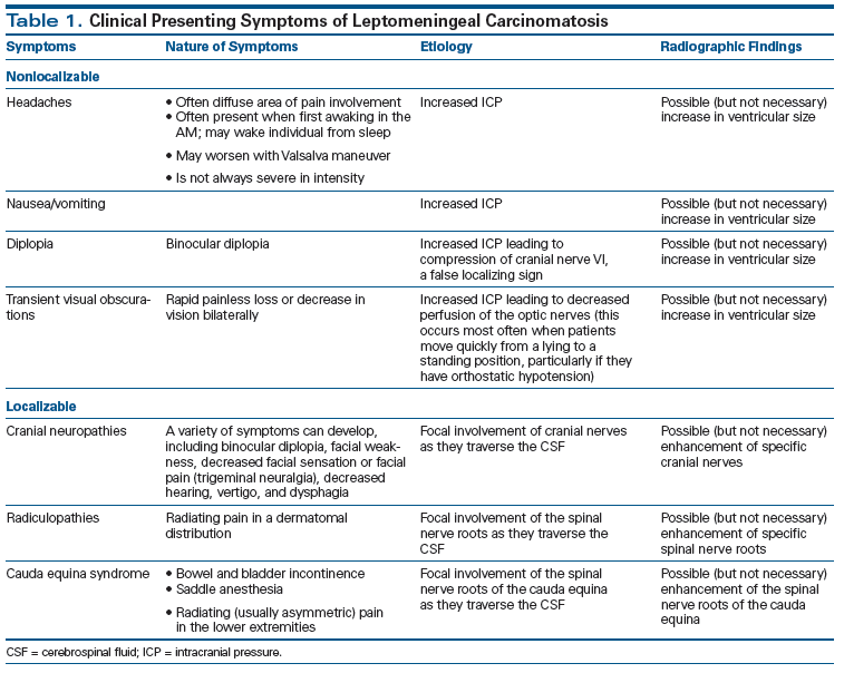
Table 1. Clinical Presenting Symptoms of Leptomeningeal Carcinomatosis

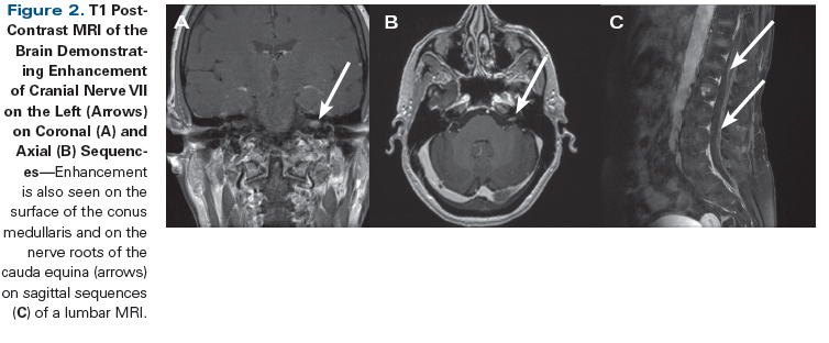
Figure 2. T1 Post-Contrast MRI of the Brain Demonstrating Enhancement of Cranial Nerve VII on the Left (Arrows) on Coronal (A) and Axial (B) Sequences

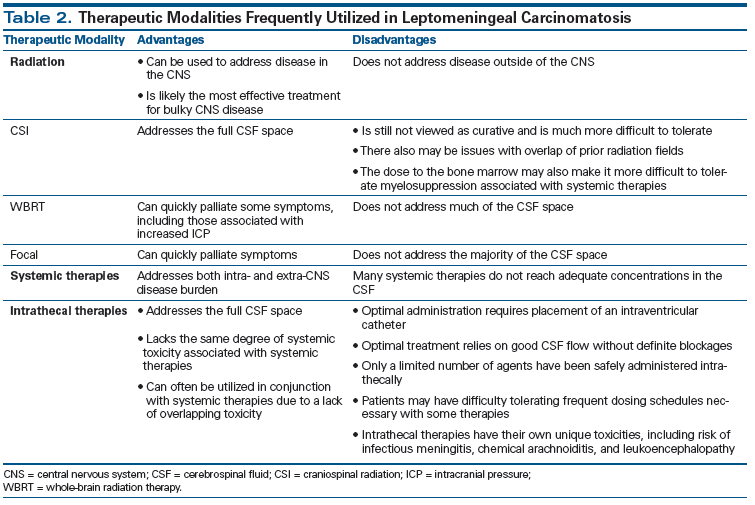
Table 2. Therapeutic Modalities Frequently Utilized in Leptomeningeal Carcinomatosis

Introduction
Leptomeningeal carcinomatosis, known by a variety of names, including leptomeningeal metastases and neoplastic meningitis, indicates spread of tumor to the cerebrospinal fluid (CSF) and invasion of the subarachnoid space. The term leptomeningeal (from the Greek lepto, meaning “fine” or “slight”) describes the thin meninges, the arachnoid and the pia mater, between which the CSF is located. Although CSF metastases are a subtype of central nervous system (CNS) metastases, it is important to differentiate them from other CNS metastases such as brain[1] or dural metastases,[2] as both prognosis and management differ significantly between these neuroanatomic locations. We will discuss key concepts in the diagnosis, prognosis, and management of leptomeningeal carcinomatosis in a case-based format.
Clinical Vignette #1
A 65-year-old woman with non−small-cell lung cancer diagnosed 11 years earlier (stage IB at diagnosis) developed bony metastases 1 year prior to the current presentation. A thoracic vertebra metastasis was treated with focal radiation therapy (RT; 60 Gy). Later on, the patient developed unilateral facial weakness. Her symptoms were initially thought to represent idiopathic Bell’s palsy, but remained refractory to treatment with steroids and acyclovir. Brain MRI at that time demonstrated ipsilateral cranial nerve VII enhancement (Figure 1). Her CSF was noninflammatory, without evidence of malignant cells. Subsequently, over a 4-month period, the patient developed bilateral facial weakness, unilateral tinnitus, dizziness, hyperacusis, and abnormal sensations in the right upper extremity. Electromyography and nerve conduction studies of facial nerves revealed an axonal degenerative process, without evidence of demyelination. Repeat CSF studies demonstrated lymphocytic pleocytosis with malignant cells on cytology, and a diagnosis of leptomeningeal carcinomatosis was made. An MRI of the spine revealed nodular enhancement at the conus medullaris.
The patient’s leptomeningeal disease was treated with whole-brain radiotherapy (WBRT) plus focal RT to the lumbosacral region (total, 28 Gy), and intrathecal (IT) liposomal cytarabine (50 mg in 5 mL preservative-free solution) was delivered every other week via an intraventricular catheter, along with oral dexamethasone (4 mg twice a day 2 days prior, day of, and 2 days post liposomal cytarabine injections). Tinnitus and sensory symptomatology improved, but the patient’s facial diplegia remained. After 8 doses (4 months) of liposomal cytarabine, follow-up CSF cytology was negative and repeat MRI imaging revealed no further evidence of leptomeningeal enhancement. Two months after starting IT chemotherapy, the patient again presented with severe headache and repetitive extremity movements that were indicative of seizures, but electroencephalogram did not show epileptiform activity. She was found to have focal edema near the intracranial catheter on imaging. She was then started on levetiracetam prophylactically and continued to receive IT liposomal cytarabine for 2 additional months, during which her functional status rapidly deteriorated, and she transitioned to hospice care.
What Are the Presenting Symptoms of Leptomeningeal Carcinomatosis?
Patients with leptomeningeal carcinomatosis may present with a wide range of clinical symptoms that can vary from barely perceptible to profoundly debilitating; they can be categorized as neuroanatomically localizable or nonlocalizable (Table 1). These symptoms may represent the initial manifestation of cancer or they may occur years after the initial diagnosis, as in Clinical Vignette #1.[3-5] The nonlocalizable symptoms arise from an increase in intracranial pressure (ICP) that manifests as headaches, which most commonly occur after prolonged time in a supine position, for example, upon awakening in the morning. This is different from many primary headache syndromes, such as migraine, which often resolve after a night’s sleep. Headaches that result from increased ICP can awaken patients from sleep, and in turn should be differentiated from primary headache disorders such as trigeminal/autonomic headaches (eg, cluster headaches) and hypnic headaches, all of which can have a similar presentation. Additional symptoms associated with trigeminal/autonomic headaches and hypnic headaches can include manifestations of autonomic dysfunction (unilateral lacrimation, rhinorrhea, and diaphoresis). However, they lack the nausea, vomiting, and diplopia which, when present, should raise concern for increased ICP.
The localizable symptoms relate to the specific neuroanatomy involved. The presence of symptoms that localize to multiple nonconfluent areas in the nervous system should raise the suspicion of a pathologic process within the CSF, which bathes the surface of the entire CNS. Symptoms can be broadly divided into two categories: intracranial and spinal. Intracranial symptomatology most commonly arises from dysfunction of cranial nerves. This can cause extraocular movement deficits leading to binocular diplopia, which resolves with single eye closure. Motor and sensory facial deficits can also develop. The sensory symptoms would be anterior to the ear, limited to the distribution of the fifth cranial nerve. The facial weakness would most likely involve the forehead in addition to the lower face, clinically resembling Bell’s palsy. Clinicians must pay careful attention to the patient’s history, and findings on the physical examination may alter one’s suspicion of leptomeningeal carcinomatosis as the underlying etiology for facial weakness. In the case described here, the known history of stage IV lung cancer should raise concern for leptomeningeal carcinomatosis as the underlying etiology of the patient’s initial facial weakness. Spinal symptomatology can manifest as weakness, due to involvement of the motor nerve roots. There may also be a loss of sensation, but more often radicular pain that radiates along a dermatomal distribution is observed. Cauda equina syndrome is another common presentation of leptomeningeal carcinomatosis. This involves sensory loss in a saddle distribution, radicular pain of the lower extremities, bladder incontinence, and constipation. In comparison to conus medullaris syndrome, which involves the lower portion of the spinal cord parenchyma, cauda equina syndromes are more likely to have asymmetric presentations and radicular pain. Electromyography and nerve conduction studies often reveal an axonal pattern of injury, as was noted in Clinical Vignette #1. This differs from immune-mediated processes, where a demyelinating pattern would more likely be expected. Once the diagnosis of leptomeningeal carcinomatosis is established, prognosis is poor and clinical deterioration can progress rapidly.
What Is the Discrepancy Between Radiologic and CSF Findings in the Setting of Leptomeningeal Carcinomatosis?
Classic radiographic findings of neoplastic meningitis include the presence of enhancement between cerebellar folia, deep in the brain sulci, and over the convexities. Enhancement can also be present on the surface of the spinal cord or on the nerve roots of the cauda equina. However, these findings are nonspecific and can also be seen in infectious diseases (tuberculosis, for example) and autoimmune conditions (sarcoidosis, for example), and oftentimes as an artifact following lumbar puncture. Thus, the timing of radiographic evaluation and lumbar puncture is important: imaging should always precede spinal tap whenever possible. It should be emphasized that radiographically evident disease may not always be symptomatic in patients with leptomeningeal carcinomatosis. The wide range of reported negative imaging in the setting of positive CSF findings (30% to 70%) arises at least in part from the varying methodologies utilized to evaluate the incidence of leptomeningeal carcinomatosis.[6] The explanation for negative CSF studies in the setting of abnormal imaging suggestive of leptomeningeal carcinomatosis may be even less clear. This may be due to some patients being diagnosed with leptomeningeal carcinomatosis radiographically and not being optimal candidates for lumbar puncture due to noncommunicating hydrocephalus, large intracranial metastases, thrombocytopenia, or other medical issues.
Clinical Vignette #2
A 40-year-old woman with a history of stage IV carcinoma of the breast with diffuse bony metastases (ypT4N3M1, infiltrating ductal carcinoma, grade 2, estrogen/progesterone receptor–positive, human epidermal growth factor receptor 2 [HER2]–negative at diagnosis) was treated with mastectomy, hormone therapy, and RT. Three years after her initial cancer diagnosis, she developed complex partial seizures (episodes of left head deviation, speech arrest, and staring lasting 1 to 2 minutes) and confusion. Her neurologic exam did not demonstrate any new focal deficit, but the patient did acknowledge shooting pains with neck flexion. Seizures were treated with the non–enzyme-inducing anti-epileptic medications (AED) levetiracetam and lacosamide. A brain MRI demonstrated ventriculomegaly and enhancement within sulci along both the frontal and parietal lobes (Figure 2). CSF cytology demonstrated metastatic cells, and a diagnosis of leptomeningeal carcinomatosis was made.
The patient was evaluated for IT chemotherapy administration but experienced rapid clinical deterioration prior to surgical placement of an intraventricular catheter. Urgent WBRT was initiated. Due to persistent seizures requiring hospitalization and an associated decline in performance status, WBRT was aborted after the patient had received 16 Gy of planned 20 Gy. Clobazam was added to her AED regimen. With better seizure control, she experienced a concomitant improvement in performance status. Follow-up brain MRI 7 months later demonstrated resolution of contrast-enhancing leptomeningeal disease. The patient has subsequently developed metastatic disease in the contralateral breast and in the bladder, and after several months she developed rapid clinical and radiographic progression of both the leptomeningeal and extra-CNS disease, leading to hospice enrollment.
Which Patients With Solid Tumors Are at Risk for Developing Leptomeningeal Carcinomatosis?
Patients with a variety of solid tumors, as well as hematologic malignancies, are at risk for developing leptomeningeal carcinomatosis. Precise data regarding the incidence of clinically significant leptomeningeal carcinomatosis are lacking. The limitations of autopsy studies[7-9] are that they may not be broadly generalizable, as they often represent data from single tertiary care referral centers, and advances in the treatment of cancer also make it difficult to compare results with the current era. Although large national or local cancer registries track the incidence of primary tumors, details regarding location of metastases are not followed in a similar manner. Thus, a range of incidence rates for leptomeningeal carcinomatosis can be found when surveying the literature, reflecting a paucity of comprehensive contemporary epidemiologic studies of this phenomenon. Incidence rates ranging from < 1% to 8% of cancer patients are frequently cited.[10] Across numerous studies, the highest incidence of leptomeningeal carcinomatosis appears to be in patients with breast cancer.[4,6,11] However, these incidence rates may not represent the unselected breast cancer population.[12] Lung cancer appears to be the second most common solid tumor to metastasize to the CSF, followed by a range of other malignancies, including gastrointestinal cancers and melanoma. The time interval between the initial cancer diagnosis and the diagnosis of leptomeningeal carcinomatosis ranges widely, from a synchronous presentation to metachronous presentations separated by multiple years.[4,5] The majority of patients are known to have metastatic disease at the time of leptomeningeal carcinomatosis diagnosis.[4] It is unclear whether the anatomic location of the primary malignancy, such as brain, dura, or vertebral bodies, significantly influences the likelihood of leptomeningeal carcinomatosis, although this has not been systematically studied. Age, performance status, and interval from initial diagnosis have not clearly been demonstrated to influence risk of the development of leptomeningeal carcinomatosis across all histologies. From a practical perspective, this forces the clinician to consider leptomeningeal carcinomatosis across multiple clinical scenarios (Table 1).
What Is the Prognosis of Leptomeningeal Carcinomatosis?
The prognosis for leptomeningeal carcinomatosis is poor. Median survival without treatment can be as short as 3 to 4 weeks. Even with treatment, survival is still usually measured in just weeks to months. However, there are some patients who experience prolonged survival and possibly clearance of disease in the CSF. Although well-established prognostication systems[1] exist for brain metastases, a similar system has not yet been codified for leptomeningeal carcinomatosis. Across histologic subtypes, performance status appears to consistently influence prognosis.[13] Survival is influenced by histology of the primary tumor, with breast cancer patients[13] faring better than most patients with other tumor histologies. Amongst breast cancer patients, it does not appear that histologic subtype provides further clear differences in prognosis.[14] One of the difficulties in counseling patients with newly diagnosed leptomeningeal carcinomatosis is the lack of randomized data demonstrating the superiority of any one therapeutic approach over others.[15]
What Management Strategies Are Employed in Solid Tumor Leptomeningeal Carcinomatosis?
Management of solid tumor leptomeningeal carcinomatosis is often tailored to the specific patient and is influenced by a number of factors, including performance status. Therapeutic options can be categorized as local and systemic; local therapy would include RT and IT therapy. Clinical management is limited by the paucity of randomized trial data for guiding decision making. Most therapeutic modalities have been evaluated in relatively small single-arm trials or case series, which results in a more “Ã la carte” (or patient-tailored) approach, where hope for efficacy is balanced against the potential toxicities of the treatments (Table 2).
RT is a local therapy that is often employed, particularly when bulky disease is present. Both locally and systemically administered chemotherapies are felt to have a limited effect on bulky disease. When radiation is utilized in this clinical setting, it is often limited to WBRT and/or focal RT to bulky and/or symptomatic disease. Craniospinal irradiation, while it addresses all of the CSF, is often not used because of the substantial toxicity without expectation for cure. This is of particular concern in patients who may have prior overlapping radiation fields or have low bone marrow reserve from prior chemotherapies. Although data are lacking in leptomeningeal disease, the utilization of proton radiotherapy is sometimes considered in this patient population; however, the optimal dose and fractionation schedule have not been defined.[16,17] In patients with poorer prognosis, a shorter course is sometimes employed. Investigations into limiting the toxicity of WBRT via hippocampal avoidance are not being actively pursued in the setting of leptomeningeal carcinomatosis.
Another means of delivering local treatment is through IT therapy, which denotes the direct administration of the therapeutic agent into the CSF. It is a means to circumvent the blood-CSF barrier and administer often small amounts of a therapeutic agent into a relatively isolated space. With this approach, systemic toxicities are, for the most part, abrogated. This provides clinicians with the flexibility to administer systemic therapies with concomitant IT therapies. However, a distinct set of toxicities are associated with IT administration of therapies. These include the potential to introduce an infection into the CSF (eg, infectious meningitis) when accessing the CSF space, marked inflammatory response to the therapeutic agent (chemical arachnoiditis), and injury to the brain and spinal cord, primarily affecting the white matter tracts (leukoencephalopathy, myelopathy). The optimal IT regimen for solid tumor leptomeningeal carcinomatosis is unknown. Methotrexate, thiotepa, cytarabine, and liposomal cytarabine are all approved for IT administration, and a number of additional agents are also utilized, including topotecan, trastuzumab, and rituximab. The potential efficacy, toxicity profile, and logistics regarding frequency of administration all influence the decision making regarding the regimen. In our experience, IT therapies are not typically associated with significant improvement in clinical symptomatology or performance status. Therefore, we often limit their use to patients with good performance status who are deemed able to tolerate both potential toxicities as well as the logistics of IT therapy administration. If possible, it is preferred that IT therapies for solid tumor leptomeningeal carcinomatosis be administered via an intraventricular catheter, such as an Ommaya reservoir. After neurosurgical placement of the intraventricular catheter, the administration of IT therapy becomes much quicker and more convenient, with a lower likelihood of inadvertent administration of the therapeutic agent into an unintended location.[18] There is also some evidence to support improved survival with administration via an intraventricular catheter as compared with lumbar puncture. This is likely most relevant when utilizing agents that have a short half-life in the CSF (eg, methotrexate).[18]
Systemic therapies in leptomeningeal carcinomatosis are hampered by the degree to which they can cross the blood-CSF barrier. CSF concentrations of systemically administered drugs have been less frequently studied than serum concentrations of these same drugs. There are some agents, however, which we know can reach substantial concentrations in the CSF and others that do not. High-dose intravenous methotrexate, the cornerstone of treatment for primary CNS lymphoma, has also been evaluated in solid tumor leptomeningeal carcinomatosis. It has demonstrated tolerability while achieving cytotoxic serum and CSF concentrations of drug and radiographic responses.[19,20] High-dose intravenous methotrexate should be considered as a component of the armamentarium in the treatment of leptomeningeal carcinomatosis. In addition to what appear to be broadly acting systemic therapies, targeted approaches can also be considered. As an example, in patients with HER2-positive breast cancer, the HER2-targeting tyrosine kinase inhibitor lapatinib has been used, often in conjunction with capecitabine, to treat brain metastases.[21,22] Although it is rational to consider the utilization of similar regimens for leptomeningeal carcinomatosis in HER2-positive breast cancer patients, this has not been evaluated in prospective trials.[13] This example serves to underscore the difficulties in clinical decision making when contemplating the role of specific systemic therapies for leptomeningeal carcinomatosis. Finally, the status of extra-CNS disease and the need for effectively treating it also enters into the decision-making process.
Conclusions
Metastasis of solid tumors to the CSF is a serious complication of cancer. Although it can occur with any type of cancer, tumors with a high propensity for CNS involvement (brain metastases) are the most likely to spread to the CSF. The clinical presentations are frequently atypical and may initially only include subtle signs or symptoms that might be missed. Some more clearly evident symptomatology that resembles common non-neoplastic clinical symptoms, such as sciatica or the facial weakness associated with Bell’s palsy, should be evaluated with particular caution in any patient with a known diagnosis of cancer. Prognosis in patients with leptomeningeal carcinomatosis is poor. In patients in whom the decision is made to treat, a number of modalities, including RT, systemic therapies, and IT therapies, or a combination of these, can be considered. At this time, there is no single management strategy that has demonstrated superiority over others.
Financial Disclosure:The authors have no significant financial interest or other relationship with the manufacturers of any products or providers of any service mentioned in this article.
References:
1. Sperduto PW, Kased N, Roberge D, et al. Summary report on the graded prognostic assessment: an accurate and facile diagnosis-specific tool to estimate survival for patients with brain metastases. J Clin Oncol. 2012;30:419-25.
2. Nayak L, Abrey L, Iwamoto FM. Intracranial dural metastases. Cancer. 2009;115:1947-53.
3. Chamberlain MC. Leptomeningeal metastasis. Curr Opin Oncol. 2010;22:627-35.
4. Clarke JL, Perez HR, Jacks LM, et al. Leptomeningeal metastases in the MRI era. Neurology. 2010;74:1449-54.
5. Lukas RV, Mata-Machado NA, Nicholas MK, et al. Leptomeningeal carcinomatosis in esophageal cancer: a case series and systematic review of the literature. Dis Esophagus. 2015;28:772-81.
6. Chamberlain M, Soffietti R, Raizer J, et al. Leptomeningeal metastasis: a response assessment in neuro-oncology critical review of endpoints and response criteria of published randomized clinical trials. Neuro Oncol. 2014;16:1176-85.
7. Posner JB. Neurological complications of systemic cancer. Med Clin N Amer. 1971;55:625-46.
8. Posner JB, Chernick NL. Intracranial metastases from systemic cancer. Adv Neurol. 1978;19:575-87.
9. Gonzalez-Vitale JC, Garcia-Bunuel R. Meningeal carcinomatosis. Cancer. 1976;37:2906-11.
10. Posner JB. Neurologic complications of cancer. Philadelphia: FA Davis Co; 1995.
11. Theodore WH, Gendelman S. Meningeal carcinomatosis. Arch Neurol. 1981;38:696-9.
12. Mittica G, Senetta R, Richiardi L, et al. Meningeal carcinomatosis underdiagnosis and overestimation: incidence in a large consecutive and unselected population of breast cancer patients. BMC Cancer. 2015;15:1021.
13. Kak M, Nanda R, Ramsdale EE, Lukas RV. Treatment of leptomeningeal carcinomatosis: current challenges and future opportunities. J Clin Neurosci. 2015;22:632-7.
14. Torrejon D, Oliveira M, Cortes J, et al. Implications for breast cancer phenotype for patients with leptomeningeal carcinomatosis. Breast. 2013;22:19-23.
15. Nabors LB, Portnow J, Ammirati M, et al. Central nervous system cancers, version 1.2015. J Natl Compr Canc Netw. 2015;13:1191-202.
16. Chang EL, Maor MH. Standard and novel radiotherapeutic approaches to neoplastic meningitis. Curr Oncol Rep. 2003;5:24-8.
17. Mehta M, Bradley K. Radiation therapy for leptomeningeal cancer. In: Abrey LE, Chamberlain M, Engelhard H, editors. Leptomeningeal metastases (Cancer treatment and research. Vol 125). New York: Springer; 2005.
18. Glantz MJ, Van Horn A, Fisher R, Chamberlain MC. Route of intracerebral spinal fluid chemotherapy administration and efficacy of therapy in neoplastic meningitis. Cancer. 2010;116:1947-52.
19. Glantz MJ, Cole BF, Recht L, et al. High-dose intravenous methotrexate for patients with non-leukemic leptomeningeal cancer: Is intrathecal chemotherapy necessary? J Clin Oncol. 1998;16:1561-7.
20. Lassman AB, Abrey LE, Shah GD, et al. Systemic high-dose intravenous methotrexate for central nervous system metastases. J Neurooncol. 2006;78:255-60.
21. Lin NU, Dieras V, Paul D, et al. Multicenter phase II study of lapatinib in patients with brain metastases from HER2-positive breast cancer. Clin Cancer Res. 2009;15:1452-9.
22. Lin NU, Eierman W, Greil R, et al. Randomized phase II study of lapatinib plus capecitabine or lapatinib plus topotecan for patients with HER2-positive breast cancer brain metastases. J Neurooncol. 2011;105:613-20.
