New ultrasound strategy helps pinpoint prostate tumors
A new technique, ultrasonic tissue-type imaging, could revolutionize the detection and treatment of prostate cancer, according to Ernest J. Feleppa, PhD, research director of the Frederic L. Lizzi Center for Biomedical Engineering at the Riverside Research Institute in New York. “The method seems to be capable of distinguishing cancerous from noncancerous tissue in the prostate,” Dr. Feleppa said
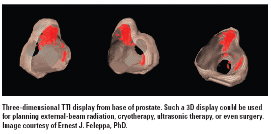
A ‘major breakthrough’ for biopsy guidance and treatment planning and delivery.
A new technique, ultrasonic tissue-type imaging, could revolutionize the detection and


treatment of prostate cancer, according to Ernest J. Feleppa, PhD, research director of the Frederic L. Lizzi Center for Biomedical Engineering at the Riverside Research Institute in New York. “The method seems to be capable of distinguishing cancerous from noncancerous tissue in the prostate,” Dr. Feleppa said. “This would be a major breakthrough for biopsy guidance and treatment planning and delivery.”
Echo signals carry sound pulses that are unique to the tissue investigated by the ultrasound probe. Much of the information present in the original echoes is currently discarded in the process of creating a conventional image. Tissue-type imaging (TTI) instead analyzes the echo-signal spectrum before it is converted into an image, he said.
In a recent study, Dr. Feleppa and coauthor Christopher Porter, MD, used TTI to analyze echo signals of 617 prostate regions under biopsy from 64 patients. The investigators combined the spectral characterization of the biopsy samples with PSA values using a computerized classification system that calculated scores for the relative likelihood of cancer. After comparing TTI-based characterization with conventional B-mode interpretations of the biopsy locations, the researchers found that TTI could successfully detect cancers missed by conventional ultrasound (RSNA 2008 abstract SSJ12-03).


Conventional imaging modalities cannot reliably detect prostate cancer. As a result, biopsies cannot be directed into suspicious regions but are, instead, systematically but “blindly” placed at predetermined sites throughout the gland, Dr. Feleppa said. Thus, treatment usually involves the entire gland because the tumor locations cannot be pinpointed accurately.
If further research validates the results obtained to date, however, biopsies would be far more accurate. TTI could bring down the false-negative biopsy rate or reduce the number of unwarranted biopsies, he said. The method could also bolster staging, reduce side effects, and provide a reliable way to follow up treated and untreated cancers. Treatment of the whole gland could potentially give way to focalized therapy approaches.
“While some advanced MR methods show similar promise, the difference in cost, ease of clinical use, and examination time make ultrasound the preferable modality by far,” Dr. Feleppa said.


Prolaris in Practice: Guiding ADT Benefits, Clinical Application, and Expert Insights From ACRO 2025
April 15th 2025Steven E. Finkelstein, MD, DABR, FACRO discuses how Prolaris distinguishes itself from other genomic biomarker platforms by providing uniquely actionable clinical information that quantifies the absolute benefit of androgen deprivation therapy when added to radiation therapy, offering clinicians a more precise tool for personalizing prostate cancer treatment strategies.


