PET/MR Could Be Effective for Staging Esophageal Cancer
Preoperative staging of esophageal cancer with PET/MR is approximately as good as with endoscopic ultrasound, and may improve slightly over ultrasound and PET/CT scans, according to a new study.
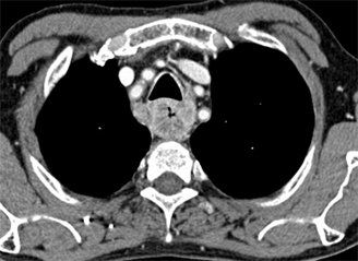
Esophageal cancer as seen on CT; source: Hellerhoff, Wikimedia Commons

Preoperative staging of esophageal cancer with the new imaging modality PET/MR is approximately as good as with endoscopic ultrasound, and may improve slightly over ultrasound and PET/CT scans, according to a new study published in the Journal of Nuclear Medicine on May 27.
“Accurate staging of esophageal cancer is critical for decisions on patient treatment,” wrote study authors led by Geewon Lee, MD, of Pusan National University Hospital in Busan, South Korea. Current guidelines recommend CT of the chest and abdomen along with endoscopic ultrasound (EUS) and PET/CT. “The recently available hybrid modality PET/MR imaging allows combination of both metabolic and anatomic information about the cancer.” To assess this new modality, the researchers compared PET/MR with EUS, CT, and PET/CT in esophageal cancer staging.
Over the course of 1 year, 19 patients with resectable esophageal cancer underwent preoperative imaging with all four of the imaging modalities, and tumor and lymph node stages were assigned. Four of the patients were subsequently treated non-surgically, and were thus excluded from the analysis.
The primary tumors were correctly staged in 13 patients (86.7%) using EUS, in 10 patients (66.7%) using PET/MR, and in five patients (33.3%) using CT scanning. EUS was significantly better than CT (P = .021), though no better than PET/MR (P = .375). PET/MR just failed to reach significance over CT imaging (P = .063).
EUS was still best with regard to determining T1 lesions, at 86.7%. PET/MR had an accuracy of 80% while CT had an accuracy of 46.7%. EUS was also better at distinguishing T3 lesions, at 93.3% accuracy; PET/MR and CT were both 86.7%.
With regard to lymph node staging, PET/MR outperformed the other modalities. Lymph node staging accuracy was 83.3% with PET/MR compared with 75% for EUS, 66.7% for PET/CT, and 50% for CT scanning. “Accurate N staging is essential, as it is a known prognostic factor associated with overall survival,” the authors wrote. “MR imaging offers the advantages of excellent soft-tissue contrast and lack of exposure to ionizing radiation.”
The authors did note the small number of patients as a limitation to the study’s interpretation, as well as the inclusion of only male patients with squamous cell carcinoma. Still, they concluded that “our results suggest that with protocol adjustments, PET/MR imaging may be used clinically for the preoperative staging of esophageal cancer in the future.”