Polypoid Lesions of the Lower Female Genital Tract
A 46-year-old multiparous (gravida 3, para 3) woman presented to her primary care provider with a palpable vulvar polypoidal mass, measuring 7 cm in greatest dimension. The mass was painless and had been growing in size over the past 2 years. Her medical history was remarkable for obesity, hypothyroidism, and an appendectomy at age 17. Her family history was significant for a sister with breast cancer, diagnosed at age 34. A core biopsy was performed.
ABSTRACT: Multidisciplinary Consultations on Challenging CasesThe University of Colorado Health Sciences Center holds weekly second opinion conferences focusing on cancer cases that represent most major sites of the disease. Patients seen for second opinions are evaluated by an oncologist. Their history, pathology, and radiographs are reviewed during the multidisciplinary conference, and then specific recommendations are made. These cases are usually challenging, and these conferences provide an outstanding educational opportunity for staff, fellows, and residents in training. The second opinion conferences include actual cases from genitourinary, lung, melanoma, breast, neurosurgery, and medical oncology. On an occasional basis, ONCOLOGY will publish the more interesting case discussions and the resultant recommendations. We would appreciate your feedback on the series; please contact us at second.opinion@uchsc.edu. E. David Crawford, MD, and Al Barqawi, MD, Guest Editors University of Colorado Health Sciences Center and University of Colorado Cancer Center Denver, Colorado
A 46-year-old multiparous (gravida 3, para 3) woman presented to her primary care provider with a palpable vulvar polypoidal mass, measuring 7 cm in greatest dimension. The mass was painless and had been growing in size over the past 2 years. Her medical history was remarkable for obesity, hypothyroidism, and an appendectomy at age 17. Her family history was significant for a sister with breast cancer, diagnosed at age 34. A core biopsy was performed.
Discussion
What did the core biopsy show?
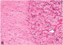
Dr. Amy Storfa: Hematoxylin and eosin-stained sections of the core biopsy showed a tumor composed of relatively small, uniform spindle cells with eosinophilic cytoplasm and bland nuclei with no appreciable cytologic atypia. The background showed various numbers of medium- to large-sized blood vessels, some with focally hyalinized walls (Figure 1A, 1B). Mitotic figures were not identified. Immunohistochemical stains for smooth muscle actin, desmin, vimentin, estrogen receptor, and progesterone receptors were positive; an immunohistochemical stain for S-100 was negative. The diagnosis was aggressive angiomyxoma, also known as deep angiomyxoma.
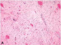
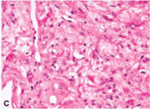
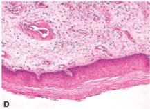
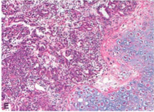
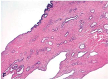
Figure 1: Polypoid Lesions of the Lower Female Genital Tract-
(A) Hematoxylin and eosin (H&E) stain of aggressive angiomyxoma showing ahypocellular tumor with numerous variable-sized blood vessels (magnification X100);
(B) H&E stain of aggressive angiomyxoma displaying proliferating capillary-sized blood vessels on the right and hypocellular stroma with spindle-shaped cells showing minimal cytologicatypia on the left (magnification X200);
(C) H&E stain of angiomyofibroblastoma composed of a proliferation of vascular spaces with relatively hypercellularstroma and spindled to ovoid cells displaying mild nuclear pleomorphism (magnification X400);
(D) H&E stain of fibroepithelial stromal polyp of the vagina covered by benign squamous epithelium. The subepithelial connective tissue is edematous and shows variable-sized blood vessels, some of which are thick-walled. The stroma ranges fromhypocellular to relatively hypercellular,and the stromal cells display mild pleomorphism (magnification X100);
(E) H&E stain of mixed malignant mllerian tumor with the left half of the photomicrograph displaying a high-grade adenocarcinoma component, and the right half displayinga chondrosarcoma component (magnification X100); (F) H&E stain of adenosarcoma showing benign glandular elements dispersed amongst a cellular stroma (magnification X40).
What were the operative findings?
Dr. Susan Davidson: A rubbery, gelatinous 10-cm mass was located in the left labia majora with extensive infiltration into the surrounding soft tissue.
What did pathologic examination of the mass show?
Dr. Meenakshi Singh: Gross examination revealed a yellow-tan ill-defined soft-tissue mass measuring 10 X 5.1 X 3.2 cm. The cut surface of the mass had a grey-pink gelatinous appearance; the edge of the lesion showed infiltration into the surrounding soft tissue. Hematoxylin and eosin-stained sections of the mass showed a poorly circumscribed tumor with extension into the surrounding connective tissue. Cytologically, the tumor showed histologic features similar to those seen in the biopsy. The surgical margin was uninvolved by tumor. The final diagnosis was aggressive angiomyxoma.
What are the clinical features of aggressive angiomyxoma?
Dr. Davidson: Aggressive angiomyxomas involve the deep soft tissue of the vulvovaginal region, pelvis, and perineum. They virtually never metastasize, but rather, infiltrate locally into these regions with potential for recurrence and localized destruction. Magnetic resonance imaging studies are often obtained to assess for local extension. Approximately 30% to 40% of the lesions recur, and lesions may grow to over 20 cm. Women in the third to fifth decade of life are almost exclusively affected. However, some cases have been reported in the inguinoscrotal area in men. Clinically, these may be mistaken for a cystic lesion like a Bartholin's cyst or a hernia.[1,3] These lesions are rare, and no specific risk factors have been described in their development.
What is the treatment of aggressive angiomyxomas?
Dr. Davidson: Careful resection of these lesions with negative surgical margins is imperative, as the mass is characteristically infiltrative. Positivity for estrogen and progesterone hormone receptors in addition to accelerated growth of these lesions during pregnancy raises the possibility that they may be hormone-responsive lesions and perhaps amenable to treatment with gonadotropin-releasing hormone (GnRH) agonists.[1]
Adjuvant treatment with GnRH agonists may be especially useful if the lesion was known to extend close to surgical margins without the option of excising more tissue. Radiation therapy has been reported in this setting but does not appear to be of proven benefit.
What other entities should be in the differential diagnosis?
Dr. Singh: The differential diagnosis for this lesion includes aggressive angiomyxoma, angiomyofibroblastoma, fibroepithelial stromal polyp, cellular angiofibroma, and superficial angiomyxoma.
Dr. Davidson: Clinically, the size of this lesion suggested aggressive angiomyxoma, but angiomyofibroblastoma and superficial angiomyxoma were also on the list of differential diagnoses in terms of presentation and location.
Dr. Singh: Angiomyofibroblastoma is a distinct neoplasm that was initially described in 1992. It occurs during reproductive age and in early postmenopausal women and is characteristically well-circumscribed with a pseudocapsule. It has also been reported in men in the inguinoscrotal region. The size of the tumor is generally smaller (< 5 cm) than an aggressive angiomyxoma. It presents clinically as a slow-growing, painless subcutaneous mass. These lesions generally behave in a benign manner and, in contrast to aggressive angiomyxomas, do not recur. When they do recur, it is usually a case of the lesion having been shelled out without a peripheral rim of nonneoplastic tissue. In any case, the recurrence is not destructive.
Histologically, the mass is well-circumscribed and has a more fibrous, less myxoid stroma. The background contains hypercellular areas admixed with hypocellular areas. Blood vessels are delicate and thin-walled and have plump epithelioid stromal cells clustered in a perivascular pattern; in contrast, aggressive angiomyxomas contain both thin- and thick-walled vessels (Figure 1C). It shares a similar immunohistochemical profile with aggressive angiomyxoma and is therefore difficult to distinguish based on immunohistochemistry alone.[7]
Fibroepithelial stromal polyps are polypoid lesions that may be pedunculated or sessile and singular or multiple. They arise in the vulva of young to middle-aged women and are benign but can recur if not completely excised. Fibroepithelial stromal polyps have an association with hormones; patients may be pregnant at the time of diagnosis or may have a history of hormone replacement therapy.[7]
Histologically the polyps are composed of a core of spindle and stellate cells and sometimes multinucleated cells in a background of fibrous stroma. The entire polyp is covered with stratified squamous epithelium with no evidence of koilocytosis (Figure 1D). There is no association with human papillomavirus infection. Immunohistochemical stains show the lesion to be immunoreactive for vimentin, desmin, and estrogen and progesterone receptors. Bizarre stromal cells have been reported but do not influence behavior, although this may raise the suspicion of sarcoma. (Hence, these lesions are sometimes referred to as pseudosarcoma botryoides.) Another consideration may be sarcoma botryoides, which can be distinguished by prepubertal presentation and immunohistochemistry positive for skeletal muscle markers.[5]
Cellular angiofibroma is a rare, well-circumscribed rubbery stromal tumor that arises in the vulva of middle-aged women. This lesion is benign, and excision is the treatment. Histologically, cellular angiofibromas are composed of uniform bland spindle cells and many thick-walled blood vessels with a mostly peripheral collection of mature adipocytes. The immunohistochemical profile of cellular angiofibroma is desmin-negative and vimentin-positive, which helps to distinguish it from the desmin- and vimentin-positive aggressive angiomyxoma.
Superficial angiomyxoma is a slow-growing painless subcutaneous mass that is usually less than 5 cm in diameter. Although this tumor does occur in the genital area, it also is found in locations such as the head and neck region and the trunk. The presence of multiple cutaneous myxomas and angiomyxomas may raise suspicion for Carney's complex. This may be an important association, as patients with Carney's complex are at increased risk of cardiac myxomas and sudden death.[3] Although superficial angiomyxoma is benign, the lesion recurs at a rate of approximately 30% in a nondestructive presentation. Excision with a clear margin is recommended. Histologically, the lesion is located in the dermis and is multilobulated; fibroblasts and thick-walled vessels in a myxoid matrix comprise the lesion.
Another lesion that may present as a polyp of the lower genital tract is malignant mllerian mixed tumor (carcinosarcoma). Although the majority of these lesions present as a uterine primary, rare carcinosarcomas of the vagina have been reported; metastases from other sites must be excluded. This malignant lesion usually occurs in elderly postmenopausal women, but cases in younger women have been reported. In the vagina, the lesions take on a polypoid configuration.
Histologically, carcinosarcoma consists of a malignant epithelial component, which is usually glandular with a sarcomatous component that can be homologous or heterologous. The mesenchymal component is typically undifferentiated sarcoma, leiomyosarcoma, or endometrial stromal sarcoma in homologous cases. If heterologous, the mesenchymal component is most commonly malignant cartilage or skeletal muscle (Figure 1E). An association between tamoxifen therapy and carcinosarcoma has been suggested. Clinically, the prognosis of primary vaginal carcinosarcomas is poor, and patients rapidly develop metastases.[5]
Adenosarcoma is also a consideration in the differential diagnosis. When arising in the vagina, it is likely to present as an exophytic lesion. But, most adenosarcomas in the vagina represent metastases from the endometrium or elsewhere in the genital tract. Histologically, it is a mixed tumor comprised of a benign epithelial glandular component and a malignant mesenchymal component (Figure 1F).[5]
How would treatment of these other entities differ from that of aggressive angiomyxoma?
Dr. Davidson: Angiomyofibroblastoma, fibroepithelial stromal polyps, cellular angiofibroma, and superficial angiomyxoma are treated with surgical excision. In all cases, recurrence is a possibility if neoplastic tissue remains. The above lesions are relatively well-circumscribed. In contrast, aggressive angiomyxomas are diffusely infiltrative into surrounding soft tissue and require close attention to surgical margins at the time of excision as well as histologic examination.[6]
Are there known cytogenetic abnormalities of aggressive angiomyxomas?
Dr. Storfa: The DNA architectural factor HMGIC, located on chromosome 12, is rearranged in aggressive angiomyxomas and results in aberrant protein expression. Expression of HMGIC is present in nuclei of aggressive angiomyxomas but absent in the other entities being considered in the differential diagnosis.[3] This knowledge may prove useful in distinguishing aggressive angiomyxoma from its histologic mimics as well as in evaluating residual disease. Of note, aberrant expression of HMGIC is reported in a variety of other benign mesenchymal neoplasms, such as endometrial polyps, uterine leiomyomas, lipomas, and pulmonary hamartomas.
What was the outcome in this case?
Dr. Davidson: The final pathology of the resection specimen showed negative surgical margins, and the patient has no evidence of recurrence at 3 years. If she did have a recurrence or had an initially unresectable tumor, I would recommend GnRH agonist therapy.
Disclosures:
The authors and participants in this conference have no significant financial interest or other relationship with the manufacturers of any products or providers of any service mentioned in this article.
References:
1. McCluggage WG: A review and update of morphologically bland vulvovaginal mesenchymal lesions. Int J Gynecol Pathol 24:26-38, 2005.
2. McCluggage WG: Recent advances in immunohistochemistry in gynaecological pathology. Histopathology 40:309-326, 2002.
3. Nucci MR, Fletcher CD: Vulvovaginal soft tissue tumours: Update and review. Histopathology 36:97-108, 2000.
4. Stoler MH, Mills SE, Frierson HF: Sternberg's Diagnostic Surgical Pathology, 4th ed, pp 2342-2343. Philadelphia, Lippincott Williams & Wilkins, 2004.
5. Tavassoli FA: World Health Organization Classification of Tumours Pathology and Genetics Tumours of the Breast and Female Genital Organs. Lyon, IARC, 2003.
6. Tochika N, Takeshita A, Sonobe H, et al: Angiomyofibroblastoma of the vulva: Report of a case. Surg Today 31:557-559, 2001.
7. Nielsen GP, Young RH: Mesenchymal tumors and tumor-like lesions of the female genital tract: A selective review with emphasis on recently described entities. Int J Gynecol Pathol 20:105-127, 2001.