Radiation Dermatitis
42-year-old Caucasian female who was in her usual state of health when her first mammogram showed suspicious calcifications and a spiculated mass in the upper outer quadrant of the right breast. An ultrasound-guided biopsy showed an invasive ductal carcinoma. She underwent a lumpectomy, with the excised tumor measuring 1.2 cm. The tumor was estrogen and progesterone positive and HER2/neu negative.
Mrs. O is a 42-year-old Caucasian female who was in her usual state of health when her first mammogram showed suspicious calcifications and a spiculated mass in the upper outer quadrant of the right breast. An ultrasound-guided biopsy showed an invasive ductal carcinoma. She underwent a lumpectomy, with the excised tumor measuring 1.2 cm. The tumor was estrogen and progesterone positive and HER2/neu negative.
Also present was an intermediate-grade ductal carcinoma in situ (DCIS) measuring approximately 4-5 cm. The DCIS margins were close in two areas, so she underwent a repeat lumpectomy 10 days later with no residual disease in the specimen. Three sentinel nodes were excised and were negative for malignancy. Oncotype DX (a multigene assay that predicts the likelihood of recurrence of invasive breast cancer) showed a low-risk lesion. Accordingly, the medical oncologist recommended adjuvant tamoxifen without chemotherapy and consultation with a radiation oncologist.
Accompanied by her husband, Mrs. O presents to the radiation oncologist 2 weeks following her second surgery. She is a homemaker who has smoked 1 pack per day (PPD) for the past 20 years and denies alcohol or drug use. Her health history includes two Cesarean sections in her 30s and no other medical conditions. She is 5 foot 10 inches in height and weighs 215 pounds. Her breasts are large and pendulous. After discussion of stage, prognosis, and treatment options, external radiation is recommended. Because the DCIS component of the tumor is greater than 3 cm, she is not a candidate for partial breast radiation with MammoSite. Baseline blood counts are within normal limits. Since she has no other health problems and did not receive chemotherapy, blood counts will not be repeated during treatment. In addition, the nurse offers consultation with a smoking-cessation specialist. Mrs. O states she is trying to quit on her own and will let staff know if she wishes to see the specialist. She also expresses interest in reducing her weight and agrees to meet with the registered dietitian at her next visit.
Treatment Summary and Nursing Management
TABLE 1

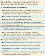
Skin Care Instructions for Patients Receiving External Radiation to the Breast
The radiation oncologist prescribes the following treatment plan: 4,500 centigray (cGy) in 25 fractions to the entire right breast and a boost to the lumpectomy site of 1,600 cGy in 8 fractions, for a total of 6,100 cGy. Radiation treatment will be given daily, Monday through Friday. At the time of consultation, the nurse provides education regarding the treatment plan, expected side effects, onset and duration, and skin care during external radiation. The nurse tells Mrs. O that she may be at increased risk of a skin reaction because of her breast size and smoking history. Patient instructions per department protocol are outlined in Table 1. Skin will be assessed at least once a week throughout treatment by the radiation oncologist and nurse. Skin toxicity is graded using the National Cancer Institute Terminology Criteria for Adverse Events Version 3.0 (Table 2).
TABLE 2

Grades of Dermatitis Associated with Radiation/Chemoradiation
The patient developed a skin reaction that progressed and resolved with treatment as follows:
• Week 1: No skin reaction, patient reports no new symptoms.
• Week 2: No skin reaction, patient reports no new symptoms.
• Week 3: Grade 1 mild erythema. Patient reports no discomfort in treatment area. She complains of mild fatigue relieved by rest periods. Continue skin care protocol.
• Week 4: Grade 2 brisk erythema. Mild discomfort and itching in the inframammary fold and upper outer quadrant of right breast. Biafine (OrthoNeutrogena, Skillman, NJ) is added twice a day to both areas of discomfort. Mrs. O continues to use 100% aloe vera applied to the entire right breast.
FIGURE 1

Radiation Dermatitis
• Week 5: Grade 2 brisk erythema with 2-cm area of dry desquamation in the upper outer quadrant. Biafine has provided relief from itching but patient is having trouble sleeping at night because of skin discomfort. Aloe and Biafine are continued, and two acetaminophen at bedtime are recommended. Radiation oncologist modified treatment by starting boost early while discontinuing radiation to whole breast to allow healing.
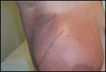
• Week 6: Grade 2 skin reaction resolving (see Figure 1). Boost completed and whole-breast radiation resumed. Aloe and Biafine are continued. Patient reports minimal discomfort not interfering with sleep, and mild fatigue.
• Week 7: Grade 2 skin reaction stable. External radiation completed. Patient instructed to continue use of aloe and Biafine for 2 weeks or until skin reaction has resolved.
• Two weeks post-treatment: Nurse calls Mrs. O to assess status. Patient reports no discomfort, skin erythema improving, skin with mild peeling, open area in upper outer quadrant healed. She continues to apply Biafine and 100% aloe vera gel.
• Four weeks post-treatment, follow-up in radiation department: Skin intact, skin reaction grade 1-mild erythema. Patient with no complaints, fatigue decreasing, no discomfort in treatment area, peeling resolved. Skin care products were discontinued, and patient was instructed to resume her usual hygiene routine.
Discussion
External radiation therapy is commonly used as part of breast conservation therapy. Some degree of skin reaction ranging from mild erythema to painful skin breakdown will occur in about 90% of women receiving external radiation following lumpectomy or mastectomy.[1] Skin care in these women varies widely among institutions. A recent review of studies conducted between 1996 and 2004 with different skin care products showed no significant difference in time to onset or severity of skin reactions based on the product used.[2] Although aluminum-based deodorant and any skin care product within 4 hours of treatment is often prohibited, two studies have shown that this practice does not prevent or decrease skin reactions.[3,4] Rather, treatment and patient factors seem to have the greatest influence on the onset and severity of radiation dermatitis.
A 1998 study showed that factors most predictive of a skin reaction included higher weight, larger breast size, lymphocele aspiration, cigarette smoking, history of skin cancer anywhere on the body, tumor stage, and radiation dose.[5] Treatment-related factors such as fraction size, type of radiation, and concurrent chemotherapy also have an impact on skin reactions.[1] If the specific skin product does not influence the outcome, the goal of nursing care should focus on maintaining the patient's quality of life by improving comfort and eliminating unnecessary restrictions. Choice of skin care product should be made based on cost, availability to patients, and effectiveness for that individual.
In the department in this case study, a mild soap and 100% aloe vera are the first-line products in the skin care protocol, provided the patient is not allergic, because it has been shown in one study to be more effective than using mild soap only,[1] is low-cost, nonprescription, and easy to obtain. Many women do not progress past a grade 1 while using 100% aloe vera alone. Mrs. O has risk factors for developing a skin reaction, including weight, large breasts, and cigarette smoking. Once Mrs. O developed a grade 2 reaction, a prescription product, Biafine, was added. Biafine was shown in a 2002 study to be more effective than 100% aloe vera at reducing dry desquamation and pain.[7]
Conclusion
Skin reactions resulting from external radiation to the breast have improved over the years, mostly as a result of advances in treatment techniques. Further improvement is anticipated with the availability of targeted or modified radiation treatment methods such as MammoSite and intensity-modulated radiation therapy (IMRT). Because many factors can influence skin reactions in these patients, best practice is a standard skin care protocol that includes a choice of skin products. Evidence from similar research indicates that minor modifications of the normal hygiene routine, such as those outlined in this case report, can have a positive impact on quality of life.
References:
1. Harper JL, Franklin LE, Jenrette JM, et al: Skin toxicity during breast irradiation: Pathophysiology and management. South Med J 97(10):989-993, 2004.
2. Aistars J: The validity of skin care protocols followed by women with breast cancer receiving external radiation. Clin J Oncol Nurs 10(4): 487-492, 2006.
3. Burch SE, Parker SA, Vann AM, et al: Measurement of 6-MV x-ray surface dose when topical agents are applied prior to external beam irradiation. Int J Radiat Oncol Biol Phys 38(2):447-451, 1997.
4. Meegan MA, Haycocks TR: An investigation into the management of acute skin reactions from tangential breast irradiation. Can J Med Radiat 28:169-173, 1997.
5. Porock D, Kristjanson L, Nikoletti S, et al: Predicting the severity of radiation skin reactions in women with breast cancer. Oncol Nurs Forum 25(6):1019-1029, 1998.
6. Olsen DL, Raub W, Bradley C, et al: The effect of aloe vera gel/mild soap versus mild soap alone in preventing skin reactions in patients undergoing radiation therapy. Oncol Nurs Forum 28(3):543-547, 2001.
7. Heggie S, Bryant GP, Tripcony L, et al: A phase III study on the efficacy of topical aloe vera gel on irradiated breast tissue. Cancer Nurs 25(6):442-451, 2002.