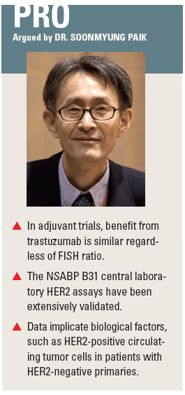
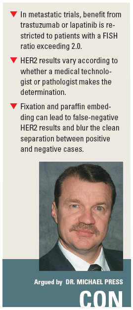
Should HER2-targeted therapies be used to treat HER2-negative breast cancers?
Some clinical trials of HER2-targeted therapies have found that patients assessed to have HER2-negative breast cancer nonetheless derive benefit. Is this a real benefit stemming from unrecognized biological factors in the adjuvant setting?
ABSTRACT: At the heart of the controversy lies the question of whether benefit in HER2-negative cases is related to complex tumor biology in the adjuvant setting or to false-negative assay results.
BALTIMORE-Some clinical trials of HER2-targeted therapies have found that patients assessed to have HER2-negative breast cancer nonetheless derive benefit. Is this a real benefit stemming from unrecognized biological factors in the adjuvant setting? Or is it an artifact of assay methodology producing false-negative HER2 results? Soonmyung Paik, MD, of the National Surgical Adjuvant Breast and Bowel Project (NSABP) and Michael Press, MD, PhD, of the Norris Comprehensive Cancer Center debated this topic at the 2008 Era of Hope meeting.
Contrasting with current dogma

Speaking in support of the use of HER2-targeted therapies in at least some cases of HER2-negative breast cancer, Dr. Paik discussed the NSABP B31 adjuvant trial, which tested the addition of trastuzumab (Herceptin) to chemotherapy among patients with HER2-positive disease. The latter was defined as 3+ staining by immunohistochemistry (IHC) or a ratio exceeding 2.0 by fluorescence in situ hybridization (FISH), as determined by any lab in the US or Canada. Central retesting showed that 10% of cases were negative for HER2 by both assays.
“Surprisingly, when we looked at efficacy or the degree of benefit from trastuzumab, we found that both HER2-negative and HER2-positive patients had the same degree of benefit,” Dr. Paik commented. Moreover, the parallel trial, NCCTG N9831, had similar findings.
A plot of the observed risk reduction in both trials as a function of FISH ratio yielded a flat line, he continued, meaning that “everybody got benefit, regardless of how much HER2 DNA or protein they had.” This result contrasts with the current dogma regarding response to HER2-targeted therapies, which is drawn from the metastatic setting and proposes an absolute threshold for response at a FISH ratio of 2.0. “If you follow that logic, patients with tumors that have a ratio of 1.99 should not respond, while [those with tumors with a ratio of] 2.01 will respond,” he commented, saying that does not intuitively make sense.
One proposed explanation for the B31 findings, according to Dr. Paik, is that the negative results obtained in the central lab were incorrect. However, these results were determined by three board-certified breast pathologists, and the assays have stood up to multiple tests scrutinizing their validity, he contended. For example, a microarray analysis of 753 HER2-positive and -negative cases from the B31 trial confirmed that there was no HER2 amplification in the negative tumors, and the specificity of the results ruled out the possibility that the results are due to degradation of mRNA in the paraffin blocks.
Furthermore, he noted, the false-positive rate for the central lab was lower than that reported by other trials, and the assay methodology used was rigorous enough that it served as the basis for the current ASCO College of American Pathologists (CAP) guidelines for HER2 testing.
Another possible explanation that the investigators considered for the response to trastuzumab in HER2-negative patients was tumor heterogeneity, with the possibility that local and central labs sampled tissue blocks from different parts of the tumor with different HER2 status. But in 67 HER2-negative cases in which both the primary tumor and a lymph node were tested by FISH, the node was positive in only two cases (3%). “That means that there is no possibility that our data can be explained by heterogeneity within the same tumor,” Dr. Paik asserted.
One might note the overlap in HER2 mRNA levels in positive and negative cases in B31 and argue that the results were due to degradation of mRNA as the result of formalin fixation and paraffin embedding, he observed. However, he pointed out, a compilation of 10 microarray studies from Oncomine looking at HER2 mRNA levels in fresh or frozen tumors showed that there is almost always overlap between amplified and nonamplified cases.
Preclinical data suggest that the correlations between level of HER2 amplification and response to trastuzumab (Emlet DR et al: Mol Cancer Ther 6:2664-2674, 2007) and lapatinib (Konecny GE et al: Cancer Res 66:1630-1639, 2006) are in fact linear, he observed. This finding, in turn, suggests that the current generation of IHC and FISH assays yielding a sharp cutoff is nonlinear, which could also be an explanatory factor.
In fact, Dr. Paik noted, HER2 expression by breast cancer is a continuous variable, with the distribution overlapping the currently used cutoffs for IHC and FISH, as shown by an RT-PCR analysis of 20,000 cases. Nevertheless, additional analyses of B31 tumors showed that HER2 content did not predict benefit from trastuzumab, regardless of whether the level was measured by a DNA, mRNA, or protein assay.

Indeed, he pointed out, none of the genes predicting trastuzumab benefit were even in the HER2 pathway. Moreover, there was no correlation between the mRNA level of these genes and that of HER2. Taken together, he said, these findings suggest that assay nonlinearity, although an issue, cannot explain the clinical results.
A final explanation, and one currently under investigation, according to Dr. Paik, is complex tumor biology in the adjuvant setting that is not yet understood. “The adjuvant setting can be very different from metastatic disease because of a lot of confounding variables,” he noted.
These could include prior removal of the primary tumor and additional therapies. One hypothesis suggests that patients with HER2-negative primaries may have circulating or disseminated tumor cells that are HER2-positive (Klein CA: Cell Cycle 3:29-31, 2004). Indeed, a study of 27 patients found that among those having a negative primary tumor and disseminated tumor cells in the bone marrow, the disseminated cells were positive 31% of the time (Schardt JA et al: Cancer Cell 8:227-239, 2005).
Similarly, a study of 24 patients with recurrent breast cancer who had a negative primary found that nine (38%) had circulating tumor cells with HER2 amplification (Meng et al: PNAS 101:9393, 2004); four cases were treated with trastuzumab, with a response in three of them. “In that setting, if you give adjuvant trastuzumab, maybe it is highly efficient in removing these circulating tumor cells or cells in the bone marrow,” he commented.
“Obviously, I am not here to argue that every patient, even if HER2-negative, should be treated with trastuzumab in the adjuvant setting,” Dr. Paik said, cautioning that some of the B31 analyses were unplanned and based on small numbers of patients. “A randomized prospective trial is needed to test the worth of HER2-targeted therapy in HER2-negative patients in the adjuvant setting,” he concluded, noting that NSABP is proposing such a trial among patients with low-level HER2 expression (1+ or 2+ by IHC and negative by FISH), with randomization to chemotherapy with or without trastuzumab.
No incremental gain
“The position I am taking is against the use of HER2-targeted therapies to treat HER2-negative breast cancer patients,” Dr. Press contended. He noted that in the pivotal trials of trastuzumab and lapatinib (Tykerb) in the metastatic setting, benefit was almost entirely restricted to patients who had a FISH ratio exceeding 2.0. “It’s not an incremental [gain] that we see in the data,” he asserted.
Dr. Press discussed the EGF100151 clinical trial, in which patients with metastatic breast cancer determined to be HER2-positive by IHC in a local lab were randomized to capecitabine (Xeloda) with or without lapatinib. Some patients were subsequently found to be HER2-negative by FISH performed in a high-volume central lab, prompting a second FISH assessment with the same method in a small academic central lab. The rate of agreement between the two central labs was 89%. He noted that progression-free survival was significantly improved by addition of lapatinib among the HER2-negative subset as determined by the high-volume lab-but not as determined by the small academic lab. The main difference between the labs was that the FISH determination was made by medical technologists in the former and by board-certified pathologists in the latter.
“Heterogeneity is not likely to account for the differences that are seen because it’s relatively infrequent,” Dr. Press further noted. For example, dual IHC and FISH analyses among patients in the lapatinib trials have shown that only 0.4% of tumors have both regions with and without amplification.
“So the primary issue, I think, has to do with the person who is performing the assessment and the qualifications of that person,” he said. In support of this, he noted that FISH results are concordant 89% to 92% of the time when comparing medical technologists with pathologists, but 99% of the time when comparing pathologists with pathologists (as found in CIRG trials).
“I would argue that one of the issues that we have has to do with the material that we are working with,” Dr. Press continued. Factors preceding the assay-tissue handling, fixation, and processing-all play a role. He noted that results obtained with frozen tissue are similar no matter what assay is used, provided the tissue is properly handled to prevent degradation of protein and RNA (Slamon D et al: Science 244:707-712, 1989). However, fixed, paraffin-embedded tissue from the same tumor shows markedly reduced antigenicity.
Dr. Press maintained that the FISH assay produces a sharp cutoff at a ratio of 2.0 between HER2-amplified and HER2-nonamplified breast cancer, as has been shown both in 44 frozen cell lines (Dering J et al: Unpublished data, 2008). “There is a clear separation in the amount of RNA that corresponds to HER2 between those two cases-there is no overlap,” he said. Similarly, in tumors from 2,502 consecutive patients screened for clinical trial entry, FISH ratios were equivocal in only a minute proportion of cases (Sauter ER et al: J Clin Oncol In press, 2008). At the same time, additional data suggest that a degradation of mRNA resulting from fixation and processing may blur that clean separation. Indeed, he noted, compared with frozen tissue from the same tumor, formalin-fixed, paraffin-embedded tissue shows substantial mRNA degradation (Chen J et al: Diagn Mol Pathol 16:61-72, 2007).
Furthermore, when the HER2 content of fixed, paraffin-embedded tissue is ascertained by both FISH and RT-PCR, although there is still some separation of amplified and nonamplified cases according to FISH ratio, there is now also considerable overlap, making it difficult to discern a cutoff.
“Based on the data that we have and have seen, HER2-negative (not amplified) breast cancer patients do not show improved outcomes relative to chemotherapy alone for either trastuzumab or lapatinib in the studies that we have participated in,” Dr. Press concluded.
“We think a pathologist evaluation of this material is quite important for assessing the HER2 status. And we think the mRNA in fixed, paraffin-embedded samples is variably degraded, not only for the HER2 gene, but for genes in general, and it will be difficult to assess in a consistent way.”