Twenty Years of Advancing Discoveries and Treatment of Mantle Cell Lymphoma
In 2023, the Lymphoma Research Foundation hosted the Mantle Cell Lymphoma Scientific Consortium and Workshop to discuss treatment advancements in the field.
Introduction
Mantle cell lymphoma (MCL) is an aggressive B-cell lymphoma most often associated with the t(11;14) translocation, leading to overexpression of cyclin D1. MCL is clinically heterogeneous, and as a result, there is no single therapeutic approach or standard of care. Furthermore, the determinants of these varying clinical phenotypes (eg, indolent disease vs more aggressive disease) are unclear and, thus far, there is no way to predict treatment response. Importantly, research results have revealed that it is not just the genetic or epigenetic profiles of individual tumor cells that affect treatment response, but also the tumor microenvironment (TME). In recent years, research has not only taken advantage of an improved ability to monitor genetic/epigenetic changes and gene expression on the single-cell level to evaluate MCL tumor biology but has expanded to include analysis of the cross talk between the tumor cells and the cells of the TME and analysis of the resultant impact on treatment response. In addition, the introduction of several new agents to treat patients with MCL has amplified the need to clearly define molecular response and clinical outcomes so that patient response may be rapidly and accurately assessed and the use of existing agents optimized. As clinical trials continue to be developed, it will be important to include biomarkers to better stratify patients and assess response in clinical trials so that these markers may eventually be used to guide clinical care.
Recognizing the need for accelerated MCL research and collaboration between clinical and scientific researchers, the Lymphoma Research Foundation has provided MCL-specific research grants and developed the Mantle Cell Lymphoma Consortium (MCLC), a working group that includes both basic scientists and translational/clinical researchers from North America and Europe. Since 2003, the MCLC has met regularly to allow researchers to share their work. It offers a unique opportunity for collaboration between investigators across a wide range of MCL areas of interest. Through this exchange, thoughts on the current and future direction of MCL research are shared and researchers are provided with a unique opportunity to develop collaborations needed to continue to drive MCL research forward. The 20th MCL Scientific Consortium and Workshop, from May 2-3, 2023, in Chicago, Illinois, included sessions on MCL genetics, mechanisms of resistance or response to treatment, personalization of therapy and prognostics using biomarkers, the TME, an international overview of clinical trials, and an open forum to establish a road map toward the cure of MCL in the next 20 years.
Keynote Speaker
MCL From the Microscope to the Genome and Beyond: A Shared Journey
The keynote speaker was Elías Campo, MD, PhD, research director and professor of anatomic pathology at the Hospital Clinic of the University of Barcelona in Spain (Institut d’ Investigacions Biomèdiques August Pi i Sunyer).
Campo discussed the history of MCL classification and reviewed the key research findings that over the years have built the current understanding of MCL molecular biology. In addition, how specific MCL features or molecular signatures have been found to affect disease clinical behavior, in particular progression and resistance, was discussed.
MCL is characterized by the t(11;14) translocation, leading to overexpression of cyclin D1 and dysregulation of the cell cycle. However, cyclin D1 has both canonical and noncanonical functions, each of which is affected by cyclin D1 overexpression. For example, cyclin D1 is a transcription factor and its overexpression in MCL induces global transcriptional downregulation, which gives rise to vulnerability against CDK9 and transcriptional inhibitors.1 Thus, transcriptional inhibitors are a potential avenue for therapeutic development. Exploration of basic MCL biology and characterization of response to therapy are critical for identifying additional therapeutic targets.
Because MCL is heterogeneous, identification of molecular differentiators to be used in disease stratification is of central importance. TP53 and CDKN2A alterations in MCL strongly affect prognosis across a range of studies but have yet to be systematically incorporated into clinical care or clinical trial planning.2,3 Using next-generation sequencing (NGS), conventional and non-nodal MCL (cMCL and nnMCL) were identified as distinct molecular subtypes, which can be differentiated based on their expression of SOX11 and other features4; however, even within cMCL and nnMCL, clinical and molecular heterogeneity persists, with indolent and aggressive subtypes that remain challenging to identify. However, researchers have discovered through mutational analysis that a higher number of genomic alterations is associated with adverse outcomes in both cMCL and nnMCL.5 Importantly, genetic mutations are not the only type of alteration that may contribute to the outcome: Epigenetic differences are also present within MCL. For nnMCL and cMCL, regions of chromatin activation differ, suggesting another possible source of differing clinical behavior.
The heterogeneity of MCL is perhaps most evident when considering the contrasting behavior of indolent MCL, which can fail to progress for years, and aggressive MCL, which progresses rapidly. Campo discussed recent research that has sought to determine the molecular drivers of disease phenotype, as well as the best clinical approach for those with indolent disease. Overall, a combination of clinical and biological factors may be used to determine treatment approach. Although there is evidence that monitoring MCL prior to initiating treatment is not associated with a worse outcome, researchers have also found that ibrutinib in combination with rituximab is associated with a high rate of complete response (CR) and minimal residual disease (MRD).6 Campo also presented several recent findings that describe the differentiation of clinical response based on MCL genetic profile, highlighting recent work from Yi et al that used both genomic and transcriptomic profiling to identify distinct molecular subsets associated with differing outcomes.7
MCL Genetics, Epigenetics, and Genomics
In MCL, ongoing research is critical not only to identify novel therapeutics but also to better understand MCL disease biology and how it relates to clinical behavior so that treatment approaches may be tailored and outcomes optimized. In the workshop proceedings described here, the very latest research and advances within these areas are shared.
To open this session, Preetesh Jain, MBBS, MD, DM, PhD, assistant professor in the Department of Lymphoma/Myeloma in the Division of Cancer Medicine at The University of Texas MD Anderson Cancer Center in Houston, discussed the impact of mutation profiling on MCL prognosis and outcomes. To date, there are no established biomarkers that are routinely used clinically to predict outcomes or to plan treatment; however, there are findings from numerous studies that have identified associations between the mutational status of individual genes and patient outcomes. A 162-gene panel was used as part of routine care for 227 patients with MCL. Within the cohort, genomic DNA was isolated from a range of sources: bone marrow aspirate; formalin-fixed, paraffin-embedded blocks; fine-needle aspirate; and peripheral blood. Patients were either treatment naive (n = 130) or pretreated (n = 97). Of the pretreated patients, 47 had received a Bruton tyrosine kinase (BTK) inhibitor and had refractory disease, and 23 had been treated with chimeric antigen receptor (CAR) T-cell therapy. The mutations present in patient samples included ATM (51.1%), TP53 (29.5%), KMT2D (21.1%), CCND1 (19.3%), BIRC3 (16.2%), NSD2 (11.4%), SMARCA4 (10.1%), UBR5 (9%), NOTCH1 (8.3%), CARD11 (7%), SAMHD1 (7%), NFKB1E (6.6%), SP140 (6.6%), S1PR1 (6.6%), DNMT3A (6%), NOTCH2 (6%), IGLL5 (6%), TRAF2 (5%), and TET2 (5%). For these patients, there was no single constellation of mutations predictive of outcome; however, patients with more than 3 mutations had significantly worse survival rate (P < .0001).
Brian Li, from Washington University School of Medicine, then presented a survey of the genomic landscape of MCL. Whole-exome sequencing (WES) on 27 tumor-normal tissue pairs (lymph node and skin, respectively) was carried out, and single-nucleotide variants, insertions/deletions, structural variants, and copy-number variants were compared. For a subset of samples, a combined analysis of whole-genome sequencing (WGS), structural analysis, and RNA fusion detection was carried out. The use of WES findings confirmed the presence of recurrent variants canonical to MCL, including t(11;14) translocation (8 of 10 WGS samples), as well as TP53 (6 of 27), CCND1 (4 of 27), NSD2 (3 of 27), and NOTCH1 (3 of 27). CCND1 and TP53 mutations were found to be co-occurring in the patient cohort (P < .05) and were associated with shorter progression-free survival (PFS; P < .05). Several novel mutations were also uncovered, including ATM (7 of 27; missense variants, frameshift variants, deletions, and duplications), ZNF804B (3 of 27), DAZAP1 (3 of 27), and ASXL1 (2 of 27). Of note, the mutational profile observed in patients following CAR T-cell therapy was unique, highlighting the need to continue research on mechanisms of relapse in this setting. Importantly, the use of multiple methods for assessment of molecular changes within this cohort of patients with MCL permitted an integrated analysis across variant types and provided some insights into the differing mechanisms by which genes are mutated. As our understanding of biomarkers expands, it will be important to include them in clinical trials for validation and to support their incorporation into clinical care.
In the next talk, Sunandini Sharma, MS, Department of Pathology and Microbiology at the University of Nebraska Medical Center, presented research results on the impact of tumor mutations on interactions with the TME, and how a combination of MCL genetics and TME composition may be used to identify prognostic subtypes in MCL. Initial research findings revealed that MCL tumors with high proliferative indices not only have different constellations of mutations but also are associated with differing populations of immune cells within the TME when compared with MCL tumors with low proliferative indices. To better understand this dynamic and how individual tumors regulate their immune landscapes,samples from 153 patients used imaging mass cytometry, a time-of-flight analysis that circumvents the need for complexing inherent in other single-cell analysis methods. The study identified at least 2 prognostic MCL subtypes dependent on ATM or TP53 mutational status. TP53-positive tumor cells (which were also found to express high levels of p-STAT3, p-NFKB, and HLA) were associated with a diminished T-cell population (CD8+ and CD4+) compared with ATM-positive tumor cells, suggesting that the clinical outcomes for these subtypes may also hinge upon TME dynamics. By including information on the TME in stratification, accuracy may be improved beyond that achievable with tumor mutational analysis alone. Future research will include assessment of expression in nearest neighbors as well as expansion of analysis to include established tumor genetic markers of disease severity and their association with effects within the TME.
MCL Mechanisms of Resistance and Response
To open the session on mechanisms of MCL therapeutic resistance and response, Jianguo Tao, MD, PhD, professor in the Department of Pathology at the University of Virginia in Charlottesville, discussed the genetic heterogeneity and plasticity of MCL and how these features give rise to therapeutic resistance and clinical progression. In MCL, plasticity manifests as adaptive remodeling of the kinome, which limits the efficacy of even combination approaches with targeted kinase inhibitors. To overcome MCL resistance to BTK inhibitor (BTKi) therapeutics, an adaptation of the global kinome must be blocked. To understand the keystone activities that permit plasticity in MCL, a molecular assessment of venetoclax-tolerant MCL persister and expander cells was carried out. This analysis suggests that chromosomal 18q21 deletion and concomitant super-enhancer remodeling drive venetoclax resistance. Although expression levels of more than 100 genes were shown to either increase or decrease, all downregulation observed is 18q21 related, indicating pressure and selection for this mutation following BTKi exposure. Importantly, persister cells from both MCL cell lines and primary patient samples are sensitive to transcription inhibition (CDK7). Thus, cotargeting CDK7 transcription reprogramming and BCL-2 may prevent and/or overcome drug resistance achieved through adaptive remodeling. In the case of CAR T-cell resistance, which eventually occurs in MCL,8 in vitro targeting of CDK7/9 transcription also overcomes TME-mediated CAR T-cell therapy resistance and enhances CAR T-cell therapy efficacy against lymphoma growth through reshaping of TME evasion, thereby enhancing CAR T-cell trafficking and efficacy. Because residual disease is the eventual cause of relapse, this research provides important insight into how MCL cells may be managed to reduce the likelihood of persistence and expansion and highlights possible therapeutic strategies to improve the efficiency of or prevent resistance to existing therapies.
Next, Tycel Phillips, MD, associate professor in the Division of Lymphoma, Department of Hematology and Hematopoietic Cell Transplantation at City of Hope in Duarte, California, provided an update on bispecific antibodies in MCL. For non-Hodgkin lymphoma, the first data on bispecific agents using a T-cell engaging strategy were generated with blinatumomab. In the original phase 1 MT103-104 dose-escalation study (NCT00274742), blinatumomab was administered as a continuous infusion with mandated hospitalization (due to the short drug half-life), using doses as high as 90 µg/m2/day.9 The study was halted due to high rates of neurological toxicity, and step-up dosing with a reduced target dose of
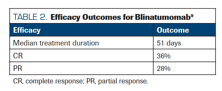
60 µg/m2/day was implemented to reduce complications. Although blinatumomab was shown to be effective, the need for inpatient infusion has limited its use for acute lymphoblastic leukemia (Tables 1 and 2).9
TABLE 1. Blinatumomab Dosing
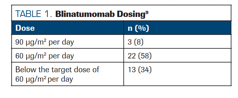
TABLE 2. Efficacy Outcomes for Blinatumomab
Thus, newer bispecifics have been modified to improve half-life and reduce the need for intensive infusion. In MCL, several CD20/CD3 bispecific studies have attempted to enroll patients with relapsed/refractory (R/R) MCL. To date, significant results have been reported with only 2 agents. The first was the phase 1/2 study of mosunetuzumab-axgb (NCT02500407), which enrolled 13 patients and had an objective response rate of 30.8% with a CR rate of 23.1%.10 Another agent, glofitamab-gxbm, has shown promising results in a phase 1/2 study (NCT03075696) in patients with R/R MCL with prior BTKi exposure.11 Thus far, glofitamab response has been positive, with most responses to therapy achieved early. Responses have also been durable, with a median duration of CR of 10.0 months (95% CI, 4.9-not eligible), and at the time of data cutoff, 74.1% of patients remained in remission. The most common glofitamab-related adverse effect was cytokine release syndrome (CRS; 73.0%). Of note, MCL has been a more difficult space for bispecific antibodies than other lymphomas, likely due to the presence of circulating tumor cells, which increases the risk of CRS/immune effector cell–associated neurotoxicity syndrome (ICANS). Beyond the efficacy of these agents in MCL, open questions include whether bispecifics can cross the blood-brain barrier as well as how these agents will be used within a landscape where CAR T-cell therapy is a treatment option.
Next, Selina Chen-Kiang, PhD, professor of pathology and laboratory medicine, and microbiology and immunology at Weill Cornell Medicine in New York, New York, discussed how CDK4/6 inhibitors’ control of T-cell surveillance may deepen or prolong the clinical response to BTKi. In MCL, overcoming drug resistance remains a significant hurdle. Due to the suspected role of transcriptional control in ibrutinib resistance, a phase 1 clinical trial (NCT02159755) exploring the effect of CD1/CDK4 inhibition, using palbociclib, on ibrutinib response in recurrent MCL was conducted in 27 patients. A CR rate of 42% was observed, and 5 patients (2 CRs and 3 partial responses) have maintained their treatment regimen for nearly 9 years.12 To understand the molecular underpinnings of these clinical results, a comprehensive longitudinal analysis was performed using single-cell RNA sequencing (RNA-seq) on sequential tissue and blood samples collected from patients before, during, and upon progression. This exploration led to the identification of 4 transcriptomically distinct clusters (Cs) within MCL cells: C1, mirroring quiescent normal B cells; C2, exhibiting characteristics of hyperactivated B cells with enhanced B-cell receptor (BCR) and cytokine signaling; C3, representing nondividing, long-lived MCL cells; and C4, comprising highly proliferative cells. Primary resistance or progression on treatment for MCL was found to correlate with a significant expansion of either C3 or C2 cell populations. The latter fuels the proliferating C4 cells. This shift was accompanied by a substantial decrease in the expression of both MHC I and MHC II on MCL cells. As disease progressed, a sharp reduction in both CD8+ and CD4+ T cells was observed, which apparently arose from 2 independent mechanisms. This suggests that T-cell surveillance is required for maintaining a prolonged response to ibrutinib. In 1 patient, progression on palbociclib plus ibrutinib in the clinical trial was specifically associated with expansion of long-lived C3 MCL cells. Treatment of venetoclax with ibrutinib restored ibrutinib sensitivity, leading to recovery of CD4+ and CD8+T cells and a complete response for 3 years and continuing. These findings highlight the importance of T-cell mediated immunity in treatment response in MCL as well as the potential for leveraging these pathways and shifts in MCL cell cluster dynamics to improve outcomes for patients.
Marcus Messmer, MD, assistant professor in the Department of Hematology/Oncology at Fox Chase Cancer Center in Philadelphia, Pennsylvania, then presented a case study lending insight into the mechanism of acquired resistance to zanubrutinib. An 81-year-old man, given zanubrutinib following prior treatment with rituximab, cyclophosphamide, doxorubicin hydrochloride, vincristine sulfate, and prednisone (R-CHOP); bendamustine and rituximab (BR); and acalabrutinib, was assessed using NGS on peripheral blood samples. The patient achieved a near-complete response on zanubrutinib but within 2 years developed a rising lymphocyte count with flow cytometry consistent with MCL. At this time in the treatment course, newly emerged mutations, BTK C481S and TP53 F134L, were uncovered. Of note, BTK C481S is rare in ibrutinib-managed MCL and so may be unique to zanubrutinib resistance. At the time of relapse, the patient was initiated on venetoclax, with normalization of lymphocyte count within 3 weeks. Circulating primary lymphoma cells were later evaluated using a novel ex vivo model incorporating the tumor microenvironment (TME), which was able to accurately predict that the MCL cells were resistant to ibrutinib, zanubrutinib, and pirtobrutinib and sensitive to venetoclax. This is of particular interest because it lends further support to the use of this ex vivo model to assess drug sensitivity, which could reduce patient exposure to therapies against which their MCL is resistant. Additionally, further studies are needed to determine whether there is a relationship between BTKi use and efficacy of pirtobrutinib at relapse.
Hilka Rauert-Wunderlich, PhD, a pathologist at the University of Würzburg in Germany, discussed research on the role of CD52 and OXPHOS in MCL. The adaptations of the transcriptome in response to ibrutinib may be assessed via time-resolved small conditional RNA-seq to reveal key features of ibrutinib-surviving cells and was used in the presented study to identify pathways that may be targeted by therapeutics.13 Following exposure to ibrutinib-sensitive MCL cells, cells that survive undergo metabolic reprogramming to reliance on oxidative phosphorylation (OXPHOS) and undergo a decrease in glycolysis and NF-B signaling, with a concomitant increase in CD52 and CD37 expression and decrease in CD40 expression.14 By combining ibrutinib with the OXPHOS inhibitor IACS-010759, MCL toxicity was significantly increased, compared with ibrutinib monotherapy. Targeting CD52 using a CD52 monoclonal antibody following ibrutinib pretreatment was also effective, leading to
complement-dependent cytotoxicity in an ibrutinib-sensitive cell line. Thus, the use of anti-CD52 therapy may be considered for consolidation therapy to achieve MRD negativity and prolong PFS in patients after ibrutinib treatment as the primary BTK. In addition, targeting OXPHOS using ibrutinib plus IACS-010759 as a coadministered combination therapy is of interest for future development.
Next, Mariusz Wasik, MD, the Donald E. and Shirley C. Morel, Stanley and Stella Bayster Chair in Molecular Diagnostics; professor and chair of pathology; and associate director of the Cancer Center at Fox Chase Cancer Center, continued the discussion of MCL mechanisms of resistance to BTKi. Although BTK or PLCG1 mutations are drivers of resistance in chronic lymphocytic leukemia (CLL), these mutations are rare in MCL, suggesting a different mechanism of resistance. Additionally, patients with MCL can exhibit varying degrees of resistance, indicating a wide range of tumor biologies underpinning resistance. In MCL, ROR1, which is absent in normal hematopoietic cells, is frequently seen, both in MCL primary cells and cell lines, where it is associated with the CD19 receptor. Clustered regularly interspaced short palindromic repeats (CRISPR)–mediated disruption of ROR1 revealed that ROR1 dependence leads to resistance to BTKi ibrutinib through BCR- and BTK-independent but CD19-dependent activation of intracellular signaling pathways PI3K-AKT and MEK-ERK. ROR1 also sustains the activity of MCL cell metabolic pathways, with both glycolysis and glutaminolysis remaining unaffected by BTK inhibition in ROR1-expressing MCL cells but not BCR/BTK-dependent MCL cells. Thus, ROR1 expression can promote MCL cell growth in a BCR/BTK-independent fashion, rendering cells insensitive to ibrutinib. Together, these findings raise the question of whether ROR1 monitoring in clinical practice would be of value and suggest a novel approach to MCL therapies that may be effective in managing BTKi-resistant MCL or even preventing the development of ROR1-dependent BTKi resistance.
Fangfang Yan, PhD, a postdoctoral fellow at MD Anderson Cancer Center, continued the discussion of resistance by presenting findings from a single-cell RNA sequencing analysis of 66 samples from 25 patients treated with BTKi and/or CAR T-cell therapy. Although CAR T-cell therapy is effective for overcoming ibrutinib resistance in MCL, many patients experienced relapse following this treatment. The analysis aimed to understand how tumor cell–intrinsic features shape resistance to BTKi therapy and CAR T-cell therapy after BTKi. Single-cell RNA was collected before and post BTKi treatment and before and post CAR T-cell treatment following ibrutinib failure. Analysis revealed that increasing MYC activity correlates with increasing, sequential resistance, relative to normal controls and BTKi-resistant samples. To permit the identification of early drivers of resistance, trajectory analysis was used to order cells based on transcriptome similarity and evaluate continuous transitions. Both HSP90AB1 and CDK9 expression, each of which is correlated with MYC activity levels, were identified as early drivers distinct from those active in ibrutinib resistance. Further, HSP90AB1 also appears to be an early driver of CAR T-cell resistance, and CDK9 was significantly upregulated in CAR T-cell and ibrutinib dual-resistant samples. Importantly, cotargeting of HSP90 and CDK9 synergistically diminished MYC activity, decreasing MCL cell viability and inducing apoptosis. Collectively, findings from the study revealed that the HSP90-MYC-CDK9 network is the primary driving force of therapeutic resistance and uncovers a possible avenue for reducing the impact of dual ibrutinib and CAR T-cell resistance.
Pradeep Kumar Gupta, PhD, a research associate in the Department of Radiology at the Perelman School of Medicine at the University of Pennsylvania in Philadelphia, discussed methods for the detection of metabolic biomarkers indicative of ibrutinib response in MCL. The primary means by which ibrutinib can successfully treat patients with MCL is through inhibition of key cell proliferation pathways; however, these same pathways also affect cell metabolism. Thus, ibrutinib-induced metabolic changes may be a valuable tool for the assessment of MCL sensitivity and response. To investigate the predictive value of these changes, MCL metabolism analysis and high-resolution hydrogen 1 magnetic resonance spectroscopy (1H MRS), performed on a 9.4T vertical bore spectrometer, were carried out on patient-derived cell lines of varying sensitivity to ibrutinib in in vitro cell growth assays (MCL-RL; high responder), REC-1 (moderate), JeKo-1 (poor), and MCL-SL (nonresponder). 1H MRS is a noninvasive modality capable of dynamic assessment that evaluates tumor response using this approach of interest to support adaptive and personalized treatment approaches. Imaging studies revealed that ibrutinib affects glycolysis (lactate), amino acid metabolism (alanine), and membrane metabolism (choline) most strongly in ibrutinib-responsive MCL-RL, followed by REC-1, and borderline in IBR poorly responsive JeKo-1. In addition, ibrutinib markedly inhibited lactate accumulation in MCL-RL and REC-1 cell lines, much less so in JeKo-1, and essentially not at all in MCL-SL cells, an effect that directly correlates to ibrutinib impact on cell growth of these MCL cell lines. These findings indicate the potential of lactate, alanine, and choline to serve as early and sensitive signatures of effective BTK inhibition.
Tumor Microenvironment in MCL
To open this session, Jain discussed findings that describe TME subtypes and their impact on BTKi response and subsequent clinical outcomes. The study, for which research is still ongoing, used multiomic profiling of the TME in tissues from patients with MCL treated with a BTKi such as ibrutinib, acalabrutinib, or zanubrutinib. Sequencing methods include WES
(n = 42) and RNA-seq (n = 42) in combination with a previously published analysis of a 122-patient MCL cohort. Based on the transcriptomic profiles obtained from patient samples, 4 distinct groups based on TME were identified: normal lymph node (n = 27), immune cell enriched (n = 46), mesenchymal (n = 44), and immune cell depleted D (n = 51). In patients with primary BTKi resistance, the D subtype was most common compared with responders and those with acquired resistance. Within the D subtype, a high tumor proliferation gene signature was observed, and Ki-67 of more than 50% of tissue biopsy results had a linear correlation with group proliferation rate signature. Somatic mutations previously reported in ibrutinib-resistant MCL and/or in patients with refractory high-risk MCL (mutations in TP53, SMARCA4, NOTCH1, NSD2) were also predominant in group D. Clinically, the D group had significantly shorter survival compared with other TME groups (P < .001). These findings may have prognostic/predictive value and suggest that the MCL TME may have a dominant role in the pathogenesis of MCL immune suppression and BTKi resistance.
Virginia Amador, PhD, group leader of the Functional Characterization of Oncogenic Mechanisms in Lymphomagenesis at the Institut d’Investigacions Biomèdiques August Pi i Sunyer in Barcelona, Spain, presented research that uncovered CD70 as a possible novel target for immunotherapy in MCL.15 SOX11 transcription factor has an established role in MCL oncogenesis.16 In cMCL, SOX11 is overexpressed, whereas in nnMCL, SOX11 is either not present or minimally expressed.17 In this study, NanoString immune cell panel-based transcriptome analysis, along with immune-cell phenotyping by immunohistochemistry of both SOX11+ and SOX11– nodal MCL, was carried out to better understand the interactions between SOX11 expression in MCL cells and the surrounding TME. These analyses showed downregulation of most of the specific immune cell subtype markers, suggesting an immunosuppressive microenvironment in SOX11+ nodal MCL. Differential expression profile analyses resulted in the identification of CD70 as a target gene of SOX11 and revealed that in SOX11+ nodal MCL, CD70 expression is activated. CD70 upregulation in MCL cells was associated with worse patient prognosis and was accompanied by TME changes, including significantly higher infiltration of effective regulatory T (eTreg) cells and lower granzyme B+ and CD8+ T cells in nodal MCL. Furthermore, CD70 upregulation in MCL and higher infiltration of eTreg cells in nodal MCLs are associated with worse survival of patients. Additionally, in a preclinical 2D coculture model of MCL and allogeneic CD3+ activated T cells, CD70-blocking antibodies increased interferon secretion and MCL cell death. Thus, expression of CD70 promotes immune evasion through inhibition of T-cell antitumor toxicity and supports increased viability and proliferation. Taken together, these findings suggest that CD70 and CD19 dual CAR T-cell therapy may be a promising therapeutic approach for MCL.
Next, Mamta Gupta, PhD, associate professor of biochemistry and molecular medicine and associate professor of dermatology at George Washington University in Washington, DC, continued the discussion of the MCL TME by sharing research on the role of macrophages in the cross talk between malignant MCL cells and intratumoral immune cells. In preclinical models, evaluation of lymphoma-associated macrophages (LAMs) showed polarization of F4/80+ LAMs into CD206+ M2 and CD80+ M1 phenotypes. Similarly, in an analysis using human MCL cell lines, coculturing monocyte-derived macrophages with MCL cells induced an M2-like phenotype by elevated CD163+ and IL-10 expression, whereas M1 markers CD80 and IL-12 were not altered. In the presented research, these findings were expanded by demonstrating that CCR1, which has established proinflammatory effects, is highly expressed in monocytes (Mo) and macrophages (M), and that pharmacologic inhibition or genetic deletion of CCR1 can block chemotactic Mo/M migration and reprogramming of M toward an MHC-II+/TNF+ immunogenic phenotype. Interestingly, MCL tumors raised in CCR1-null (CCR1 KO) mice showed significantly smaller tumors with decreased infiltration of peritoneal-M, compared with wild-type CCR1. In addition, CCR1 KO mice exhibited increased T-cell infiltration in MCL-TME and an antitumor CD8+ T-cell response. Collectively, these data highlight the importance of LAMs reprogramming in MCL progression and CCR1 antagonists as a potential therapeutic strategy against MCL.
To continue the discussion of the MCL TME, Dylan McNally, a graduate student at Weill Cornell Medicine, discussed findings from a study on TME structural and compositional patterns. First, multiparameter imaging of 44 proteins in 155 treatment-naive tumor samples from the prospective Lymphoma Epidemiology of Outcomes cohort study was carried out. A total of 5.5 million single cells were analyzed for established B-cell, NK, stromal, myeloid, T-cell, and mitotic markers. This analysis identified 46 cell types in MCL TME, including 5 malignant MCL states, 12 T-cell populations, 8 monocyte/macrophage populations, and 6 stromal populations. Next, composition-based clustering of protein expression revealed distinct TMEs, or structural patterns. “Cold” TMEs were largely depleted of infiltrating immune and stromal cells; “follicular” TMEs presented extensive follicular dendritic cell networks intermixed with malignant cells; “T-cell regulated” TMEs were highly enriched for CD4+ T cells, including FOXP3+ Tregs and only residual myeloid cell infiltration; “inflammatory” TMEs were enriched with plasma cells, neutrophils, cytotoxic lymphocytes, and CD163+/S100A9+ macrophages; and “atypical myeloid” TMEs were enriched with CD16+ monocytes and CD16+/CD206+ macrophages. These 5 MCL TME subtypes were subjected to a survival analysis, which revealed that follicular TME was significantly associated with superior outcome (OS; log rank P < .01). Importantly, the imaging technology used in this study allows for the assessment of spatial interactions and community analysis, which are important next steps.
Michael Wang, MD, and His Treatment Approach to MCL
To open the second day of the workshop, Michael Wang, MD, professor in the Department of Lymphoma and Myeloma at MD Anderson Cancer Center, presented clinical pearls for treating patients with MCL along with several case studies illustrating preferred approaches to disease management. Wang began by discussing considerations for approaching the newly diagnosed patient with MCL, including features of both typical and atypical patient presentation, approaches to risk stratification, and important clinical and nonclinical factors for treatment selection. He then discussed his treatment approach for patients older than 65 years and younger than 65 years, including his approach to incorporating trial therapies and novel off-trial therapies and the rationale for considering or selecting each option. The presentation included a discussion of ibrutinib withdrawal and a review of open questions in MCL with regard to treatment selection and clinical management. Wang also provided a review of recent scientific findings that suggest alternative pathways in MCL for which novel therapeutics may be developed, including those that have been recently demonstrated to be critical for metabolic reprogramming and ibrutinib resistance. For example, the 17q gain and BIRC5 may cause clonal evolution and disease progression in ibrutinib-venetoclax nonresponders. Additionally, TIGIT overexpression has been recently shown to be a critical part of T-cell exhaustion and CAR T-cell resistance in MCL.
Advances in MCL Epidemiology: Prognostications, Predictive Biomarkers, and Precision Medicine
To begin the session, Max J. Gordon, MD, from the Department of Lymphoma/Myeloma of MD Anderson Cancer Center, presented a validated comorbidity score associated with survival in patients with MCL. First, to efficiently and accurately assess patient risk in CLL, the Three-Factor Risk Estimate Scale (TRES) comorbidity score was developed in CLL from the Cumulative Illness Rating Scale (CIRS).18 In the presented study, the utility of the TRES was investigated for MCL. A multicenter retrospective cohort of patients with MCL from 4 US sites (n = 413), with a median age of 63 years (range, 37-86), was used. Of these patients, 361 were previously untreated; most treated patients received bendamustine-rituximab (n = 120) and 173 received autologous stem cell transplants. Findings were then validated using a 1565-patient cohort from SEER-Medicare. The TRES score grades risk based on 3 categories to generate a measure of risk: low (0), intermediate (1), and high (2-3). A single point is given based on the presence of (1) vascular comorbidities (any CIRS grade; venous insufficiency/lymphedema, aortic stenosis, deep vein thrombosis or pulmonary embolism, symptomatic atherosclerosis, etc), (2) upper gastrointestinal comorbidities (documented opioid use disorder, acute or chronic pancreatitis, melena, history of perforated ulcer, or gastric cancer), and (3) moderate to severe endocrine disorders (eg, diabetes with oral agents or insulin, hyperthyroidism, obesity, adrenal insufficiency, etc). In this study’s primary cohort, the TRES comorbidity risk score was low in 51% of patients, intermediate in 31%, and high in 18%. The median event-free survival (EFS) was 77, 56, and 42 months (P = .019) and 5-year OS was 81%, 78%, and 61% in patients with low, intermediate, and high TRES scores, respectively. TRES was also associated with EFS (P = .004) and OS (P = .002) in frontline MCL. Following a similar pattern, in the validation cohort, from time of diagnosis, 5-year OS was 52%, 41%, and 31% (P <.001) in low, intermediate, and high TRES, respectively. Because TRES is an easy-to-use comorbidity score independently associated with survival in older patients with MCL, it may be used in addition to age to stratify patients for clinical trials or in clinical practice to identify patients who are at high risk. Importantly, some of the identified comorbidities are treatable and/or modifiable, highlighting the importance of proactive management of non-MCL comorbidities as part of clinical care.
Yucai Wang, MD, PhD, a hematologist/oncologist at Mayo Clinic in Rochester, Minnesota, then presented findings from a prospective cohort study on treatment patterns and associated outcomes in patients with R/R MCL in the Mayo/Iowa Molecular Epidemiology Resource in a prospective cohort treated with second-line therapy. Patients in the study were diagnosed with MCL between 2002 and 2015.19 During this time, the landscape of frontline therapy evolved, and several new agents for both first- and second-line therapy became available. To understand if these changes have given rise to changes in second-line treatment choice, response, and/or associated survival, 183 patients with R/R MCL who received second-line therapy were analyzed (Figure 1). Sixty-one patients from Era 1 (2003-2009), 73 from Era 2 (2010-2014), and 49 from Era 3 (2015-2021) were included in the analysis. There were no statistical differences in age, sex, stage, or simplified MCL International Prognostic Index (MIPI) between eras. Second-line treatment was clearly different between eras, indicating that second-line MCL therapy use has evolved. For example, second-line BTKi use was largely limited to Era 3 (44%), due to availability. Second-line bendamustine-rituximab use was minimal in Era 1 (3%) and less common in Era 3 (17%) than in Era 2 (37%). Second-line stem cell transplant use was similar across eras, ranging from 9% to 14%. Observed treatment advances appear to be associated with improved outcomes. The 2-year EFS rate was 21% in Era 1, 39% in Era 2, and 58% in Era 3. The 5-year OS rate was 31%, 38%, and 69%, respectively. The overall response rate (ORR) to second-line therapy was 56%, 80%, and 88%, respectively (CR, 31%, 54%, and 53%, respectively).
FIGURE 1. Timeline of Second-Line Therapy in the MER Model

Kavindra Nath, PhD, research assistant professor of radiology at the University of Pennsylvania, continued the discussion by reviewing data on in vivo detection of BTK inhibition using MCL xenograft models. Currently, MCL and other lymphomas are staged by 18F fludeoxyglucose FDG PET/CT imaging, and therapeutic response is measured primarily by changes in tumor volume. However, BTKi activity is more cytostatic than cytotoxic, and changes in tumor burden are delayed. In contrast, metabolic changes indicative of BTKi response may occur within hours. Thus, to rapidly distinguish between responders and nonresponders, alternative methods are needed. In the presented study, MCL-derived cell lines REC-1 (ibrutinib responsive in in vitro studies), JeKo-1 (poorly responsive), and SL (resistant) were xenotransplanted into mice and examined for metabolic changes induced by treatment with ibrutinib using 1H MRS on days 0, 2, and 7 following initiation of BTKi therapy. In this model, ibrutinib produced an early and profound reduction in REC-1 tumor concentrations of lactate (biomarker of glycolysis), alanine (biomarker of amino acid metabolism), and, to a lesser degree, choline (biomarker of membrane metabolism) tumors. In JeKo-1 tumors, this reduction was less profound. Conversely, ibrutinib-resistant tumor cells showed no significant reduction of the above-listed metabolomic biomarkers. Thus, inhibition of metabolic pathways and resultant reduction of intratumoral concentrations of lactate, alanine, and possibly choline measured by 1H MRS hold potential as early biomarkers of BTK inhibition in MCL.
MCL Clinical Trials to Know
To begin a discussion of MCL clinical trials, Timothy Fenske, MD, MS, a professor at Froedtert & the Medical College of Wisconsin in Milwaukee, discussed MRD. MRD has been used in MCL to assess prognoses and guide the direction of therapy, as a clinical trial end point and sensitive measure for response to treatment, and for MCL surveillance. There is a range of methods for assessing MRD, including multicolor flow cytometry, quantitative polymerase chain reaction (PCR), immunoglobulin gene high throughput sequencing (Ig-HTS), and other NGS approaches (CAPP seq, PhasED seq). To date, most first-line and second-line clinical studies that have included MRD have used PCR-based measures and most often the end point used is the rate of MRD. In these studies, as well as in studies using Ig-HTS (clonoSEQ), MRD is consistently shown to be predictive of PFS. Despite its potential to accelerate clinical trial readout, there have been no MRD-driven trials. To meet this need, the phase 3 ECOG-ACRIN EA4151 trial (NCT03267433) was designed to assess the benefit from autologous hematopoietic cell transplantation in patients achieving MRD-negative CR in the first line, with a primary outcome of OS.20 At the time of this presentation, 573 patients had been enrolled, with a target of 689 patients, across 4 arms; results are pending. Fenske also presented data on MRD as a surveillance tool that may be considered to identify patients who are at high risk for early interventions.
In the next presentation, Jia Ruan, MD, PhD, professor of clinical medicine in the Division of Hematology and Medical Oncology at Weill Cornell Medicine, presented the results of a phase 2 trial (NCT03863184) assessing acalabrutinib, lenalidomide, and rituximab with real-time monitoring of MRD in 24 patients with treatment-naive MCL.21 Given the past success of lenalidomide plus rituximab in first-line MCL, as well as the positive effect of acalabrutinib, this study of the combination was not only designed to assess the viability of this triplet therapy but also to explore the feasibility of MRD response-adapted strategy during maintenance.22,23 Patients received 12 cycles of the combination, followed by maintenance with real-time MRD using adaptive clonoSEQ of peripheral blood. At the end of induction, 83.3% of patients had a CR and there was a 100% ORR. After 24 cycles, therapy was de-escalated in those who achieved MRD negativity, and at the time of the consortium presentation, 10 patients with MRD-negative CR had discontinued acalabrutinib and lenalidomide. At a median follow-up of 26 months, all 22 patients had completed induction and remained in remission, and 2 patients had progressed during maintenance. The 2-year OS rate was 100%, and the 2-year PFS rate was 90%. Peripheral blood MRD was undetectable (less than 10–6) in 50% of patients after 6 cycles, 67% after 12 cycles, and 80% after 24 cycles. Overall, preliminary data demonstrated that this combination is well tolerated and highly effective, and it produces high rates of MRD-negative CR as initial treatment, even in patients with TP53 mutations. In addition, these findings suggest that real-time MRD has the potential to guide response-adapted treatment de-escalation, especially during maintenance where it is critical to minimize toxicity.
Nikesh Shah, MD, a hematologist/oncologist at Tampa General Hospital in Florida, discussed recent data on frontline treatment approaches in TP53-aberrant MCL. Patients with MCL who had TP53 alterations (present in approximately 15% of cases) have poor outcomes, even when they receive intensive frontline chemotherapy.23 Lenalidomide plus rituximab has been shown to have activity in MCL; however there are limited data available on activity specifically in TP53-mutated MCL.24 Shah presented findings from a retrospective review of 89 patients with MCL with TP53 mutations, deletions, or both who were diagnosed between 2005 and 2019 at 2 academic cancer centers in Florida. In the study cohort, TP53 aberration was detected either at diagnosis (n = 54) or at relapse/progression (n = 35). Compared with MCL that did not have TP53 mutations, TP53-altered MCL was associated with more aggressive disease at presentation and worse OS. Of the patients who had TP53 alterations, 83.1% had 1 or more high-risk features and 58.3% had a high MIPI score. Frontline therapies received by these patients included bendamustine plus rituximab (BR) or R-CHOP (n = 28), a high-intensity regimen (cytarabine-based or autologous stem cell transplant consolidation; n = 23), lenalidomide plus rituximab (n = 14), BTKi plus rituximab (n = 2), palliation (n = 14), or observation (n = 8). There was no significant difference in EFS among patients with TP53 mutation, deletion, or both (P = .86). For patients with TP53 alterations detected at diagnosis, median EFS was higher for those receiving lenalidomide plus rituximab vs R-CHOP/BR or high-intensity regimens (85 vs 7.5 vs 19 months; P < .001), respectively. Median OS was not significantly different among regimens (lenalidomide plus rituximab [74 months], R-CHOP/BR [27.5 months], and high-intensity [28.5 months]; P = .24). Performance status was associated with better survival across treatment lines. Together, these data support consideration of lenalidomide plus rituximab as a chemotherapy-free first-line option for patients with TP53-mutated MCL, in particular in patients unfit for transplant and/or cytotoxic chemotherapy. There remains a substantial need for prospective trials to further explore immunomodulatory therapies for TP53-mutated MCL as well as CAR T-cell and bispecific antibodies.
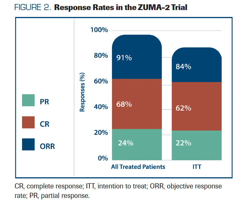
Wang shared the US Lymphoma CAR T Consortium experience of brexucabtagene autoleucel (brexu-cel) for R/R MCL as standard of care. Brexu-cel was approved by the FDA for R/R MCL based on the pivotal phase 2 ZUMA-2 trial (NCT02601313), which showed a 91% ORR and 68% CR rate (Figure 2) and durable responses, with a median PFS of 25.8 months and a median OS of 46.6 months at 3-year follow-up.24 After FDA approval, there is a need to continue to evaluate outcomes in real-world patients and to compare this experience with trial data. In the presented retrospective study, 189 patients who underwent leukapheresis between August 2020 and December 2021 at 16 US institutions that had an intent to manufacture commercial brexu-cel were included, and data were analyzed for treatment response, outcome, and toxicities.25 Of the 189 enrolled patients, 168 (89%) received brexu-cel infusion. Of the 189 patients who received leukapheresis, 86% were BTKi refractory, and 68% received bridging therapy. Of note, 79% would not have met ZUMA-2 eligibility criteria (61% would have been ineligible due to disease status). After a median postinfusion follow-up of 14.3 months, the 6-month PFS was 69% and the 12-month PFS was 59%. The 6-month OS was 86% and the 12-month OS was 75%. CRS occurred in 90% of patients, with 8% having grade 3 or higher and 61% of patients had neurotoxicity, with 32% having grade 3 or higher. CRS and ICANS incidences were similar to the clinical trial experience. In a univariable analysis, high-risk simplified MIPI, high Ki-67, TP53 aberration, complex karyotype, and blastoid/pleomorphic variant were associated with shorter PFS after brexu-cel infusion. Patients with bendamustine exposure within 24 months before leukapheresis had shorter PFS and OS in the intention-to-treat univariable analysis. Taken together, these data show that the efficacy and toxicity of brexu-cel in SOC practice are consistent with that reported in the ZUMA-2 trial. Importantly, the study results highlight that tumor-intrinsic features of MCL, and possibly recent bendamustine exposure, may be associated with inferior efficacy outcomes.
FIGURE 2. Response Rates in the ZUMA-2 Trial
To close the session on MCL clinical trials, Brad Kahl, MD, a professor in the Division of Oncology at the Washington University School of Medicine in St Louis, Missouri, and Martin Dreyling, MD, PhD, professor of medicine in the Department of Medicine and head of the Medical Clinic III at the University of Munich-Grosshadern in Germany, shared an update of US and European clinical trials, respectively. In the US, the National Clinical Trials Network continues to make significant contributions to MCL treatment, supporting multiple intergroup studies involving SWOG, ECOG-ACRIN, ALLIANCE, and BMT CTN. These trials are aimed at optimizing treatment approaches specifically for older or less fit patients, younger or fit patients, and those with relapsed/refractory disease, or treatment approaches that include novel drug combinations. Kahl first presented information on the phase 2 EA4181 trial (NCT04115631), which is comparing 3 chemotherapy regimens consisting of bendamustine, rituximab, high-dose cytarabine, and acalabrutinib in newly diagnosed MCL. Trial parameters were shared, and Kahl provided the update that the trial is now fully enrolled and primary end point analysis was expected by the end of 2023. Next, developments in EA4151, a phase 3 trial of rituximab after stem cell transplant comparing rituximab alone in MRD-negative MCL in first complete remission, were also presented. Although more than the 412 patients needed to be randomly assigned and have been enrolled, almost 10% of patients have refused treatment assignment and the study has enrolled additional patients to compensate. For the phase 2 E1411 trial (NCT01415752), data confirm BR as effective induction in patients who are older and show that rituximab, bendamustine hydrochloride, and bortezomib plus rituximab-based consolidation yield a median PFS of more than 5 years. Another phase 2 study, PrE0405 (NCT03834688), which is looking at bendamustine and rituximab plus venetoclax in patients with untreated MCL who are older than 60 years, has been fully enrolled since March 2022, and data were presented at the 2023 American Society of Hematology Annual Meeting & Exposition.25 Finally, A052101 (NCT05976763), a randomized phase 3 trial comparing continuous vs intermittent maintenance therapy with zanubrutinib as up-front treatment in patients older than 70 years or those 60 years or older with comorbidities, is expected to enroll in 2024.
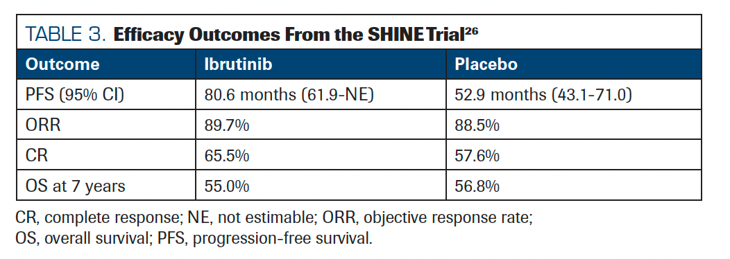
Dreyling then discussed updates to European MCL Network studies, starting with the SHINE trial (NCT01776840), a phase 3 study of ibrutinib in combination with BR and rituximab maintenance as first-line treatment for patients with MCL who are 65 years or older.26 Findings from this study demonstrated that ibrutinib plus BR with rituximab maintenance significantly improved PFS compared with standard chemoimmunotherapy, with a median PFS of 6.7 years (Table 3). TRIANGLE, a phase 3 trial (NCT02858258) of ibrutinib with SOC without autologous transplantation in first-line MCL, was also reviewed.27 Adding ibrutinib improved PFS and OS, begging the question of whether autologous transplant is needed first line in MCL. The final analysis is currently underway, and questions remain about how these findings will impact SOC. Finally, building on results of the phase 2 AIM study (NCT02471391) of venetoclax plus ibrutinib in MCL, the phase 2 OAsIs trial (NCT02558816) investigated if adding venetoclax to ibrutinib plus obinutuzumab will result in improved outcomes in patients with relapsed MCL.28,29
TABLE 3. Efficacy Outcomes From the SHINE Trial
Together, these studies not only evaluate a range of new therapeutic approaches in MCL but also contribute to the development of biomarkers and disease stratification. They present an opportunity to better understand the molecular, cellular, and tumor microenvironment changes that occur during treatment response and/or development of resistance.
Road Map to the Cure for MCL in the Next 20 Years: Panel Discussion
In the final session of the workshop, a panel of MCL expert academic researchers and industry representatives from various pharmaceutical companies discussed opportunities and challenges in MCL clinical trials. As our understanding of molecular drivers of disease continues to evolve, collaboration between regulatory and industry stakeholders will be critical for rapidly incorporating biomarkers and other molecular measures of response into clinical trials as end points or stratification tools. It is important to be aware that the adoption of surrogate end points can drastically reduce the costs of clinical trials, accelerate readout, and lead to improved patient care. Especially in an era of incremental progress, surrogate measures are needed because effect sizes are smaller, and therefore trials must include more patients. Furthermore, as more MCL subgroups are recognized, trials specific to disease subsets will be increasingly more difficult to enroll. In addition to innovation in the execution of clinical trials so that trials are smaller and smarter, the use of biomarkers to further reduce the time to readout and the number of patients needed will be critical.
Other challenges facing research in MCL are low enrollment and a lack of diversity in clinical trial populations. Although this represents a complex and multifaceted issue, one preliminary step could be to adjust exclusion criteria so that real-world patients may be more easily enrolled. Physicians emphasized that ongoing partnership and communication between industry and physicians throughout clinical trial planning can improve feasibility (especially around enrollment criteria and end point selection). As trials are designed, it is critical to involve clinicians who care for patients because they are better able to identify designs that are unnecessarily burdensome to patients (eg, frequent follow-ups for long periods). In addition, adequate support for sites (eg, study coordination) and industry support for rapid enrollment are critical. Clinicians agreed that enrollment is bolstered by including a comparative arm that includes a regimen appealing to patients. Patients with MCL tend to be well educated and savvy and largely uninterested in enrolling in trials where the comparator is clearly going to underperform, compared with the experimental arm.
In addition to traditional safety measures, there is a need to use efficient patient-reported outcome tools to assess quality of life as well as short- and long-term outcomes. Measures should also be age adjusted so that outcomes are relevant for the given age group.
Panelists agreed that the poor outcomes of patients with TP53 mutations, even with immunotherapy, highlights that there is some aspect of disease pathology that remains to be uncovered and targeted. Incorporating exploratory measures that facilitate investigation into these mechanisms is needed.
Finally, the impact of out-of-pocket costs for patients continues to grow. These increasing costs are problematic for all patients, but also worsen access and increase health care disparity. The cost of care is especially burdensome for patients who may not be able to take time off work, live some distance from the center, or are underinsured or uninsured.
Summary
The 2023 MCL Scientific Consortium and Workshop covered recent advances in our understanding of MCL biology and how these advances have uncovered several potential avenues for drug development. Continuing efforts are needed to better understand the basic biology of MCL so that novel therapeutics can be developed to target MCL, especially BTKi-resistant MCL. As our ability to evaluate disease metabolism, molecular changes, and the TME continues to improve, it will be critical to incorporate these findings into clinical studies and treatment paradigms so that adjustments to therapy may be made in real time. As clinical trials continue to be developed, it is important that biomarkers of disease risk are applied for stratification and novel measures of response are incorporated as end points to increase the efficiency of clinical trials and to decrease the time to readout.
Acknowledgments
This meeting, in addition to several projects presented, was supported by grants from the Lymphoma Research Foundation Mantle Cell Lymphoma Initiative and the MCL Consortium. Each presenter whose work is included herein reviewed and approved the summary of that work.
References
- Albero R, Enjuanes A, Demajo S, et al. Cyclin D1 overexpression induces global transcriptional downregulation in lymphoid neoplasms. J Clin Invest. 2018;128(9):4132-4147. doi:10.1172/JCI96520
- Eskelund CW, Dahl C, Hansen JW, et al. TP53 mutations identify younger mantle cell lymphoma patients who do not benefit from intensive chemoimmunotherapy. Blood. 2017;130(17):1903-1910. doi:10.1182/blood-2017-04-779736
- Delfau-Larue MH, Klapper W, Berger F, et al; European Mantle Cell Lymphoma Network. High-dose cytarabine does not overcome the adverse prognostic value of CDKN2A and TP53 deletions in mantle cell lymphoma. Blood. 2015;126(5):604-611. doi:10.1182/blood-2015-02-628792
- Puente XS, Jares P, Campo E. Chronic lymphocytic leukemia and mantle cell lymphoma: crossroads of genetic and microenvironment interactions. Blood. 2018;131(21):2283-2296. doi:10.1182/blood-2017-10-764373
- Nadeu F, Martin-Garcia D, Clot G, et al. Genomic and epigenomic insights into the origin, pathogenesis, and clinical behavior of mantle cell lymphoma subtypes. Blood. 2020;136(12):1419-1432. doi:10.1182/blood.2020005289
- Giné E, de la Cruz F, Jiménez Ubieto A, et al. Ibrutinib in combination with rituximab for indolent clinical forms of mantle cell lymphoma (IMCL-2015): a multicenter, open-label, single-arm, phase II trial. J Clin Oncol. 2022;40(11):1196-1205. doi:10.1200/JCO.21.02321
- Yi S, Yan Y, Jin M, et al. Genomic and transcriptomic profiling reveals distinct molecular subsets associated with outcomes in mantle cell lymphoma. J Clin Invest. 2022;132(3):e153283. doi:10.1172/JCI153283
- Locke FL, Ghobadi A, Jacobson CA, et al. Long-term safety and activity of axicabtagene ciloleucel in refractory large B-cell lymphoma (ZUMA-1): a single-arm, multicentre, phase 1-2 trial. Lancet Oncol. 2019;20(1):31-42. doi:10.1016/S1470-2045(18)30864-7
- Dufner V, Sayehli CM, Chatterjee M, et al. Long-term outcome of patients with relapsed/refractory B-cell non-Hodgkin lymphoma treated with blinatumomab. Blood Adv. 2019;3(16):2491-2498. doi:10.1182/bloodadvances.2019000025
- Budde LE, Sehn LH, Matasar M, et al. Safety and efficacy of mosunetuzumab, a bispecific antibody, in patients with relapsed or refractory follicular lymphoma: a single-arm, multicentre, phase 2 study. Lancet Oncol. 2022;23(8):1055-1065. doi:10.1016/S1470-2045(22)00335-7
- Bacac M, Colombetti S, Herter S, et al. CD20-TCB with obinutuzumab pretreatment as next-generation treatment of hematologic malignancies. Clin Cancer Res. 2018;24(19):4785-4797. doi:10.1158/1078-0432.CCR-18-0455
- Martin P, Bartlett NL, Blum KA, et al. A phase 1 trial of ibrutinib plus palbociclib in previously treated mantle cell lymphoma. Blood. 2019;134(11):1201-1204. doi:10.1182/blood-2018-11-886457
- Fuhr V, Vafadarnejad E, Dietrich O, et al. Time-resolved scRNA-seq tracks the adaptation of a sensitive MCL cell line to ibrutinib treatment. Int J Mol Sci. 2021;22(5):2276. doi:10.3390/ijms22052276
- Fuhr V, Heidenreich S, Srivastava M, et al. CD52 and OXPHOS-potential targets in ibrutinib-treated mantle cell lymphoma. Cell Death Discov. 2022;8(1):505. doi:10.1038/s41420-022-01289-7
- Balsas P, Veloza L, Clot G, et al. SOX11, CD70, and Treg cells configure the tumor-immune microenvironment of aggressive mantle cell lymphoma. Blood. 2021;138(22):2202-2215. doi:10.1182/blood.2020010527
- Beekman R, Amador V, Campo E. SOX11, a key oncogenic factor in mantle cell lymphoma. Curr Opin Hematol. 2018;25(4):299-306. doi:10.1097/MOH.0000000000000434
- Soldini D, Valera A, Solé C, et al. Assessment of SOX11 expression in routine lymphoma tissue sections: characterization of new monoclonal antibodies for diagnosis of mantle cell lymphoma. Am J Surg Pathol. 2014;38(1):86-93. doi:10.1097/PAS.0b013e3182a43996
- Rotbain EC, Gordon MJ, Vainer N, et al. The CLL comorbidity index in a population-based cohort: a tool for clinical care and research. Blood Adv. 2022;6(8):2701-2706. doi:10.1182/bloodadvances.2021005716
- Mayo Clinic. Molecular Epidemiology Resource. Accessed January 9, 2024. https://mayocl.in/3Hht4xS
- ECOG-ACRIN Cancer Research Group. Rituximab with or without stem cell transplant in treating patients with minimal residual disease-negative mantle cell lymphoma in first complete remission. Accessed January 9, 2024. https://bit.ly/3vle2Vd
- Ruan J, Leonard JP, Chen GZ, et al. Phase 2 trial of acalabrutinib-lenalidomide-rituximab (ALR) with real-time monitoring of MRD in patients with treatment-naïve mantle cell lymphoma. Blood. 2022;140(suppl 1):175-177. doi.10.1182/blood-2022-158656
- Ruan J, Martin P, Christos P, et al. Five-year follow-up of lenalidomide plus rituximab as initial treatment of mantle cell lymphoma. Blood. 2018;132(19):2016-2025. doi:10.1182/blood-2018-07-859769
- Wang M, Munoz J, Goy A, et al. Three-year follow-up of KTE-X19 in patients with relapsed/refractory mantle cell lymphoma, including high-risk subgroups, in the ZUMA-2 study. J Clin Oncol. 2023;41(3):555-567. doi:10.1200/JCO.21.02370
- FDA approves brexucabtagene autoleucel for relapsed or refractory mantle cell lymphoma. News release. FDA. July 24, 2020. Accessed January 9, 2024. https://bit.ly/3NUDUhc
- Portell CA, Jegede O, Bennani NN, et al. Primary analysis and results of bendamustine, rituximab, and venetoclax (BR-VEN) for initial treatment of mantle cell lymphoma in subjects over 60 years of age (PrE0405). Blood. 2023;1429suppl 1):733. doi.10.1182/blood-2023-182374
- Wang ML, Jurczak W, Jerkeman M, et al. Ibrutinib plus bendamustine and rituximab in untreated mantle-cell lymphoma. N Engl J Med. 2022;386(26):2482-2494. doi:10.1056/NEJMoa2201817
- Dreyling M, Doorduijn JK, Gine E, et al. Efficacy and safety of ibrutinib combined with standard first-line treatment or as substitute for autologous stem cell transplantation in younger patients with mantle cell lymphoma: results from the randomized Triangle trial by the European MCL Network. Blood. 2022;140(suppl 1):1-3. doi.10.1182/blood-2022-163018
- Handunnetti SM, Anderson MA, Burbury K, et al. Three year update of the phase II ABT-199 (Venetoclax) and ibrutinib in mantle cell lymphoma (AIM) study. Blood. 2019;134(suppl 1):756. doi.10.1182/blood-2019-126619
- Le Gouill S, Morschhauser F, Chiron D, et al. Ibrutinib, obinutuzumab, and venetoclax in relapsed and untreated patients with mantle cell lymphoma: a phase 1/2 trial. Blood. 2021;137(7):877-887. doi:10.1182/blood.2020008727
Highlighting Insights From the Marginal Zone Lymphoma Workshop
Clinicians outline the significance of the MZL Workshop, where a gathering of international experts in the field discussed updates in the disease state.