Sentinel Node Evaluation in Gynecologic Cancer
A review of sentinel node mapping in vulvar and cervical cancer, a technique intended to reduced lymphadenectomy-associated morbidity, and the related controversies.
ABSTRACT: The sentinel node evaluation has revolutionized the modern surgical management of cutaneous melanoma and breast cancer. In gynecologic oncology, sentinel node mapping has been mainly studied in vulvar and cervical cancer. In vulvar cancer, data from 12 studies including 353 cases indicate that the sentinel node detection rate is 92% and the negative-predictive value is 99%. Three groin recurrences have been documented so far (< 1%). The technique has more recently been studied in cervical cancer. Data from 12 studies including 323 cases indicate a lower sentinel node detection rate of 80% to 86% and a negative- predictive value of 99%. Three false-negative cases have been reported so far (< 1%). Review of the literature suggests that the combined approach with blue dye and lymphoscintigraphy is superior to the blue dye alone for sentinel node detection. It also suggests that the sentinel node mapping technique is feasible in vulvar and cervical cancer and that it may become a valuable alternative to the traditional groin and pelvic lymphadenectomy. However, results have not been duplicated in large multi-institutional trials, and the technique should still be performed in the context of clinical trials. Complications of the sentinel node mapping technique are rare and usually benign but physicians should be aware of the serious risk of anaphylactic reaction to the blue dye (1% to 2%). Before this technique becomes a standard approach in the management of gynecologic malignancies, more data will be needed to clarify some of the related controversies.
The concept of sentinel node mapping was proposed by Cabanas in 1977 for the management of patients with penile cancer, using lymphangiography of the dorsal lymphatics of the penis.[1] Fifteen year later, Morton applied the same concept to the management of cutaneous melanoma using an intraoperative blue dye injection for localization of the sentinel node.[2] Lymphoscintigraphy using a radioactive tracer (technetium [Tc]-99) was added to the blue dye technique by Krag et al, to help localize sentinel nodes located in unusual or aberrant lymphatic basins such as in melanoma of the trunk.[3] Experience in melanoma and breast cancer has shown that this combined technique is superior to the use of blue dye alone for the detection of the sentinel node,[4-6] and it has become the standard approach in the clinical management of those malignancies in several cancer centers.
The general objectives of nodal staging are threefold: (1) establish the prognosis, (2) obtain regional control, and (3) improve survival. The traditional, complete lymphadenectomy is associated with potentially significant morbidity. Thus, the goal of sentinel node mapping is to accurately determine the status of the regional lymph nodes while reducing the morbidity of the procedure. In this article, we will review the technique and literature on sentinel node mapping in vulvar and cervical cancer and emphasize some of the controversies.
Vulvar Cancer
Vulvar cancer represents only about 5% of gynecologic cancers. The International Federation of Gynecology and Obstetrics (FIGO) surgical staging is determined after the removal of the primary vulvar lesion and regional lymph node dissection. Unfortunately, the majority of patients with clinical stage I/II vulvar cancer do not clearly benefit from the inguinofemoral dissection, because only about 10% to 15% have lymph node metastasis.[7]
In addition, nodal staging is associated with several potential complications such as the formation of lymphocysts and seromas, wound infection and breakdown, pain and paresthesia, vascular and neural injuries, and adhesion formation.[8] The longterm and often debilitating lymphedema is particularly troublesome. This condition occurs more frequently in patients who receive adjuvant radiation therapy, and these patients are at higher risk of secondary lymphangitis and cellulitis.
Technique
Currently, two different methods are available to locate the sentinel node: the blue dye technique and lymphoscintigraphy. Data suggest that the two methods are complementary and the best results are obtained by combining them.
• Lymphoscintigraphy-Lymphoscintigraphy-The principle of lymphoscintigraphy is to inject a weakly radioactive tracer at the interface between the tumor and the normal tissue, where the lymphatic network is rich and abundant. It is important to avoid intratumoral injection. Usually 1.0 to 1.5 mL of filtered Tc-99 radiocolloid, with an activity of 0.4 to 0.8 mCi, is injected very superficially under the dermis with a 27-gauge needle. The tracer is picked up by the microlymphatics surrounding the tumor and is transported via the lymphatic channels to the regional lymph nodes, where it accumulates. In vulvar cancer, it is worthwhile applying an anesthetic cream locally, 30 to 60 minutes before the injection, because the Tc-99 injection burns and is very painful.
Only one anterior lymphoscintigram with a posterior transmission flood can be performed as early as 15 minutes after the injection to locate the sentinel node, which appears as a black dot. The nuclear medicine specialist can mark the skin to locate the sentinel node to be removed by the surgeon, as it is usually located superficially in the node-bearing area of the groin. However, this part is not absolutely essential.
• Blue Dye Technique-Conversely, the blue dye is injected immediately before surgery. After induction of general anesthesia, 2 mL of isosulfan blue dye (Lymphazurin 1%) is injected at the periphery of the vulvar lesion, also using a 27-gauge needle. Surgery begins with the sentinel node mapping. A small incision is made over the inguinal crease, and the surgeon carefully looks for blue channels that normally lead to a blue lymph node. Care is taken to avoid disruption of the lymphatic channels and bleeding.
The handheld gamma probe (eg, Navigator, Autosuture) is then passed along the area in a search for areas of high radioactivity. A count is performed on the hottest area, which usually corresponds to the blue node. The blue/hot node is removed separately, and a radioactive count is done again ex vivo for correlation. The sentinel node is sent to the pathologist for frozen section. The gamma probe is passed along the area again, looking for other hot areas. Additional nodes may be considered secondary sentinel nodes if their radioactivity count is 10 times the background count. A complete superficial and deep node dissection should then be completed, at least until data from large cooperative group studies confirm the accuracy and reproducibility of the sentinel node mapping technique. Without such confirmation, the technique cannot be considered a standard of care. Depending on the location of the lesion, sentinel node mapping can be done unilaterally or bilaterally, and the vulvar surgery itself is completed afterwards.
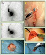
FIGURE 1

Sentinel Node Mapping in Vulvar Cancer
• Assessment-Removed sentinel nodes are sent for immediate frozen section. If they look grossly involved by tumor, only one section is performed to confirm the diagnosis. If they look normal, a random section is examined to look for the presence of cancer cells. For the final pathologic analysis, "ultrastaging" of the sentinel node is performed, which involves three sections per node, perpendicular to the long axis of the node (every 40 to 50 μm). Each level is stained for hematoxylin and eosin (H&E) and one is also stained for immunohistochemical analysis with cytokeratins (CAM 5.2 and AE1:AE3) to look for the presence of micrometastasis.
• Procedure Overvie-Figure 1 shows an example of sentinel node mapping in vulvar cancer. First, Tc-99 is injected superficially at the periphery of the lesion. A lymphoscintigram is peformed 20 minutes later, showing in this case a unilateral uptake on the right side of the groin (Figure 1A). Just prior to surgery, blue dye is injected again superficially at the periphery of the tumor. Note the onset of blue lymphatic tracking under the skin (Figure 1B). The right groin is then explored with the handheld gamma probe. Like others, we have noticed that the sentinel node is often located quite medially in the groin (Figure 1C).
Once a hot spot has been detected, the groin is further explored searching for a blue node, with particular attention to meticulous surgical technique to avoid bleeding in the field and disruption of the blue lymphatics. Note the blue lymphatics entering the bluish node (Figure 1D). Sometimes, more than one blue node is removed (Figure 1E). Extracorporeal count is always performed on the sentinel node removed to make sure that the hot node detected in the groin is really the one that has been removed. The extracorporeal count also helps determine, when more than one sentinel node is removed, which one is the primary sentinel node (it should be the one with the highest count, Figure 1F). The issue of secondary sentinel node is not clarified at this point.
Review of the Literature
Clinical examination or imaging techniques cannot reliably detect the presence of lymph node metastasis in vulvar cancer. Even the use of newer noninvasive imaging techniques such as the positron-emission tomography (PET) scan are relatively insensitive in predicting lymph node metastasis in vulvar cancer.[9] Up until now, the standard of care has been a complete superficial and deep groin node dissection, either unilateral or bilateral, which is unfortuantely associated with significant morbidity.
Recently, the concept of sentinel node biopsy has been performed in other malignancies such as breast cancer. Although the long-term morbidity of sentinel node mapping has not been formally reported yet in vulvar cancer, a comparative study in breast cancer confirms a reduced hospital stay and postoperative morbidity in patients who underwent sentinel node biospy as opposed to complete axillary node dissection.[10] The potential of sentinel node mapping in reducing the morbidity of a traditional complete inguinofemoral lymph node dissection in vulvar cancer is thus enormous.
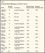
TABLE 1

Sentinel Node Mapping in Vulvar Cancer
In vulvar cancer, accumulating data indicate that the sentinel node mapping technique is feasible and highly accurate in predicting the status of the inguinofemoral lymph nodes; most studies have also confirmed that the combined technique is superior to the blue dye alone in identifying the sentinel node.[11-22] Puig-Tintor et al recently summarized 12 series published since 1997, totaling 353 cases (Table 1).[22] The negative predictive value was 100%, except in one series (96%). The review also indicates that the sensitivity of the blue dye technique alone in detecting the sentinel node is only 56% to 88% and that the addition of lymphoscintigraphy significantly improves the sentinel node detection rate to nearly always 100%. Their conclusion is that the available data are strong enough to recommend the routine use of sentinel node mapping in the management of vulvar cancer.[22]
Conversely, one published multiinstitutional study using the blue dye alone reported a sentinel node identification rate of only 56% with two false-negative cases.[13] Of note, three of the five participating centers contributed six cases or fewer, suggesting that efficacy and reproducibility of the technique may be lower when applied at centers with less experience. Also, the sensitivity of the technique may not be as good in the presence of clinically suspicious groin nodes, such that the mapping technique should probably be limited to the detection of subclinical metastasis.[20]
Learning Curve
The sentinel node mapping technique is relatively easy to learn in vulvar cancer, because vulvar lesions are usually readily visible and accessible for the injection. As stated above, it may be worthwhile to apply an anesthetic cream on the vulva 20 minutes before the injection, to avoid pain locally.[ 15] de Hullu et al suggested that the learning curve for properly performing the sentinel node technique in vulvar cancer is reached with approximately 10 cases, unless the surgeon has significant expertise with the technique in other sites (ie, breast cancer).[17]
Recurrence
Recurrences following sentinel node biopsy have been reported by three groups, although most published series have had a short follow-up. In the report by Terada et al, the sentinel node was, in fact, retrospectively found to harbor micrometastasis upon serial sectioning when the patient experienced a recurrence in the groin.[16]
In the series from Rodier et al, one patient suffered a medial groin recurrence 6 months after sentinel node biopsy and died 2 months later. The sentinel node was not identified using the blue dye technique alone, and a complete inguinofemoral node dissection was performed.[23] This case suggests that a positive groin node was probably missed by the blue dye and not removed by the groin dissection. Perhaps lymphoscintigraphy would have helped localize a more medial groin node-a localization found more frequently than anticipated by others.[14] It is also interesting that the patient was the first of the series, which emphasizes the importance of the learning curve in relation to the accuracy of sentinel node identification.
Finally, in the report by Tamussino et al, the patient had a positive microscopic implant in one of seven sentinel nodes removed, and adjuvant treatment was not given in light of the patient's age.[24] A groin recurrence occurred 16 months later.
This report was criticized by de Hullu et al, who considered that the removal of up to seven sentinel nodes was in fact a lymphadenectomy and no longer a conservative procedure, defeating the purpose of reducing morbidity.[ 25] However, in some patients, several nodes are either blue, hot, or both, frequently representing a chain of nodes rather than nodes originating from distinct locations.
So how many sentinel nodes should be removed? The question was studied by McMasters et al who concluded in their study that if only the most radioactive sentinel node had been removed, 13% of the nodal basins with positive sentinel node would have been missed. They recommend that all blue lymph nodes and all lymph nodes with a 10% or higher radioactive count compared to the hottest node be removed and pathologically assessed.[26]
Pathologic Evaluation
A great deal of uncertainty surrounds the pathologic evaluation of sentinel nodes. The concept of ultrastaging, initially developed in melanoma patients, involves serial sectioning of the sentinel node and immunohistochemistry staining with anticytokeratin antibodies (such as CAM 5.3, AE1:AE3) to detect the presence of micrometastasis. In the series reported by deHullu et al, 25% of the sentinel nodes considered negative using conventional techniques were found to be positive after serial sectioning.[ 17] In the series by Puig-Tintor, 38% of sentinel node metastases were identified by serial sectioning and immunohistochemistry.[ 22] They were all less than 2 mm in size. In the series by Molpus et al, 11% of negative sentinel nodes by routine H&E evaluation were found to contain micrometastasis after serial sectioning and immunohistochemistry.[20]
It remains to be determined whether in vulvar cancer, for instance, the presence of microscopic tumor implants is associated with a poorer survival and whether these patients should be treated with adjuvant therapy. It is also unclear if the size of the lymph node metastasis itself represents a risk factor and affects the prognosis of patients, and whether patients with micrometastasis should be assigned to a separate subclassification in the FIGO or TNM system.
Tjan-Heijnen et al reviewed the issue of micrometastasis and outcome in breast cancer and found that, in most cases, occult metastasis was not an independent risk factor for survival.[ 27] On the other hand, the clinical significance of sentinel node micrometastasis, although controversial, should not be totally overlooked, in light of the groin recurrence reported in two patients with microscopic sentinel node metastasis who did not receive adjuvant treatment.[16,24] Terada et al concluded that the presence of a micrometastasis in a sentinel node requires additional treatment- either formal complete lymph node dissection or adjuvant radiation therapy.[ 16] This issue needs to be further clarified as more data accumulate.
Lymphoscintigraphy vs Blue Dye
The blue dye technique has several technical limitations. Indeed, the dissection and search of the blue channels and nodes has to be atraumatic and bloodless; otherwise, the lymphatic vessels are damaged or the surgical field is obscured by blood. This can make identification of blue nodes very difficult or even impossible, particularly if the blue node is located deep or in an unusual area. In a multicenter trial using only blue dye in vulvar cancer, the sentinel node detection rate was only 56%, suggesting that this technique alone is not very reliable.[13]
Levenback et al, a group with extensive experience in sentinel node mapping, reported a sentinel node detection rate of 75% with the dye alone.[14] Sliutz et al have stopped using the blue dye because of an inability to trace the afferent lymphatics in three of eight cases.[21] However, the blue dye may be advantageous in tumors located near the inguinal region, where the high background radioactivity from the injected Tc-99 in the vulva may obscure the radioactive search for nodes in the groin.[28] Moreover, the blue dye technique is a simple and inexpensive method, but on the other hand, most of the serious complications reported to date have been secondary to the dye.
Conversely, the Tc-99 radiocolloid has an extremely good therapeutic index with essentially no side effects reported to date. Fewer technical problems are associated with the detection of radioactive nodes, compared to the blue dye. Intraoperative lymphoscintigraphy is very accurate and has the advantage of identifying unusual lymphatic drainage sites. De Ciccio et al reported a 100% sentinel node detection rate using lymphoscintigraphy alone.[15] Lymphoscintigraphy can also help select patients who require unilateral vs bilateral groin dissection in cases of midline lesions where the lympatic drainage clearly goes to one side only.[15,17] Several authors have noted that a sentinel node can readily be identified 15 to 30 minutes after Tc-99 injection, which makes planning of the surgery easier.[15,20,28] However, lymphoscintigraphy is definitely more cumbersome and costly to organize.
TABLE 2

Advantages and Disadvantages of Sentinel Node Identification Techniques
Further studies should clarify which of the two techniques is superior and if, in fact, the addition of the blue dye technique is really necessary, considering its potential serious side effects. Levenback et al have nicely outlined the advantages and disadvantages of both techniques, concluding that, in the end, the choice of technique may rest primarily on the surgeon's preference and expertise (Table 2).[14]
Vulvar Melanoma
A few authors have reported their experience with sentinel node mapping in vulvar or vaginal melanoma.[ 29-31] The technique is feasible, and the sensitivity of such mapping is 100%. However, de Hullu et al noted that in their series of nine cases, there was a trend toward more frequent groin recurrences in patients undergoing a sentinel node mapping procedure compared to historic controls, suggesting that the procedure may increase the risk of in-transit metastasis especially in patients with thick melanomas (> 4 mm).[31] The authors recommend that the sentinel node mapping technique be performed only in the context of clinical trials and be limited to melanomas with intermediate thickness (1-4 mm) until more data are available.
Abramova et al concluded that the Tc-99 injection should probably be done the night before surgery because of the proximity between the vulva and the groin nodes. This allows time for the background radioactivity to dissipate, which may facilitate identification of the regional lymph nodes.[30]
Summary
Available data from 12 series totaling more than 350 cases of vulvar cancer, mostly from single institutions, indicate a sensitivity (ability to locate a sentinel node) of essentially 100% using the combined dye and lymphoscintigraphy technique, a false-negative rate < 1% and essentially no recurrence directly related to the technique.[ 22] The Gynecologic Oncology Group is currently conducting a multi-institutional feasability trial (GOG 173) in vulvar cancer to determine the negative predictive value of sentinel node identification. However, as stated by de Hullu, the standard of care should remain the complete superficial and deep inguinofemoral dissection until results from large and adequately powered randomized trials confirm the safety of the sentinel node mapping.[25] While awaiting those results, sentinel node localization alone should be conducted in the context of clinical trials only.
As mentioned by Ansink et al, the outcome of patients with early-stage vulvar cancer is excellent. Therefore, to be acceptable, the false-negative rate for sentinel node mapping will have to be very low.[13] Outcome should not be jeopardized by the use of a lesser surgical procedure to reduce morbidity. In a recent survey, de Hullu et al showed that women would not accept a sentinel node false-negative rate of 5% and were willing to accept greater morbidity, although gynecologic oncologists considered a false-negative rate of 5% to 20% acceptable.[32]
Before sentinel node mapping in vulvar cancer becomes the standard of care, clear algorithms will need to be defined with regard to controversial issues such as a positive sentinel node discovered on final pathology, indications for complete inguinofemoral dissection, indications for adjuvant therapy in relation to the size of the sentinel node metastasis, and consensus on the pathologic evaluation of the sentinel node.[33]
Cervical Cancer
Cervical cancer is the third most common gynecologic malignancy, with almost 13,000 newly diagnosed cases per year in the United States and 4,400 related deaths.[34] In patients with stage IA2-IB1 disease, the rate of lymph node metastases averages 15%. Thus, as in vulvar cancer, the majority of cervical cancer patients subjected to a complete lymph node dissection do not derive clear benefit from it yet the procedure is associated with potentially serious complications (eg, lymphocysts and adhesion formation, pain, paresthesia, vascular damage, and lymphedema), particularly if it is followed by adjuvant radiation therapy.
Technique
The technique for sentinel node mapping in cervical cancer is similar to that described for vulvar cancer.The Tc-99 is injected very superficially at the periphery of the tumor, or in the four quadrants of the cervix, using a 25-gauge spinal needle, 3½- in. long. To facilitate the injection and render the spinal needle more stiff, a second spinal needle (20- gauge, 2-in. long) is slid over the first one and secured with Steri-Strips. Alternatively, a rigid needle extender such as those used for pudendal blocks can be used. Care is taken to avoid injecting the radiocolloid into the tumor itself.
A lymphoscintigram is performed 30 minutes after the injection. (In our experience, dynamic studies have shown that sentinel nodes can readily be seen as early as 15 minutes after the injection). Because the nodes are located deep in the pelvis, skin marking of the sentinel node is not reliable. However, it is useful to have the hard-copy lymphoscintigram available in the operating room to help search for the sentinel node, particularly when it appears to be in an aberrant location.
The blue dye is injected just before the surgery. After induction of general anesthesia, 2 to 4 mL of isosulfan blue dye is injected superficially in the four quadrants of the cervix. After completing the dye injection, a regular rectovaginal examination is performed. The patient is then prepped and draped, and the sentinel node exploration begins immediately. It can be performed via an abdominal incision (laparotomy) or by laparoscopy. Only the laparoscopic approach will be described here.
After insertion of the four trocars, the retroperitoneum on one side is opened. Blue lymphatics emerging from the lateral parametrium, usually crossing over the obliterated umbilical artery, are followed all the way to their ending in a blue node, which is considered to be the sentinel node. Occasionally, more than one sentinel node is identified per side. Before removing the blue nodes, the laparoscopic gamma probe is inserted through the suprapubic 10-mm port (eg, Versaport 5-10, Autosuture). Counts are first obtained on the blue node and recorded. The node is then retrieved, and extracorporeal counts are performed again on that node for confirmation. The laparoscopic probe is reintroduced, and the pelvic walls are scanned in a search for other signals.
The same procedure is performed on the contralateral side. In cases where the dye injection did not work well and no blue nodes are identified at laparoscopy, a detailed exploration of the pelvic sidewalls with the laparoscopic gamma probe is performed along the major blood vessels, including the common iliac, presacral, and lower para-aortic areas, to search for a radioactive signal. The preoperative lymphoscintigram is useful in these cases, to help identify the approximate location of the sentinel node.
• Assessment-All sentinel nodes are sent for immediate frozen section. If they are negative, a complete bilateral pelvic node dissection is completed laparoscopically, as sentinel node mapping is not considered the standard of care. Radical surgery follows (either abdominal or vaginal radical hysterectomy or radical trachelectomy). If the sentinel nodes are positive, a bilateral para-aortic node dissection is performed, but radical surgery is abandoned in favor of combined chemoradiation.
The sentinel node mapping performed via an abdominal incision is identical, except that a handheld gamma probe is used.
Review of the Literature
Sentinel node mapping was developed more recently in cervical cancer. Twelve studies have been published since 1999, totaling 323 cases,[12,35-45] but most of these series have included a small number of patients. Moreover, the technique used for sentinel node mapping, patient selection criteria, surgical approach, and sentinel node pathologic evaluation vary among the studies. Nevertheless, these preliminary studies indicate that sentinel node mapping is feasible in cervical cancer.
TABLE 3

Sentinel Node Mapping in Cervical Cancer
With the exception of the first study, the overall sentinel node detection rate ranges from 60% to 100% (Table 3). Several studies convincingly demonstrate the superiority of the combined blue dye/preoperative lymphoscintigraphy technique to improve the sentinel node detection rate.[36,41,43,45] Indeed, in Malur's study, the sentinel node detection rate with the blue dye alone was only 55%; with the radiolabeled Tc-99, it increased to 76%, and with the combined technique, it reached 90%.[41] Levenback et al reported a 100% detection rate with the combined technique, whereas in their preliminary report using the blue dye alone, the detection rate was only 60%.[38,43] In the study by Verheijen et al, the detection rate improved from 40% to 80% with the addition of lymphoscintigraphy compared to the blue dye alone.[36] In our own experience, the sentinel node detection rate increased from 79% to 93% with the combined technique.[45]
Lymphatic Drainage and Sentinel Node Localization
The lymphatic drainage of the cervix is much more complex than that in vulvar or breast cancer. Indeed, studies by Plentl and Friedman showed that there are three main trunks arising on each side of the cervix: the lateral, anterior, and posterior collecting trunks.[46] The lateral trunk, which is the main lymphatic pathway, further divides into the upper, middle, and lower branches. Therefore, the search for the sentinel node must be very meticulous, and because the lymphatic drainage of the cervix is more haphazard, lymphoscintigraphy is particularly useful.
Because the cervix is a midline structure, it would seem logical that the lymphatic drainage should be bilateral in most cases. This assumption is based on the pioneering work of Leveuf and Godard, who showed that when the cervix is injected anteriorly and posteriorly, the lymphatic drainage is bilateral.[47] Others refute the sentinel node concept in cervical cancer altogether, concluding that the pattern of lymphatic drainage in early-stage cancer of the cervix is completely random.[48]
• Common Sites-The localization of the sentinel node in cervical cancer is more variable than in vulvar cancer for the reasons mentioned above. The three most frequent sentinel node sites for cervical cancer are the external iliac nodes (Leveuf and Godard's "intermediate"), followed by the obturator nodes ("caudal"), and the bifurcation nodes ("cephalad").[ 36,38-43,45,47] Parametrial nodes, considered in-transit nodes between the cervix and the pelvic nodes, are particularly difficult to identify because of the proximity of the cervix, where the blue coloration is intense and the radioactivity high.[45,47]
Occasionally, sentinel nodes are identified at the common iliac, at the level of the internal iliac artery alongside the ureter, or even in the lower para-aortic region. These more uncommon patterns of spread can easily be explained by the lymphatic drainage arising from the lower branch of the lateral collecting trunk and/or from the posterior collecting trunk.[46] Unusual sites of metastasis such as the groin-probably secondary to retrograde lymphatic drainage-have also been reported by two groups.[42,49]
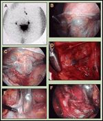
FIGURE 2

Various Sentinel Node Localizations in Cervical Cancer
Figure 2 shows examples of various sentinel node localizations. First, a lymphoscintigram is done 20 minutes after an intracervical Tc-99 injection, showing in this case a bilateral sentinel node uptake with continued uptake on the left side toward the left para-aortic region (Figure 2A). A right external iliac blue sentinel node with the blue lymphatic entering it is easily visible (Figure 2B). A right obturator blue sentinel node is discovered after opening the obturator fossa (Figure 2C). In addition, a right parametrial blue sentinel node is found, with a blue lymphatic vessel continuing under the superior vesical artery and ending in the obturator fossa (Figure 2D). The ureter can be seen just on the left of the parametrial sentinel node. The gamma probe also confirmed a very hot signal.
Figure 2E shows an unusual sentinel node location in the left presacral area. In that case, no sentinel nodes were identifed upon thorough exploration of the left retroperitoneal area; however, scanning of the lower paraaortic region with the laparoscopic gamma probe identified a hot area with a faint blue stain in the left presacral area (Figure 2E). Detailed exploration confirmed the presence of a sentinel node medial to the common iliac vein, which was pale blue and very hot. Note the complete dissection of the common iliac artery (Figure 2F). This case illustrates the importance of a thorough sentinel node search and the advantage of the gamma probe when no blue nodes are identified. Unusual lymphatic drainage sites probably explain some situations where all the pelvic nodes removed in a patient are negative, yet the patient develops a recurrence in the lower para-aortic region sometime later.
Lymphoscintigraphy
Given the complex lymphatic drainage of the cervix, the addition of lymphoscintigraphy to the blue dye procedure appears to contribute valuable information in cervical cancer.[ 41,43,45] The same has been observed in melanoma of the trunk, where the lymphatic drainage is also less predictable. and the status of an individual nodal basin does not always reflect reliably the status of other draining basins.[50] Malur et al have questioned whether a lymphoscintigram is in fact necessary, as it cannot precisely predict the location of the sentinel node.[41] It adds complexity and cost to the procedure, and places more pressure on the nuclear medicine ward. Nevertheless, it is useful to have the hard copy lymphoscintigram available in the operating room to confirm technetium uptake (unilaterally or bilaterally) and guide the intraoperative sentinel node search, particularly if the blue dye technique did not work well (cf Figures 2E-F).
Bilateral Sentinel Node Identification
In our study, the rate of bilateral sentinel node detection was 55%- identical to the rate reported by Levenback et al.[43,45] However, in our subgroup of patients who had the combined blue dye/lymphoscintigraphy, the bilateral sentinel node detection rate increased to 72% (P < .01). Given that the cervix is a midline structure, we believe that, ideally, at least one sentinel node should be identified per side of dissection, because one cannot assume that the status of the sentinel node on one side also reflects the status of the nodes on the other side.
This assumption has significant implications from a clinical point of view because if in the future we wish to rely on the status of the sentinel node only to avoid a complete lymphadenectomy, the rate of sentinel node identification by side of dissection will have to be high; otherwise, too many patients will have to undergo at least an ipsilateral complete lymphadenectomy (on the side where the sentinel node is not identified). This would significantly reduce the benefits of sentinel node mapping, ie, avoidance of the morbidity associated with complete lymphadenectomy. It will thus be important, in future trials, to report the sentinel node detection rate per patient and by side of dissection as well as the number of sentinel nodes detected per patient and by side of dissection.
False-Negative Rate
As in vulvar cancer, the true falsenegative rate-defined as a case in which the sentinel node is negative but other nonsentinel nodes are found to be positive-is very low for cervical cancer. Sentinel node mapping has been reported in 323 patients with cervical cancer, and only three falsenegative cases have been reported for an overall rate of less than 1%. In one case, it is not clear if the positive nonsentinel node was on the same side as the negative sentinel node or on the contralateral side. Additionally, the sentinel node was evaluated using the standard H&E method and not with serial sections and immunohistochemistry.[ 41] In another falsenegative case, four sentinel nodes were found to be negative, yet four small parametrial nodes removed en bloc with the hysterectomy specimen contained microscopic metastasis, illustrating the difficulty in identifying parametrial nodes.[43]
In the last case, the sentinel node tested false-negative on frozen section and not on final pathology.[42] The accuracy of frozen section analysis of the sentinel node is an interesting concept because, from a clinical point of view, this is the information we would eventually rely on to determine, at the time of surgery, whether a more complete lymph node dissection needs to be done (if the sentinel node is positive) or not (if the sentinel node is negative). Conversely, if the frozen section analysis of the sentinel node is inaccurate, too many patients would need to be reoperated to complete the node dissection or receive adjuvant radiation therapy. This concept of frozen section analysis of the sentinel node has been applied to breast cancer, and two-thirds of patients were spared the need for reoperative axillary lymphadenectomy.[ 51]
Macroscopic Nodes
As in vulvar cancer, macroscopic nodes may not be well detected by the blue dye or lymphoscintigraphy technique in cervical cancer patients. In our series, the sentinel node detection rate for positive but normal appearing sentinel nodes was 75%, whereas it was only 56% in patients with macroscopically suspicious nodes at laparoscopy.[ 45] Malur et al also reported that patients with macroscopic lymph nodes are not well detected by the sentinel node mapping technique.[41]
This phenomenon may be explained by the fact that lymphatic vessels may be blocked by tumor cells, thus preventing the migration of the injected dye/technetium, or the lymph node capsule may be blocked by tumor cell emboli, again preventing the dye/technetium from entering the node. However, the objective of the sentinel node technique clearly is not to identify macroscopically involved lymph nodes that are readily visible at surgery, but rather, to identify the sentinel node potentially involved by subclinical micrometastasis.
Learning Curve
It is difficult to determine the optimal number of cases before a surgeon is considered competent in performing the procedure (cf discussion of the learning curve in vulvar cancer section). This issue of surgeons' experience has been discussed extensively in breast cancer and melanoma, and 20 to 30 is often quoted as the number of cases required before a surgeon can enter patients in clinical trials.[ 52,53] In cervical cancer, the learning curve is likely to be closer to 20 because the intracervical injection is technically more difficult and, as stated above, the lymphatic drainage of the cervix is much more complex.[46]
The published series indeed demonstrate an overall lower sentinel node detection rate in cervical cancer (80%) compared to the results obtained in vulvar cancer (92%). In our own experience, we identified a sentinel node bilaterally in 14 of the last 15 cases (97%), which also clearly suggests that with time and experience the rate of sentinel node identification improves significantly.[45] However, gynecologic oncologists are gaining expertise with the sentinel node technique in vulvar cancer, so this may reduce the learning curve in cervical cancer.
Pathologic Evaluation
As mentioned in the vulvar cancer section, there is currently no standardized protocol for the evaluation of the sentinel node in cervical cancer, but most centers do serial sectioning of the sentinel node and immunohistochemistry with anticytokeratin antibodies to detect the presence of microscopic tumor cells. A recent study in breast cancer patients reported that serial sectioning and immunohistochemistry detected 16% more occult micrometastasis in sentinel nodes than was detected with standard H&E staining.[54]
Reverse transcriptase-polymerase chain reaction (RT-PCR) detection of micrometastasis at a molecular level using primers designed to amplify cytokeratin 19 has been studied in cervical cancer; the authors reported that 50% of early-stage cervical cancer patients were found to harbor evidence of tumor cells in their pelvic lymph nodes.[55] The molecular detection of micrometastasis may thus help explain recurrences in some patients with negative lymph nodes. The clinical significance of occult metastasis and recommendations for adjuvant treatment remain unclear and highly controversial at this point. Further studies are needed to clarify the issue.
Complications
As with any new technology, complications can occur. Few side effects have been reported in patients undergoing the procedure for cervical cancer, and all have been secondary to the blue dye. The most frequent side effect is the change in urine color. Since the blue dye is excreted by the kidneys, the urine may be blue-green for 24 to 48 hours. Patients and staff should be aware of this to avoid unnecessary concerns.
• Hypoxia-Decreases in oxygen saturation as measured by pulse oximetry have been reported.[45,56] In those cases, the blue dye gets absorbed into the general circulation and interferes with the pulse oximetry readings.[ 57] True hypoxia must be ruled out by taking an immediate arterial blood gas measurement; co-oximetry is also useful to rule out methemoglobinemia as a cause of hypoxia. The duration and magnitude of the effect is variable. Usually the effect is transient and lasts a few minutes, but it may be more prolonged in patients with impaired liver or biliary function, as the blue dye is excreted by the biliary tract.[57]
• Allergic Reactions-Allergic reactions to the blue dye have been reported. The most frequent reaction is called blue urticaria, which is characterized by the appearance of classic blue patches on the skin.[45] An immediate type 1 (immunoglobulin [Ig]E-dependent) hypersensitivity reaction, it is usually self-limited and rarely requires medication. It may occur alone or in association with an anaphylactic reaction.[58]
• Anaphylaxis-Few authors have reported a severe anaphylactoid reaction to the blue dye. Verheijen et al reported one anaphylactic reaction that lead to cancellation of the surgery,[36] and O'Boyle's group reported a potential anaphylactic reaction, although they could not exclude an allergic reaction to the antibiotics given 5 minutes prior to the event.[38] We had one case of severe anaphylactoid reaction that required intensive resuscitation maneuvers and ICU monitoring, and the surgery had to be cancelled.[ 45] The patient recovered uneventfully, and the surgery was rescheduled for 2 weeks later. She was injected with Tc-99 alone at that time, and no side effects occurred.
Characteristically, the anaphylactic reaction is sudden and profound, usually occurring 10 to 20 minutes after the injection, with severe circulatory collapse, hypotension, and pulmonary edema requiring aggressive resuscitation maneuvers.[59] Most patients will require the use of vasopressors and ICU monitoring. The reaction may be biphasic, with a delayed reaction that can occur hours later, so it is recommended that patients be kept under close observation for at least 24 hours. In the series reported by Albo et al, two patients had a second episode 6 and 8 hours, respectively, after the first one.[60]
The reaction appears to be IgEmediated, leading to a massive mast cell histamine release. Prior exposure to blue dyes, which are prevalent in the pharmaceutical, cosmetic, textile, and food industries, may cross-sensitize with the blue dye used in sentinel node mapping.[58,59] Overall, the reported rate of severe anaphylactic reaction to the blue dye in large series of breast cancer and melanoma patients ranges from 1% to 2%, and the effect appears to be dose-related, with most reactions occurring when 4 mL or more of dye is injected.[61] It may therefore be worthwhile to limit the dose to 2 mL. We have recently been diluting the blue dye with saline (50:50) and have still obtained excellent blue staining of the nodes. Because of the rarity of the severe reaction to the blue dye, routine skinprick testing has not been recommended to date.[58] However, now that sentinel node mapping with the blue dye is becoming increasingly common, severe reactions are more likely to occur. This raises the question of whether it would be safe to perform sentinel node mapping procedures under local anesthesia in outpatient clinics. The key point is that surgeons and anesthetists need to be aware of the possibility, and intervention must be rapid. As no side effects have yet been reported from the Tc-99 injection, it should also be questioned whether the blue dye technique adds accuracy to the procedure and whether the apparently safer lymphoscintigraphy should be the technique of choice. Further studies will need to clarify this issue.
• Tumor Spread-One last potential complication of the sentinel node mapping technique is the spreading of tumor cells. Vigorous or prolonged massage of the tumor after injection should probably be avoided because of the potential risk of driving tumor cells into the lymphatics, although this effect has never actually been demonstrated.[53]
Laparoscopic Approach
Few groups have reported their experience with the laparoscopic approach to sentinel node detection in cervical cancer.[37,39,41,44,45] Kamprath et al removed the nodes laparoscopically but performed the radioactive counts extracorporeally using a handheld gamma probe.[37] Dargent et al used the blue dye technique to visualize the blue channels and remove the nodes laparoscopically.[ 39] Malur et al were the first to report on the use of the miniaturized laparoscopic gamma probe for laparoscopic sentinel node detection.[41]
In cervical cancer, it would seem perfectly logical to perform the sentinel node mapping laparoscopically now that a laparoscopic gamma probe is available. The advantages are several: First, the laparoscopic surgical approach allows a more delicate and bloodless dissection of the retroperitoneum. Second, the laparoscope allows magnification of the surgical field, which facilitates the visualization of the blue lymphatic vessels. Third, if positive nodes are identified, the surgeon has the opportunity to end the procedure and offer patients chemoradiation with minimal delay and reduced morbidity compared to a laparotomy.
Summary
The objective of sentinel node mapping is to offer adequate staging while reducing the morbidity associated with a complete lymph node dissection. Preliminary published results from 12 studies totaling 323 patients indicate that the technique is feasible in cervical cancer. The overall sentinel node detection rate is 86% (excluding the study by Echt et al), and the negative predictive value is 99%. The technique combining blue dye and lymphoscintigraphy seems to be superior to the dye alone, probably because of the complexity of the lymphatic drainage of the cervix. For the same reason, the learning curve to reach a high and consistent sentinel node identification rate will probably be longer in cervical cancer than in vulvar cancer.
There are no ongoing multi-institutional trials of sentinel node mapping in cervical cancer, but the Gynecologic Oncology Group is developing one. However, before this new technology becomes clinically attractive, the rate of sentinel node localization by side of dissection will have to be consistently over 95%; otherwise, too many lymphadenectomies will still need to be done, significantly reducing the benefits of sentinel node mapping. Standardization of the technique and certification are other important issues to be clarified. More data are necessary, but sentinel node mapping appears promising for the future conservative management of early-stage cervical cancer.
Financial Disclosure: The authors have no significant financial interest or other relationship with the manufacturers of any products or providers of any service mentioned in this article.
References:
1.
Cabanas RM: An approach for the treatmentof penile carcinoma. Cancer 39: 456-466,1977.
2.
Morton DL, Wen D-R, Wong JH, et al:Technical details of intraoperative lymphaticmapping for early stage melanoma. Arch Surg1:247-259, 1992.
3.
Alex JC, Krag DN: Gamma-probe guidedlocalization of lymph nodes. Surg Oncol 2:137-143, 1993.
4.
Morton DL, Thompson JF, Essner R, etal:Validation of the accuracy of intraoperativelymphatic mapping and sentinel lymphadenectomyfor early-stage melanoma: A multicentertrial. Multicenter Selective LymphadenectomyTrial Group. Ann Surg 230:453-463, 1999.
5.
Krag D, Weaver D, Ashikaga T, et al: Thesentinel node in breast cancer: A multicentervalidation study. N Engl J Med 339:941-946,1998.
6.
Cserni G, Rajtar M, Boross G, et al: Comparisonof vital dye-guided lymphatic mappingand dye plus gamma probe-guided sentinelnode biopsy in breast cancer. World J Surg26:592-597, 2002.
7.
Sedlis A, Homesley H, Bundy BN, et al:Positive groin lymph nodes in superficial squamouscell vulvar cancer. A Gynecologic OncologyGroup Study. Am J Obstet Gynecol156:1159-1164, 1987.
8.
Burke TW, Stringer CA, Gershenson DM,et al: Radical wide excision and selective inguinalnode dissection for squamous cell carcinomaof the vulva. Gynecol Oncol 38:328-332, 1990
9.
Cohn DE, Dehdashti F, Gibb RK, et al:Prospective evaluation of positron emissiontomography for the detection of groin nodemetastases from vulvar cancer. Gynecol Oncol85:179-184, 2002.
10.
Haid A, Koberle-Wuhrer R, Knauer M,et al: Morbidity of breast cancer patients followingcomplete axillary dissection or sentinelnode biopsy: A comparative evaluation.Breast Cancer Res Treat 73:31-36, 2002.
11.
Decesare SL, Fiorica JV, Roberts WS, etal: A pilot study utilizing intraoperativelymphoscintigraphy for identification of thesentinel lymph nodes in vulvar cancer. GynecolOncol 66:425-428, 1997.
12.
Echt ML, Finan MA, Hoffman MS, etal: Detection of sentinel lymph nodes withlymphazurin in cervical, uterine, and vulvarmalignancies. South Med J 92:204-208, 1999.
13.
Ansink AC, Sie-Go DM, van der VeldenJ, et al: Identification of sentinel lymph nodesin vulvar carcinoma patients with the aid of apatent blue V injection: A multicenter study.Cancer 86:652-656, 1999.
14.
Levenback C, Coleman RL, Burke TW,et al: Intraoperative lymphatic mapping andsentinel node identification with blue dye inpatients with vulvar cancer. Gynecol Oncol83:276-81, 2001.
15.
De Cicco C, Sideri M, Bartolomei M, etal: Sentinel node biopsy in early vulvar cancer.Br J Cancer 82:295-299, 2000.
16.
Terada KY, Shimizu DM, Wong JH: Sentinelnode dissection and ultrastaging in squa-mous cell cancer of the vulva. Gynecol Oncol76:40-44, 2000.
17.
de Hullu JA, Hollema H, Piers DA, et al:Sentinel lymph node procedure is highly accuratein squamous cell carcinoma of the vulva.J Clin Oncol 18:2811-2816, 2000.
18.
Sideri M, De Cicco C, Maggioni A,et al: Detection of sentinel nodes bylymphoscintigraphy and gamma probe guidedsurgery in vulvar neoplasia. Tumori 86:359-363, 2000.
19.
Tavares MG, Sapienza MT, Galeb NAJr, et al: The use of 99mTc-phytate for sentinelnode mapping in melanoma, breast cancer andvulvar cancer: A study of 100 cases. Eur J NuclMed 28:1597-604, 2001.
20.
Molpus KL, Kelley MC, Johnson JE, etal: Sentinel lymph node detection andmicrostaging in vulvar carcinoma. J ReprodMed 46:863-869, 2001.
21.
Sliutz G, Reinthaller A, Lantzsch T, etal: Lymphatic mapping of sentinel nodes inearly vulvar cancer. Gynecol Oncol 84:449-452, 2002.
22.
Puig-Tintore LM, Ordi J, Vidal-Sicart S,et al: Further data on the usefulness of sentinellymph node identification and ultrastaging invulvar squamous cell carcinoma. GynecolOncol 88:29-34, 2003.
23.
Rodier JF, Janser JC, Routiot T, et al:Sentinel node biopsy in vulvar malignancies:A preliminary feasibility study. Oncol Rep6:1249-1252, 1999.
24.
Tamussino KF, Bader AA, Lax SF, et al:Groin recurrence after micrometastasis in a sentinelnode in a patient with vulvar cancer.Gynecol Oncol 86:99-101, 2002.
25.
de Hullu JA, van der Zee AG:Tammussino et al: Groin recurrence aftermicrometastasis in a sentinel lymph node in apatient with vulvar cancer.Gynecol Oncol89:189-189, 2003.
26.
McMasters KM, Reintgen DS, Ross MI,et al: Sentinel lymph node biopsy for melanoma:How many radioactive nodes should beremoved? Ann Surg Oncol 8:192-197, 2001.
27.
Tjan-Heijnen VC, Buit P, de Widt-EvertLM, et al: Micro-metastases in axillary lymphnodes: An increasing classification and treatmentdilemma in breast cancer due to the introductionof the sentinel lymph node procedure.Breast Cancer Res Treat 70:81-88, 2001.
28.
Makar AP, Scheistroen M, van denWeyngaert D, et al: Surgical management ofstage I and II vulvar cancer: The role of thesentinel node biopsy. Review of literature. IntJ Gynecol Cancer 11:255-262, 2001.
29.
Nakagawa S, Koga K, Kugu K, et al:The evaluation of the sentinel node successfullyconducted in a case of malignant melanomaof the vagina. Gynecol Oncol 86:387-389, 2002.
30.
Abramova L, Parekh J, Irvin WP Jr, et al:Sentinel node biopsy in vulvar and vaginal melanoma:Presentation of six cases and a literaturereview. Ann Surg Oncol 9:840-846, 2002.
31.
de Hullu JA, Hollema H, Hoekstra HJ, etal: Vulvar melanoma: Is there a role for sentinellymph node biopsy? Cancer 94:486-491, 2002.
32.
de Hullu JA, Ansink AC, Tymstra T, etal: What doctors and patients think about falsenegativesentinel lymph nodes in vulvar cancer.J Psychosom Obstet Gynaecol 22:199-203,2001.
33.
Coleman RL: Vulvar lymphatic mapping:Coming of age? Ann Surg Oncol 9:823-825, 2002.
34.
Greenlee RT, Hill-Harmon MB, MurrayT, et al: Cancer statistics, 2001. CA Cancer JClin 51:15-36, 2001.
35.
Medl M, Peters-Engl C, Schutz P, et al:First report of lymphatic mapping withisosulfan blue dye and sentinel node biopsy incervical cancer. Anticancer Res 20:1133-1134,2000.
36.
Verheijen RHM, Pijpers R, van Diest PJ,et al: Sentinel node detection in cervical cancer.Obstet Gynecol 96:135-138, 2000.
37.
Kamprath S, Possover M, Schneider A:Laparoscopic sentinel lymph node detection inpatients with cervical cancer (letter). Am JObstet Gynecol 182:1648, 2000.
38.
O’Boyle JD, Coleman RL, Bernstein SG,et al: Intraoperative lymphatic mapping in cervixcancer undergoing radical hysterectomy:A pilot study. Gynecol Oncol 79:238-243,2000.
39.
Dargent D, Martin X, Mathevet P:Laparoscopic assessment of the sentinel nodein early stage cervical cancer. Gynecol Oncol80:254-257, 2001.
40.
Lantzch T, Wolters M, Grimm J, et al:Sentinel node procedure in Ib cervical cancer:A preliminary series. Br J Cancer 85:791-794,2001.
41.
Malur S, Krause N, Kohler C, et al: Sentinellymph node detection in patients withcervical cancer. Gynecol Oncol 80:254-257,2001.
42.
Rhim CC, Park JS, Bae SN, et al: Sentinelnode biopsy as an indicator for pelvic nodesdissection in early stage cervical cancer. J KoreanMed Sci 17:507-511, 2002.
43.
Levenback C, Coleman RL, Burke TW,et al: Lymphatic mapping and sentinel nodeidentification in patients with cervix cancerundergoing radical hysterectomy and pelviclymphadenectomy. J Clin Oncol 20:688-693,2002.
44.
Van Dam PA, Hauspy J, VanderheydenT, et al: Intraoperative sentinel node identificationwith technetium-99m-labelednanocolloid in patients with cancer of the uterinecervix: A feasibility study. Int J GynecolCancer 13:182-186, 2003.
45.
Plante M, Renaud M-C, Tetu B, et al:Laparoscopic sentinel node mapping in earlystagecervical cancer. Gynecol Oncol 91:494-503, 2003.
46.
Plentl A, Friedman EA (eds): The lymphaticsystem of the female genitalia, in Lymphaticsof the Cervix Uteri, vol 2, pp 75-84.Philadelphia, WB Saunders, 1971.
47.
Leveuf J, Godard H: Les lymphatiquesde l’utérus. Rev Chir 219-248, 1923.
48.
Metcalf KS, Johnson N, Calvert S, et al:Site specific lymph node metastasis in carcinomaof the cervix: Is there a sentinel node?Int J Gynecol Cancer 10:411-416, 2000.
49.
Hauspy J, Verkinderen L, De Pooter C,et al: Sentinel node metastasis in the groin detectedby technetium-labeled nannocolloid ina patient with cervical cancer. Gynecol Oncol86:358-360, 2002.
50.
Porter GA, Ross MI, Berman RS, et al:Significance of multiple nodal basin drainagein truncal melanoma patients undergoing sentinellymph node biopsy. Ann Surg Oncol7:256-261, 2000.
51.
Chao C, Wong SL, Ackermann D, et al:Utility of intraoperative frozen section analysisof sentinel lymph nodes in breast cancer.Am J Surg 182:609-615, 2001.
52.
Ross G, Shoaib T, Scott J, et al:The learningcurve for sentinel node biopsy in malignantmelanoma. Br J Plast Surg 55:298, 2002.
53.
McMasters KM, Wong SL, Chao C, etal, for the University of Louisville Breast CancerStudy Group: Defining the optimal surgeonexperience for breast cancer sentinel lymphnode biopsy: A model for implementation ofnew surgical techniques. Ann Surg 234:292-299, 2001.
54.
Motomura K, Komoike Y, Inaji H, et al:Multiple sectioning and immunohistochemicalstaining of sentinel nodes in patients with breastcancer. Br J Surg 89:1032-1034, 2002.
55.
Van Trappen PO, Gyselman VG, LoweDG, et al: Molecular quantification and mappingof lymph-node micrometastases in cervicalcancer. Lancet 357:15-20, 2001.
56.
Coleman RL, Whitten CW, O’Boyle J,et al: Unexplained decrease in measured oxygensaturation by pulse oximetry following injectionof lymphazurin 1% (isosulfan blue)during a lymphatic mapping procedure. J SurgOncol 70:126-129, 1999.
57.
Hoskin RW, Granger R: Intraoperativedecrease in pulse oximeter readings followinginjection of isosulfan blue. Can J Anaesth48:38-40, 2001.
58.
Sadiq TS, Burns WW 3rd, Taber DJ, etal: Blue urticaria: A previously unreported adverseevent associated with isosulfan blue. ArchSurg 136:1433-1435, 2001.
59.
Leong SP, Donegan E, Heffernon W,et al: Adverse reactions to isosulfan blue duringselective sentinel lymph node dissectionin melanoma. Ann Surg Oncol 7:361-366,2000.
60.
Albo D, Wayne JD, Hunt KK, et al: Anaphylacticreactions to isosulfan blue dye duringsentinel lymph node biopsy for breast cancer.Am J Surg 182:393-398, 2001.
61.
Montgomery LL, Thorne AC, Van ZeeKJ, et al: Isosulfan blue dye reactions duringsentinel lymph node mapping for breast cancer.Anesth Analg 95:385-388, 2002.