ACR Appropriateness Criteria® Localized Nodal Indolent Lymphoma
The present guidelines review epidemiology, pathology, presentation, workup, staging, prognostic factors, and treatment options for patients with localized nodal indolent lymphoma, with an emphasis on radiation guidelines, including radiation dose, field design, and radiation techniques.
The present guidelines review epidemiology, pathology, presentation, workup, staging, prognostic factors, and treatment options for patients with localized nodal indolent lymphoma, with an emphasis on radiation guidelines, including radiation dose, field design, and radiation techniques. Following a discussion of the current literature and available data for treatment and outcomes of patients with indolent lymphoma, several different example cases are reviewed to help physicians make appropriate treatment decisions. The American College of Radiology Appropriateness Criteria are evidence-based guidelines for specific clinical conditions that are reviewed every 2 years by a multidisciplinary expert panel. The guideline development and review include an extensive analysis of current medical literature from peer-reviewed journals and the application of a well-established consensus methodology (modified Delphi) by which the panel rates the appropriateness of imaging and treatment procedures. In those instances where evidence is lacking or not definitive, expert opinion may be used to recommend imaging or treatment.
Summary of Literature Review
Introduction
Patients with indolent lymphomas are predominantly found to have advanced-stage disease (stage III and IV) at diagnosis. Although effective therapies exist for patients found to have early-stage disease (stage I or II, nonbulky), the patients with advanced disease are incurable. Fortunately, the typical slow progression of indolent lymphomas, even with observation alone, and their high responsiveness to a number of different therapeutic agents, translates into longer survival times compared with most other malignancies.
Epidemiology
An estimated 70,000 people were diagnosed with non-Hodgkin lymphoma (NHL) in the United States in 2012, which will result in close to 20,000 deaths.[1,2] Indolent lymphomas make up over 35% of NHL cases, of which two-thirds are follicular lymphoma and the remainder include small lymphocytic lymphoma/chronic lymphocytic leukemia (SLL/CLL) and marginal zone lymphoma.[3]
The average age at diagnosis is approximately 60 years, and indolent lymphoma is rarely diagnosed in children.[4] There is a slight male predominance (1.2:1) of follicular lymphoma and a higher predominance among whites (white-to-black, 2.5:1; white-to-Asian, 2.4:1).[5] Additionally, a higher incidence has been noted in families of patients with follicular lymphoma.[6]
Pathology, Immunophenotype, and Genetics
Follicular lymphoma is graded according to the extent to which large centroblasts are visible in the biopsy:
• Grade 1: 0 to 5 centroblasts/high-power field (hpf); ~50% of cases.
• Grade 2: 6 to 15 centroblasts/hpf; ~30% of cases.
• Grade 3: > 15 centroblasts/hpf; ~20% of cases.
Grade 3 can be divided into grade 3a, with centrocytes present, and grade 3b, with solid sheets of centroblasts. Grade 3 follicular lymphoma behaves more like aggressive NHL, and its treatment therefore follows the same guidelines as those used for a diffuse large B-cell lymphoma.[7]
Follicular lymphomas express pan–B-cell antigens (CD19, CD20, CD22) and lack T-cell antigens (CD5, CD43). Approximately 85% of follicular lymphoma patients have a chromosomal translocation, t(14;18)(q32;q21), which results in an upregulation of the BCL-2 oncogene.[8]
Presentation and Staging
Patients typically present with painless peripheral adenopathy that may wax and wane in size. Classical B symptoms, including fevers, drenching night sweats, or unintentional weight loss of > 10% in the 6 months before presentation, occur in about 20% of patients.
Follicular lymphoma is staged by the Ann Arbor system used for other lymphomas.[9] Distribution by stage is approximately 17% for stage I, 15% for stage II, 30% for stage III, and 37% for stage IV. It is critical to perform a careful physical examination of all the lymph node regions, including the liver (involved in up to 50% of cases) and spleen (involved in up to 40% of cases). Important tests to order include a complete blood count, liver function tests, lactate dehydrogenase (LDH) level, bone marrow biopsy (involved in up to 60% to 70% of cases),[10] and a computed tomography (CT) scan of the neck, chest, abdomen, and pelvis. Additional fluorodeoxyglucose (FDG) positron emission tomography (PET)/CT scans may be useful in confirming localized involvement (stage I and II) and for radiotherapy (RT) treatment planning. In a study by Wirth et al,[11] 42 patients with stage I/II follicular lymphoma staged by physical examination, CT, and bone marrow biopsy underwent FDG-PET imaging to evaluate the additional impact it would have on staging. Of these patients, 31% were upstaged to either stage III or IV, which would have led to a change in management from involved-field radiation therapy (IFRT) to either expectant management or systemic therapy. An additional 14% of patients were found to have more sites of nodal disease without having stage III or IV disease, and thus required more IFRT than previously anticipated. In a similar study by Le Dortz et al,[12] 45 patients with follicular lymphoma underwent CT and PET/CT staging. PET imaging increased the Ann Arbor staging in 18% of patients, including 11% who were upstaged from stage I or II disease to stage III or IV and were thus not eligible for curative treatment with RT. Upstaging patients with PET imaging can impact not only current and future studies but also how we evaluate older pre–PET-era studies that report on treatment outcomes in patients with stage I and II disease.
Follicular Lymphoma International Prognostic Index (FLIPI)
Follicular lymphoma has its own prognostic index: the Follicular Lymphoma International Prognostic Index, or FLIPI. The FLIPI includes five risk factors and comprises a low-risk group (no more than one factor), an intermediate-risk group (two factors), and a high-risk group (three or more factors). The five factors are: age ≥ 60 years, stage III or IV, hemoglobin level < 12 g/dL, serum LDH > upper limit of normal, and number of involved nodal sites (not per Ann Arbor) ≥ 5. For the low-, intermediate-, and high-risk groups, the 10-year overall survival (OS) rates are 71%, 51%, and 26%, respectively.[13] The FLIPI has been validated in patients diagnosed with early-stage disease, where it was demonstrated that patients with two factors had a worse outcome than those with no more than one factor, including median failure-free survival time (11.1 years vs 2.4 years; P = .02) and median OS time (not reached vs 7.4 years; P = .01).[14] Additionally, the FLIPI has been validated in patients undergoing rituximab and chemotherapy,[15] and has been validated for predicting risk of transformation.[16] Recently a newer prognostic index was developed, FLIPI2. The risk factors it includes are: beta-2 microglobulin level, longest diameter of lymph node involvement (> 6 cm), bone marrow involvement, hemoglobin level < 12 g/dL, and age > 60 years; FLIPI2 has been shown to be predictive of progression-free survival (PFS) and OS times.[17]
VARIANT 1
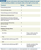
58-year-old woman with stage IA follicular lymphoma (grade 1 or 2) involving multiple 2-cm lymph nodes in the right cervical neck
Transformation Into Aggressive Non-Hodgkin Lymphoma
Histologic transformation from an indolent lymphoma into an aggressive NHL is most commonly seen in follicular lymphoma transforming into diffuse large B-cell lymphoma. Studies have reported incidences between 15% and 30% at 10 years following initial diagnosis of the indolent lymphoma.[16,18,19] Little is currently understood about the events leading to transformation. Treatment is directed based on newly transformed histology, with median survival times of 1 to 2 years.[16]
Early-Stage Follicular Lymphoma
Late relapses and the rarity of early-stage follicular lymphoma make phase III studies difficult to conduct. Therefore, the array of treatments delivered in both academic and community practices varies widely. In a recent report from the National LymphoCare Study,[4] a multicenter, longitudinal observational study including both community and academic practices, of 474 patients with stage I follicular lymphoma, 29% underwent observation alone, 23% received RT alone, 35% received rituximab alone or with chemotherapy, and 8% received rituximab with or without chemotherapy followed by RT. Preliminary analysis of patients with stage I disease in this study (median follow-up, 4.75 years) suggests that similarly excellent early outcomes can be achieved with a variety of treatment approaches (see Variant 1, Variant 2, and Variant 3).[20]
VARIANT 2
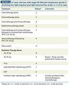
67-year-old man with stage IIA follicular lymphoma (grade 2) involving the right inguinal and right internal iliac nodes (2–3 cm in size)
Surveillance
Although typically used to manage patients with incurable stage III or IV disease, surveillance has been used in some cases of curable stage I and II disease. One retrospective study by Advani et al[21] reported on 43 patients with stage I or II follicular lymphoma diagnosed between 1969 and 2000 who underwent surveillance due to concerns regarding toxicity from RT alone (n = 16), advanced age or comorbidities (n = 10), or patient or physician preference (n = 17). Of note, these patients were not part of a prospective surveillance study, and patients who progressed in the 3 months following diagnosis were excluded from this study. Of the study patients, 63% never required treatment; however, 37% did require treatment (median time to treatment, 22 months) due to progressive disease (n = 12) or transformed disease (n = 4). All 9 deaths occurred among the 16 patients who required treatment. Although this study demonstrated that not everyone with early-stage disease requires treatment, there are currently no clear guidelines to distinguish these patients from those who progress during observation. Considering the high death rate among those who progressed and required treatment, active surveillance should be reserved for select patients who are not eligible for definitive treatment with RT.
Surgery
In patients with limited stage I disease, diagnostic surgery may eradicate all evidence of gross disease. Soubeyran et al[22] reported on 26 patients who had complete eradication of their lymphoma from the diagnostic surgery and who underwent close observation. During follow-up, 13 of the patients relapsed, with a 5-year PFS rate of 53% and a 10-year PFS rate of 27%. Six of the relapses occurred locally, but only half were salvaged with RT. No local failures occurred among 17 additional patients who received IFRT and were studied in parallel with those who had undergone close observation. Additionally, only 2 patients among the 17 treated died during follow-up, while 7 among those who underwent close observation died.
VARIANT 3
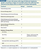
35-year-old woman with stage IA follicular lymphoma (grade 1) involving a single 2.8-cm lymph node in the axilla that was removed at the biopsy with no evidence of residual adenopathy
Radiotherapy
Several studies have described the outcomes for patients with early-stage follicular lymphoma treated with RT alone. Appendix 1 reviews the outcomes for several of these studies, demonstrating 10-year PFS rates of approximately 50% and OS rates of 60% to 70%. When evaluating these studies, one must keep in mind that they were done prior to the use of PET imaging for staging. PET imaging may facilitate disease control after RT alone by improving patient selection and target volume delineation.
In one of the largest studies, Stanford University (Palo Alto, California) reported on 177 patients treated with RT between 1961 and 1994 for stage I and II low-grade follicular lymphoma.[23] Various radiation techniques and doses were used during the different time periods covered in the study. Patients received IFRT, extended-field radiation therapy (EFRT), total lymphoid irradiation (TLI), or sub-TLI (STLI) to doses of 35 Gy to 50 Gy at 1.5 Gy to 2.5 Gy per fraction. With a median follow-up of 7.7 years, the median OS time was 13.8 years, with a 10-year freedom from recurrence rate of 44% and an OS rate of 64%. In the experience of Princess Margaret Hospital (Toronto, Ontario) [24] treating 190 patients with stage I or II follicular lymphoma, definitive treatment with RT (with variations in radiation field designs and doses) led to a 12-year relapse-free survival rate of 53%, a cause-specific survival rate of 67%, a local relapse-free survival rate of 87%, a distant relapse-free survival rate of 62%, and an OS rate of 58%. Additionally, it was noted that the relapses usually occurred at unirradiated sites. Lastly, a large study from the British National Lymphoma Investigation[25] reported on the outcomes of 208 patients with low-grade stage I disease treated with RT alone between 1974 and 1991 (radiation fields were not described, but a dose of 35 Gy was suggested). The OS rate at 10 years was 64% and the cancer-specific survival rate was 71%.
Considering the infrequency of early-stage follicular lymphoma and the need for single institutions to include 20 to 30 years’ worth of patients in their studies to provide an adequate cohort, studies that include large databases can be helpful. Pugh et al[26] recently reported on the outcomes of 6,568 patients with stage I and II, grade 1 and 2 follicular lymphoma diagnosed between 1973 and 2004 who were registered in the Surveillance Epidemiology and End Results (SEER) registry. During the 30-year study period, approximately one-third of patients received upfront RT. Due to limitations of the SEER database, information regarding radiation field design, radiation dose, and the use of chemotherapy or immunotherapy is not known. Researchers found that the 10-year disease-specific and OS rates were higher in patients who received RT as part of their treatment than in patients who did not receive RT (79% vs 66%, P < .0001; and 62% vs 48%, P < .0001; respectively). Although any SEER analysis has a number of weaknesses for the reasons given, these outcomes do coincide with those seen in other studies. Nevertheless, the magnitude of difference between the two groups may, in part, be due to likely selection bias of observation or chemotherapy alone in patients with significant comorbidities compared with patients without comorbidities, who would be more likely to receive RT (see Appendix 1).
VARIANT 4
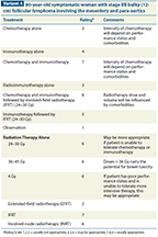
80-year-old symptomatic woman with stage IIB bulky (12-cm) follicular lymphoma involving the mesentery and para-aortics
Chemotherapy and radiotherapy
Studies regarding treatment regimens for localized low-grade NHL that combine chemotherapy and RT have generally included few patients and have shown conflicting results. Still, two studies did have reasonable patient numbers for evaluation. In a randomized study by the British National Lymphoma Investigation group in which 148 patients were randomly assigned to RT either with or without adjuvant chlorambucil, with a minimum follow-up of 11 years, no differences were seen in OS (P > .1) or relapse-free survival (P = .14).[27] In another study of 102 patients with stage I/II low-grade lymphoma, treatment consisted of 10 cycles of cyclophosphamide, vincristine, prednisone, and bleomycin, with or without doxorubicin, followed by 30 Gy to 40 Gy of IFRT.[28] After a median follow-up of 10 years, the relapse-free and OS rates at 10 years were 76% and 82%, respectively. Late toxicities, however, did include 2 patients with leukemia and 12 patients with solid cancers (4 with cancers within the RT field). In the LymphoCare database, 26 patients who were rigorously staged and found to have stage I disease received systemic therapy and RT, demonstrating that this regimen has been adopted by some oncologists, especially in patients with grade 3 histology and those with B symptoms.[20]
VARIANT 5
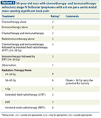
50-year-old man with chemotherapy- and immunotherapy-refractory stage IV follicular lymphoma with a 6-cm para-aortic nodal mass causing significant back pain
Radiotherapy dose
Studies evaluating the use of RT in patients with early-stage indolent lymphoma have typically used radiation doses between 20 Gy and 50 Gy. Although retrospective studies have demonstrated that lower radiation doses appeared to be as effective as higher doses, a recent phase III randomized study from the United Kingdom tested the results of 24 Gy vs 40 to 45 Gy.[29] Two hundred eighty-nine patients with 361 sites of indolent lymphoma were randomized. The results demonstrated no difference in overall response rate between the standard-dose and lower-dose arms (93% vs 92%, respectively). With a median follow-up of 5.6 years, there was no significant difference in the rate of within–radiation field progression (hazard ratio [HR] = 1.09; 95% confidence interval [CI], 0.76–1.56; P = .64) nor in the PFS or OS. In a subset analysis of 248 patients who received radical RT as first-line therapy, there was no difference in PFS, with 54% in the high-dose arm and 64% in the 24-Gy arm remaining alive without recurrence at any site at last follow-up. Thus, lower doses of 24 Gy to 30 Gy are generally preferred due to the anticipated lower risk of radiation-induced acute and late toxicity.
APPENDIX 1

Outcomes of Definitive Radiotherapy for Follicular Lymphoma
Radiotherapy field size
Treatment fields for the management of indolent lymphoma have ranged from total body irradiation (TBI) to TLI, to EFRT, to IFRT, and most recently to a tailored field (see Appendix 1). Owing to improvements in staging and concern for radiation toxicity, the standard field size for the last 20 years has been IFRT. A recent retrospective study evaluated the impact of an involved regional RT field (142 patients; 60%) compared with a tailored field that included the involved node with up to a 5-cm margin (95 patients; 40%).[30] At 10 years, the PFS rate was 49% and the OS rate was 66%, with no differences between the two groups. Furthermore, after receiving radiation to the tailored field, 1% of patients had a regional-only recurrence.
APPENDIX 2

Outcomes of Low-Dose Radiotherapy for Indolent Lymphoma
Recently, a new set of field designs, the involved-site radiation therapy (ISRT) fields, have been developed and endorsed by the steering committee of the International Lymphoma Radiation Oncology Group (ILROG). Led by experienced radiation oncologists specializing in lymphoma who initially organized the standardization of IFRT fields in the two-dimensional (2D) era a decade ago, the ISRT fields are a “modernized” version of IFRT fields. These new field designs were developed to take into consideration modern technology, including the use of staging PET/CT scans, three-dimensional and four-dimensional treatment planning with CT scanners, and conformal treatment techniques, as well as the use of image guidance, in order to replace the antiquated IFRT fields that were based on 2D treatment planning and bony anatomy. These new fields are expected to be somewhat smaller than the traditional IFRT fields, but bigger than the involved node radiation therapy (INRT) fields. A more detailed description of the ISRT field design is expected to be published in the near future; early descriptions can be found in the 2013 National Comprehensive Cancer Network (NCCN) guidelines for Hodgkin lymphoma.[31]
Advanced-Stage Follicular Lymphoma
The management of patients with more advanced disease, including stage IIB bulky, stage III, and stage IV, is quite controversial. Options that have been used and studied in the past include surveillance, radiation (low-dose TBI or TLI), immunotherapy (rituximab), chemotherapy (eg, bendamustine, CHOP [cyclophosphamide, doxorubicin, vincristine, predisone], CVP [cyclophosphamide, vincristine, prednisone], fludarabine), or some combination of the agents.
Indications for treatment may include symptomatic disease, threatened end-organ function, cytopenia due to lymphoma involvement, bulky disease, progressing disease, patient preference, or a combination of these factors. Currently, systemic therapies are preferred to EFRT in the management of advanced-stage disease out of concerns for acute and late RT toxicity; however, localized palliative RT may be helpful in patients unsuitable for systemic therapy (see Variant 4).
Relapsed and Refractory Disease
The management of relapsed and refractory disease is similar to that used for advanced-stage disease and includes a combination of immunotherapy with chemotherapy. In addition to systemic chemotherapy, options may include radioimmunotherapy (ibritumomab, tiuxetan, or tositumomab), high-dose therapy with autologous stem cell rescue, an allogeneic stem cell transplant, or palliative RT, all of which confer reasonable response rates (see Variant 5).
Summary
• Localized indolent nodal lymphoma is a rare diagnosis.
• FDG-PET/CT scans can demonstrate more disseminated disease and upstage patients thought to have localized involvement. Additionally, they can be useful in RT treatment planning.
• Based on a number of prospective and retrospective studies of patients treated with RT alone, the 10-year PFS and OS rates are expected to be approximately 50% and 65%, respectively.
• Definitive treatment with RT should include the use of IFRT and doses between 24 and 30.6 Gy.
• Advanced radiation techniques, such as IMRT and proton therapy, may be considered depending on the clinical scenario and on whether an improvement in the therapeutic ratio is expected.
• Observation can be considered in specialized cases where the risk from treatment outweighs the benefits.
• For patients with more advanced stage II disease, such as bulky disease and B symptoms, a more aggressive approach, including chemotherapy, immunotherapy, and RT, should be considered.
• Patients with advanced-stage disease refractory to chemotherapy and immunotherapy can sustain long intervals of local control with low-dose palliative RT of 4Gy.
Palliative Radiation Therapy
The radiosensitivity of indolent lymphoma has been recognized for years. A prospective study of patients with advanced follicular lymphoma randomized patients to single-alkylating-agent chemotherapy (cyclophosphamide or chlorambucil), CVP chemotherapy, or whole-body irradiation (WBI) at 30 cGy per day, 2 to 3 times per week, to a total dose of 150 cGy (with a boost of 20 Gy in 2 to 3 weeks to sites of initial involvement). The investigators reported a 5-year relapse-free survival rate of 35% following WBI.[32] In Europe, after several patients responded early in their treatment course, either after a few fractions of radiation or when they prematurely stopped their planned RT, investigators became interested in prospective studies of low-dose palliative RT to localized sites. In the largest multicenter phase II study evaluating palliative RT for indolent lymphoma, 109 patients with a total of 304 symptomatic sites of recurrent indolent lymphoma were irradiated with 4 Gy either over 2 fractions or in a single fraction to either the entire lymph node region or the lymph node with a 1.5-cm margin. The overall response rate was 92%, with a median duration time until local relapse of 25 months and time to any progression of 14 months.[33] Other single-institution studies have also demonstrated similar results following low-dose palliative RT, with median local control times of 1 to 2 years (see Appendix 2).[33-41]
Radiation Therapy Techniques
Considering the involved sites at presentation and the relatively low dose of RT needed for disease control, the most basic treatment planning is acceptable, using a CT simulation for three-dimensional conformal radiotherapy (3D-CRT) planning with an anterior-posterior/posterior-anterior field arrangement. For some patients, however, a more complex 3D-CRT plan, an intensity-modulated radiation therapy (IMRT) plan, or even a proton therapy plan should be considered to minimize dose to critical normal structures. If using more conformal techniques, one should keep in mind that data about them are limited, and further clinical studies are needed. Furthermore, one should consider the impact of radiation dose uncertainty and the potential impact of irradiating more tissue with low-dose radiation.
Financial Disclosure:Christopher R. Flowers, MD, MS, reports: “I provide consultation for the Prescription Solutions Pharmacy & Therapeutics Division. My consultation involves advice on FDA regulated drugs only. None of my consultation overlaps with existing or planned ACR guidelines.” Anas Younes, MD, reports: “Honoraria from Novartis, Seattle Genetics, Millennium, Celgene, Curis, Sanofi, Pharmacyclics, and Incyte; research support from Novartis, Johnson & Johnson, Seattle Genetics, Merck, Genentech, Infinity, and Gilead.”
The American College of Radiology (ACR) seeks and encourages collaboration with other organizations on the development of the ACR Appropriateness Criteria® through society representation on expert panels. Participation by representatives from collaborating societies on the expert panel does not necessarily imply individual or society endorsement of the final document.
For additional information on ACR Appropriateness Criteria®, refer to www.acr.org/ac.
References:
REFERENCES
1. National Cancer Institute. Comprehensive Cancer Information. Available at: http://www.cancer.gov/cancertopics/types/non-hodgkin. Accessed April 12, 2013.
2. Siegel R, Naishadham D, Jemal A. Cancer statistics, 2013. CA Cancer J Clin. 2013;63:11-30.
3. A clinical evaluation of the International Lymphoma Study Group classification of non-Hodgkin's lymphoma. The Non-Hodgkin’s Lymphoma Classification Project. Blood. 1997;89:3909-18.
4. Friedberg JW, Taylor MD, Cerhan JR, et al. Follicular lymphoma in the United States: first report of the national LymphoCare study. J Clin Oncol. 2009;27:1202-8.
5. Morton LM, Wang SS, Devesa SS, et al. Lymphoma incidence patterns by WHO subtype in the United States, 1992-2001. Blood. 2006;107:265-76.
6. Goldin LR, Bjorkholm M, Kristinsson SY, et al. Highly increased familial risks for specific lymphoma subtypes. Br J Haematol. 2009;146:91-4.
7. Wahlin BE, Yri OE, Kimby E, et al. Clinical significance of the WHO grades of follicular lymphoma in a population-based cohort of 505 patients with long follow-up times. Br J Haematol. 2012;156:225-33.
8. Tsujimoto Y, Cossman J, Jaffe E, Croce CM. Involvement of the bcl-2 gene in human follicular lymphoma. Science. 1985;228:1440-3.
9. Carbone PP, Kaplan HS, Musshoff K, et al. Report of the Committee on Hodgkin's Disease Staging Classification. Cancer Res. 1971;31:1860-1.
10. Canioni D, Brice P, Lepage E, et al. Bone marrow histological patterns can predict survival of patients with grade 1 or 2 follicular lymphoma: a study from the Groupe d'Etude des Lymphomes Folliculaires. Br J Haematol. 2004;126:364-71.
11. Wirth A, Foo M, Seymour JF, et al. Impact of [18f] fluorodeoxyglucose positron emission tomography on staging and management of early-stage follicular non-hodgkin lymphoma. Int J Radiat Oncol Biol Phys. 2008;71:213-9.
12. Le Dortz L, De Guibert S, Bayat S, et al. Diagnostic and prognostic impact of 18F-FDG PET/CT in follicular lymphoma. Eur J Nucl Med Mol Imaging. 2010;37:2307-14.
13. Solal-Celigny P, Roy P, Colombat P, et al. Follicular lymphoma international prognostic index. Blood. 2004;104:1258-65.
14. Plancarte F, Lopez-Guillermo A, Arenillas L, et al. Follicular lymphoma in early stages: high risk of relapse and usefulness of the Follicular Lymphoma International Prognostic Index to predict the outcome of patients. Eur J Haematol. 2006;76:58-63.
15. Buske C, Hoster E, Dreyling M, et al. The Follicular Lymphoma International Prognostic Index (FLIPI) separates high-risk from intermediate- or low-risk patients with advanced-stage follicular lymphoma treated front-line with rituximab and the combination of cyclophosphamide, doxorubicin, vincristine, and prednisone (R-CHOP) with respect to treatment outcome. Blood. 2006;108:1504-8.
16. Montoto S, Davies AJ, Matthews J, et al. Risk and clinical implications of transformation of follicular lymphoma to diffuse large B-cell lymphoma. J Clin Oncol. 2007;25:2426-33.
17. Federico M, Bellei M, Marcheselli L, et al. Follicular lymphoma international prognostic index 2: a new prognostic index for follicular lymphoma developed by the international follicular lymphoma prognostic factor project. J Clin Oncol. 2009;27:4555-62.
18. Bastion Y, Sebban C, Berger F, et al. Incidence, predictive factors, and outcome of lymphoma transformation in follicular lymphoma patients. J Clin Oncol. 1997;15:1587-94.
19. Conconi A, Ponzio C, Lobetti-Bodoni C, et al. Incidence, risk factors and outcome of histological transformation in follicular lymphoma. Br J Haematol. 2012;15):188-96.
20. Friedberg JW, Byrtek M, Link BK, et al. Effectiveness of first-line management strategies for stage I follicular lymphoma: analysis of the National LymphoCare Study. J Clin Oncol. 2012;30:3368-75.
21. Advani R, Rosenberg SA, Horning SJ. Stage I and II follicular non-Hodgkin's lymphoma: long-term follow-up of no initial therapy. J Clin Oncol. 2004;22:1454-9.
22. Soubeyran P, Eghbali H, Trojani M, Bonichon F, Richaud P, Hoerni B. Is there any place for a wait-and-see policy in stage I0 follicular lymphoma? A study of 43 consecutive patients in a single center. Ann Oncol. 1996;7:713-8.
23. Mac Manus MP, Hoppe RT. Is radiotherapy curative for stage I and II low-grade follicular lymphoma? Results of a long-term follow-up study of patients treated at Stanford University. J Clin Oncol. 1996;14:1282-90.
24. Gospodarowicz MK, Bush RS, Brown TC, Chua T. Prognostic factors in nodular lymphomas: a multivariate analysis based on the Princess Margaret Hospital experience. Int J Radiat Oncol Biol Phys. 1984;10:489-97.
25. Vaughan Hudson B, Vaughan Hudson G, MacLennan KA, et al. Clinical stage 1 non-Hodgkin's lymphoma: long-term follow-up of patients treated by the British National Lymphoma Investigation with radiotherapy alone as initial therapy. Br J Cancer. 1994;69:1088-93.
26. Pugh TJ, Ballonoff A, Newman F, Rabinovitch R. Improved survival in patients with early stage low-grade follicular lymphoma treated with radiation: a Surveillance, Epidemiology, and End Results database analysis. Cancer. 2010;116:3843-51.
27. Kelsey SM, Newland AC, Hudson GV, Jelliffe AM. A British National Lymphoma Investigation randomised trial of single agent chlorambucil plus radiotherapy versus radiotherapy alone in low grade, localised non-Hodgkins lymphoma. Med Oncol. 1994;11:19-25.
28. Seymour JF, Pro B, Fuller LM, et al. Long-term follow-up of a prospective study of combined modality therapy for stage I-II indolent non-Hodgkin's lymphoma. J Clin Oncol. 2003;21:2115-22.
29. Lowry L, Smith P, Qian W, et al. Reduced dose radiotherapy for local control in non-Hodgkin lymphoma: a randomised phase III trial. Radiother Oncol. 2011;100:86-92.
30. Campbell BA, Voss N, Woods R, et al. Long-term outcomes for patients with limited stage follicular lymphoma: involved regional radiotherapy versus involved node radiotherapy. Cancer. 2010;116:3797-806.
31. NCCN Clinical Practice Guidelines in Oncology. Hodgkin Lymphoma. Version 1.2013. Available at: http://www.nccn.org/professionals/physician_gls/pdf/hodgkins.pdf. Accessed April 12, 2013.
32. Hohloch K, Delaloye AB, Windemuth-Kieselbach C, et al. Radioimmunotherapy confers long-term survival to lymphoma patients with acceptable toxicity: registry analysis by the International Radioimmunotherapy Network. J Nucl Med. 2011;52:1354-60.
33. Haas RL, Poortmans P, de Jong D, et al. High response rates and lasting remissions after low-dose involved field radiotherapy in indolent lymphomas. J Clin Oncol. 2003;21:2474-80.
34. Chan EK, Fung S, Gospodarowicz M, et al. Palliation by low-dose local radiation therapy for indolent non-Hodgkin lymphoma. Int J Radiat Oncol Biol Phys. 2011;81:e781-6.
35. Ganem G, Lambin P, Socie G, et al. Potential role for low dose limited-field radiation therapy (2 × 2 Grays) in advanced low-grade non-Hodgkin's lymphomas. Hematol Oncol. 1994;12:1-8.
36. Girinsky T, Guillot-Vals D, Koscielny S, et al. A high and sustained response rate in refractory or relapsing low-grade lymphoma masses after low-dose radiation: analysis of predictive parameters of response to treatment. Int J Radiat Oncol Biol Phys. 2001;51:148-55.
37. Haas RL, Poortmans P, de Jong D, et al. Effective palliation by low dose local radiotherapy for recurrent and/or chemotherapy refractory non-follicular lymphoma patients. Eur J Cancer. 2005;41:1724-30.
38. Luthy SK, Ng AK, Silver B, et al. Response to low-dose involved-field radiotherapy in patients with non-Hodgkin's lymphoma. Ann Oncol. 2008;19:2043-7.
39. Murthy V, Thomas K, Foo K, et al. Efficacy of palliative low-dose involved-field radiation therapy in advanced lymphoma: a phase II study. Clin Lymphoma Myeloma. 2008;8:241-5.
40. Rossier C, Schick U, Miralbell R, et al. Low-dose radiotherapy in indolent lymphoma. Int J Radiat Oncol Biol Phys. 2011;81:e1-6.
41. Sawyer EJ, Timothy AR. Low dose palliative radiotherapy in low grade non-Hodgkin's lymphoma. Radiother Oncol. 1997;42:49-51.
42. Kamath SS, Marcus RB, Jr., Lynch JW, Mendenhall NP. The impact of radiotherapy dose and other treatment-related and clinical factors on in-field control in stage I and II non-Hodgkin's lymphoma. Int J Radiat Oncol Biol Phys. 1999;44:563-8.
43. Lawrence TS, Urba WJ, Steinberg SM, et al. Retrospective analysis of stage I and II indolent lymphomas at the National Cancer Institute. Int J Radiat Oncol Biol Phys. 1988;14:417-24.
44. Pendlebury S, el Awadi M, Ashley S, et al. Radiotherapy results in early stage low grade nodal non-Hodgkin's lymphoma. Radiother Oncol. 1995;36:167-71.
45. Taylor RE, Allan SG, McIntyre MA, et al. Low grade stage I and II non-Hodgkin's lymphoma: results of treatment and relapse pattern following therapy. Clin Radiol. 1988;39:287-90.
46. Wilder RB, Jones D, Tucker SL, et al. Long-term results with radiotherapy for Stage I-II follicular lymphomas. Int J Radiat Oncol Biol Phys. 2001;51:1219-27.