Nonseminomatous Germ Cell Tumor of Testis and Interrupted Treatment
A 22-year-old man presented to the emergency department with a 5-cm painful testicular mass that had increased in size over the previous month. Tumor markers were drawn and an inguinal orchiectomy was performed.
The Case:A 22-year-old man presented to the emergency department with a 5-cm painful testicular mass that had increased in size over the previous month. Tumor markers were drawn and an inguinal orchiectomy was performed. The beta human chorionic gonadotropin (β HCG) level was 40 mIU/mL (reference range, 0–5 mIU/mL), the alpha-fetoprotein (AFP) level was 841 ng/mL (reference range, 0–9 ng/mL), and the lactate dehydrogenase (LDH) level was 157 U/L (reference range, 98–192 U/L). A computed tomography (CT) scan of the chest, abdomen, and pelvis was negative. The pathology report showed a mixed nonseminomatous germ cell tumor (NSGCT) measuring 5.0 × 4.7 × 4.6 cm and confined to the testis without evidence of invasion of the tunica albuginea or rete testis; the spermatic cord margin was negative as well. No vascular/lymphatic invasion was seen. Components consisted of 60% teratoma, 30% yolk sac tumor, and 10% embryonal carcinoma. He was referred to a medical oncologist who reviewed the options of surveillance, retroperitoneal lymph node dissection (RPLND), or adjuvant chemotherapy.
The patient did not return to the medical oncology team, but 3 months later he presented to the emergency department with left lower back pain. A repeat CT scan now showed pulmonary nodules, as well as retroperitoneal and mediastinal adenopathy, with nodes measuring up to 2.7 cm in the short axis. Tumor markers had increased, with the AFP level now 2,602 ng/mL. A brain magnetic resonance imaging (MRI) scan was negative for metastatic disease. The patient reported anxiety, depression, and no insurance coverage. He denied suicidal ideation. Examination was negative for peripheral adenopathy, organomegaly, or focal neurologic signs. The patient was admitted to the hospital, and cisplatin, etoposide, and bleomycin (PEB) therapy was initiated at standard doses. The AFP level initially increased to 4,209 ng/mL, then decreased to 3,413 ng/mL. The βHCG level decreased from 12 mIU/mL to 6 mIU/mL. A second cycle of PEB was administered on schedule 3 weeks later. On his third admission, the patient checked out of the hospital against medical advice, and he was lost to follow-up.
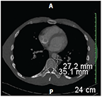
Four months later, he presented to the emergency department with a history of a 30-lb weight loss and increasing back pain. He was immediately sent to the oncology clinic, where complete laboratory tests were ordered and a repeat CT scan of his chest/abdomen/pelvis was scheduled. Examination revealed a thin, anxious man with multiple tattoos, but no organomegaly, abdominal mass, lymphadenopathy, or other evidence of disease. His remaining testis was palpably normal. All of his tumor markers had returned to the normal range. The CT scan now revealed worsening of retroperitoneal and mediastinal adenopathy compared with the scan obtained before the initiation of treatment. Small sub-centimeter pulmonary nodules were again noted, some of which were smaller and others larger, with some new nodules present. Many of the nodes revealed evidence of central necrosis. A repeat brain MRI scan remained negative. The diffusing capacity of the lungs for carbon monoxide was 79% of predicted value.
The patient was admitted to the hospital, and treatment with PEB chemotherapy was reinstituted.
FIGURE 1

CT Scan Showing Persistent Mediastinal Adenopathy at the Time of Re-Presentation.
Discussion
This case presents multiple challenges. In the beginning, the patient had low-risk NSGCT, but his psychosocial history was already of concern. In counseling such patients, their ability to comply with recommended treatment or surveillance must be carefully considered. Although surveillance is increasingly recommended to avoid short- and long-term complications of chemotherapy or RPLND, in many cases surveillance is much less attractive because a patient is either unwilling or unable to follow recommended guidelines. Examples include individuals with a record of poor compliance due to financial or social issues, as well as patients who must travel extensively in developing countries and are unable to access recommended healthcare facilities in a timely manner (eg, peace corps volunteers and some military personnel).
When this patient reappeared in our oncology clinic, the expectation was that he would have progressive disease; however, we were surprised to discover that his markers had completely normalized. At odds with his normal markers was the increasing disease on imaging studies, but since no imaging had been carried out since his initial evaluation, it was difficult to determine whether his disease was actually progressing or perhaps had stabilized. In our tumor board review, some of the participating clinicians advocated reassessment of the patient’s abdominal disease prior to reinitiation of PEB chemotherapy, with the rationale that he might have only teratoma and that surgical intervention would provide a more accurate assessment of his status. Consideration was also given to changing his therapy to a salvage regimen, such as paclitaxel, ifosfamide, cisplatin, since he might have higher risk of resistant disease. A review of the available literature does not provide guidance in such a situation, and ultimately, the clinicians must use their best judgment in deciding on a treatment plan. Clearly, regardless of the plan selected or its outcome, significant effort will be required to provide this young man with the support he needs to comply with further treatment. This will involve consultation with social services, palliative care, and possibly the chaplain if appropriate.
Outcome
A single cycle of PEB chemotherapy was administered at standard doses. A CT scan demonstrated no change in retroperitoneal, mediastinal, and right hilar adenopathy. There were no new pulmonary nodules and minimal changes in existing ones. The patient was taken to surgery, where a right thoracoabdominal approach allowed resection of all known lymphatic disease in the mediastinum, retroperitoneum, and abdomen. Three pulmonary nodules were also resected. The pathology results demonstrated mature teratoma, with areas of fibrosis in all of the resected tissue. No immature teratoma or germ cell tumor elements were seen. He is now entering the surveillance phase of care.