The Approach to the Patient With Synchronous Bilateral Germ Cell Tumors: A Lesson in Oncologic Prioritization
Hammerich et al. report a case of synchronous bilateral germ cell tumors (GCT) of different histologies occurring in a patient with a history of cryptorchidism. There are several interesting aspects of this case and the authors’ management and discussion that warrant commentary.
Hammerich et al. report a case of synchronous bilateral germ cell tumors (GCT) of different histologies occurring in a patient with a history of cryptorchidism. There are several interesting aspects of this case and the authors' management and discussion that warrant commentary.
Cryptorchidism and Risk of Testicular Cancer
Cryptorchidism is the most well-defined risk factor for the development of testicular GCT; if uncorrected, the relative risk is 5 to 9 times that of healthy age-matched controls.[1] Approximately 10% of all cases of testicular GCT are associated with cryptorchidism and 10% of patients with cryptorchidism develop testicular GCT.[2,3] When testicular GCT develops in patients with unilateral cryptorchidism, the ipsilateral testis is affected in 80% to 90% of cases, but the contralateral testis can also be affected. Therefore, bilateral testicular GCT can also be observed in these patients, as illustrated by this case.
Bilateral Testicular GCT
Approximately 2% of men with testicular GCT will have the contralateral testis affected at some point during their lifetime.[4] In the largest study in the United States, including 29,515 testicular cancer patients, the 15-year risk of contralateral GCT was 1.9%, with 62% occurring metachronously and 38% synchronously.[4] The ratio of metachronous to synchronous cases is underestimated by these numbers since the follow-up time for patients varied.[4] The risk of contralateral GCT appears to be higher for younger patients and those with seminomas.[4]
How Rare Are Divergent Histologies in Synchronous GCT?
In addition to the patient described by Hammerich and colleagues, a review of the literature reveals several prior case reports[5-8] of synchronous bilateral GCT of divergent histologies. In fact, two large series[4,9] found differing histologies to be more common than bilateral nonseminomatous GCT (NSGCT). In one study of 175 patients with synchronous tumors, 50% had bilateral seminomas-34% with discordant histol-ogies and 16% with bilateral NSGCT.[4] Therefore, although uncommon, con-
comitant presentation with histologically distinct bilateral GCT is not as rare as the authors suggest.
Unusual Sites of Metastases
Lymphatic spread from testicular cancer is highly predictable, with the left para-aortic and interaortocaval regions representing the landing zones for left- and right-sided tumors, respectively. Inguinal nodal metastases are rare and primarily develop in the setting of retrograde flow from bulky retroperitoneal adenopathy. Therefore, the authors were surprised that their patient had a right inguinal metastasis without right-sided retroperitoneal adenopathy. However, it is important to recognize that violation of scrotal integrity can allow atypical patterns of lymphatic spread[10-12] and is a major reason that inguinal rather than transcrotal orchiectomy is recommended for the management of testicular cancer. In addition, spermatic cord (SC) involvement has been implicated in the development of inguinal metastases through direct tumor extension from the upper SC.[10] Therefore, either the patient's prior orchiopexy or SC involvement could explain his isolated right inguinal metastasis.
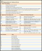
TABLE 1

AJCC Staging System for Testicular Cancer
Staging of Synchronous Bilateral GCT
It is important to emphasize that bilateral testicular GCT not be viewed as a metastasis from one testis to the other (M1 disease), but rather as the development of two independent primary tumors. Therefore, the authors correctly chose to independently stage both tumors. The major difficulty in staging posed by synchronous bilateral primary tumors is an inability to determine noninvasively from which tumor a patient's metastases are derived. This distinction is only important when the primary tumors are of differing histologies and treatment would change based on which tumor is the source of the metastases. Clues to accurately identify the histology of metastatic foci in these cases include tumor marker levels (for example, seminomas do not produce alpha fetoprotein (AFP), and the location of retroperitoneal nodes.
The American Joint Committee on Cancer staging system for testicular GCT is provided in Table 1.[13] An important distinguishing feature of this tumor type is the inclusion of serum tumor markers ("S" stage). For patients who undergo orchiectomy prior to starting chemotherapy, a common mistake is to assign an S stage based on marker values obtained pre- rather than post-operatively. Moreover, S staging is critical for NSGCT patients since, unlike seminoma, it also affects prognosis and chemotherapy selection. NSGCT patients with S2 or S3 marker levels require four cycles of bleomycin, etoposide, and cisplatin (BEP) whereas those with S1 markers can be treated with either three cycles of BEP or four cycles of EP.[14,15] Unfortunately, in the case report, the authors provide only the pre-orchiectomy marker levels, leaving his true S and overall stage unknown.
Prioritization in the Management of Synchronous Tumors
Simply stated, when two neoplasms present simultaneously, treatment should be tailored to the more aggressive disease. For example, if a patient presented concurrently with metastatic lung cancer and localized prostate cancer, treatment would be directed at the former, since the latter is unlikely to be life-threatening. The principle is especially true when, as in our patient's case, therapy for the more aggressive tumor (NSGCT) is also sufficient for the less aggressive neoplasm (seminoma), eliminating the need for two different treatment strategies.
So, How Would I Have Staged and Managed the Patient in the Vignette?
Similar to the authors, I would have staged the right-sided seminoma as pT1pN3MOSx, or stage IIC. The Sx designation results from the tumor markers being drawn preoperatively (rather than postoperatively) and because it is unknown whether the elevated B-hCG or LDH were the result of metastatic seminoma or NSGCT (S1 vs S0). In contrast, the AFP elevation is presumed to derive from the NSGCT since seminomas do not produce this marker. Importantly, I disagree with the authors' statement that stage IIC seminoma should be treated with radiation therapy. Most modern guidelines recommend systemic chemotherapy with either EP 3 4 or BEP 3 3 for these patients as well as for some with bulky stage IIB seminoma.[16-18]
The left-sided NSGCT is best staged as pT2cN1M0Sx. The designation cN1 assumes the NSGCT to be the etiology of the left para-aortic adenopathy. The markedly elevated AFP suggests spread of the NSGCT beyond the testis and the left para-aortic node is in the landing zone for a left testicular tumor. Again, Sx indicates the unknown postoperative marker levels. As previously mentioned, staging and prognostication are based on values obtained after any interventions (eg, orchiectomy) leading up to the initiation of chemotherapy. Declining markers should be followed until they rise, plateau, or it becomes obvious that they will fail to normalize, based on a slow rate of decline compared with the expected half-life.
Therefore, the correct NSGCT stage would have been either pT2cN1M0S2 (stage IIIB) or pT2cN1M0S1 (stage IIA) depending on his post-orchiectomy AFP level (≥1,000 vs <1,000). This is important since in the latter case, he would have been considered good- rather than intermediate-risk, and required less intensive chemotherapy (EP 3 4 or BEP 3 3 instead of BEP 3 4). The benefit of reducing chemotherapy intensity is not trivial since acute and chronic toxicities may impair both the quality and longevity of life of cancer survivors.
As stated, treatment should be targeted towards the more aggressive tumor. In the present case, chemotherapy is necessary for both neoplasms, but regimen selection should be based on the more advanced stage left-sided NSGCT. If the patient's post-orchiectomy AFP remained in the S2 range (≥1,000), then the preferred treatment would be BEP 3 4, which is also more than sufficient for the stage IIC seminoma.
Conclusions
Bilateral synchronous GCT are rare, affecting less than 1% of testicular cancer patients in the United States. The presence of discordant histologies (seminoma and NSGCT) in the primary tumors poses a unique set of challenges to staging and management as illustrated by the case presentation. However, independent of the ability to accurately stage both tumors, the most important oncologic lesson highlighted by this case is that treatment should be directed toward the most aggressive histology/presentation, particularly when it will also suffice for the less aggressive histology.
Financial Disclosure:The author has no significant financial interest or other relationship with the manufacturers of any products or providers of any service mentioned in this article.
References:
References
1. Wood HM, Elder JS: Cryptorchidism and testicular cancer: Separating fact from fiction. J Urol 181:452-461, 2009.
2. Kanto S, Hiramatsu M, Suzuki K, et al: Risk factors in past histories and familial episodes related to development of testicular germ cell tumor. Int J Urol 11:640-646, 2004.
3. Prener A, Engholm G, Jensen OM: Genital anomalies and risk for testicular cancer in Danish men. Epidemiology 7:14-9, 1996.
4. Fossa SD, Chen J, Schonfeld SJ, et al: Risk of contralateral testicular cancer: A population-based study of 29,515 U.S. men. J Natl Cancer Inst 97:1056-1066, 2005.
5. Dieckmann KP, Hamm B, Due W, et al: Simultaneous bilateral testicular germ cell tumors with dissimilar histology. Case report and review of the literature. Urol Int 43:305-309, 1988.
6. Gasent Blesa JM, Laforga Canales J, Romero Perez P, et al: Young male patient with bilateral synchronous testicular germ cell tumour. Considerations for partial orchiectomy. Clin Transl Oncol 10:850-852, 2008.
7. Reinberg Y, Manivel JC, Zhang G, et al: Synchronous bilateral testicular germ cell tumors of different histologic type. Pathogenetic and practical implications of bilaterality in testicular germ cell tumors. Cancer 68:1082-1085, 1991.
8. Coli A, Bigotti G, Dell’Isola C, et al: Synchronous bilateral testicular germ cell tumor with different histology. Case report and review of the literature. Urol Int 71:412-417, 2003.
9. Holzbeierlein JM, Sogani PC, Sheinfeld J: Histology and clinical outcomes in patients with bilateral testicular germ cell tumors: The Memorial Sloan Kettering Cancer Center experience 1950 to 2001. J Urol 169:2122-2125, 2003.
10. Daugaard G, Karas V, Sommer P: Inguinal metastases from testicular cancer. BJU Int 97:724-726, 2006.
11. Klein FA, Whitmore WF, Jr., Sogani PC, et al: Inguinal lymph node metastases from germ cell testicular tumors. J Urol 131:497-500, 1984.
12. Mason MD, Featherstone T, Olliff J, et al: Inguinal and iliac lymph node involvement in germ cell tumours of the testis: Implications for radiological investigation and for therapy. Clin Oncol (R Coll Radiol) 3:147-150, 1991.
13. Greene FL, Fritz AG, Balch CM, et al: AJCC Cancer Staging Handbook, ed 6. New York, Springer-Verlag, 2002.
14. Feldman DR, Bosl GJ, Sheinfeld J, et al: Medical treatment of advanced testicular cancer. JAMA 299:672-684, 2008.
15. International Germ Cell Cancer Collaborative Group: International Germ Cell Consensus Classification: A prognostic factor-based staging system for metastatic germ cell cancers. J Clin Oncol 15:594-603, 1997.
16. Motzer RJ, Agarwal N, Beard C, et al: NCCN clinical practice guidelines in oncology: Testicular cancer. J Natl Compr Canc Netw 7:672-693, 2009.
17. Schmoll HJ, Jordan K, Huddart R, et al: Testicular seminoma: ESMO clinical recommendations for diagnosis, treatment and follow-up. Ann Oncol 20 (suppl 4):83-88, 2009.
18. Krege S, Beyer J, Souchon R, et al: European consensus conference on diagnosis and treatment of germ cell cancer: A report of the second meeting of the European Germ Cell Cancer Consensus Group (EGCCCG): Part II. Eur Urol 53:497-513, 2008.
Late Hepatic Recurrence From Granulosa Cell Tumor: A Case Report
Granulosa cell tumors exhibit late recurrence and rare hepatic metastasis, emphasizing the need for lifelong surveillance in affected patients.