Combined PET/CT Aids in Head and Neck Cancer Management
TORONTO, Canada-The combination of positron emission tomography (PET) and computed tomography (CT) has proved particularly advantageous in the diagnosis and treatment of cancer of the head and neck, Carolyn Cidis Meltzer, MD, said at the 48th Annual Meeting of the Society of Nuclear Medicine (abstract 133).
TORONTO, CanadaThe combination of positron emission tomography (PET) and computed tomography (CT) has proved particularly advantageous in the diagnosis and treatment of cancer of the head and neck, Carolyn Cidis Meltzer, MD, said at the 48th Annual Meeting of the Society of Nuclear Medicine (abstract 133).
Dr. Meltzer reported on the application of a combined PET/CT scanner developed at the University of Pittsburgh School of Medicine by David Townsend, PhD, professor of radiology, Dr. Meltzer, and their colleagues. Dr. Meltzer is associate professor of radiology and psychiatry, and medical director of the University of Pittsburgh Medical Center’s PET facility.
With the new instrument, the CT scan can be followed immediately by a PET scan in the same imaging session. When the images are combined, the results are superior to those obtained by using either imaging technique on its own. The Pittsburgh prototype PET/CT machine has received FDA approval for marketing as the Biograph (CTI/Siemens).
PET measures the rate of metabolic activity in different regions of the body, by detecting the rate of accumulation of radiolabeled fluorodeoxyglucose (FDG). Because malignant cells often show enhanced glucose metabolism, the resulting PET scan can differentiate between cancer cells and normal tissue, and can even distinguish between benign and malignant tumors.
The interpretation of PET images can sometimes be complicated, however, since enhanced FDG uptake can also occur in a variety of normal tissues, including striated muscle, bone marrow, lymphoid tissue, salivary glands, and brain.
Not only are these various tissues in close proximity to each other in the head and neck, but variable uptake in normal tissues can occur asymmetrically, giving rise to the sort of localized signal that could be misinterpreted as coming from a malignant tumor.
Limitations of CT
Conversely, CT images, derived from a series of x-rays, provide much better anatomic definition than PET, but cannot assess the physiologic state of tissue and thus are unable to measure one of the key distinguishing markers of cancer cells. This problem is of particular concern in assessing lymph nodes for metastases, since the only distinguishing character on CT images is size. Nodes greater than 1 cm in diameter on CT scan are considered highly suspect, but this criterion is by no means definitive.
As a result of these limitations, clinicians have looked for ways to combine the two imaging modalities.
"One approach to combining the two methodologies has involved performance of separate PET and CT scans, with retrospective fusion of the images to display registered metabolic and anatomic information," she said.
This technique can help improve the accuracy of tumor detection, Dr. Meltzer said, but since the images are taken at different times, with concomitant repositioning of the patient, the co-registration is often insufficiently precise.
"The combined PET/CT scanner obviates many of these concerns, and allows us to evaluate both anatomy and physiology, so that we can assess the stage of the tumor and the degree of any metastasis," Dr. Meltzer said.
70 Head and Neck Patients
More than 300 patients have been imaged on the combined scanner, 70 of them with head and neck malignancies. The new technique has proved useful both in the initial detection of tumors, as well as in post-therapeutic monitoring for potential recurrence, Dr. Meltzer said.
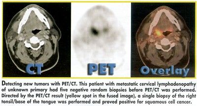
The images below show an example of detection of new tumors, in this case a squamous cell carcinoma of unknown primary origin that had been missed in five biopsies taken prior to the PET/CT scan. In the CT image, there is no evidence of anatomic derangement, but enhanced FDG uptake is clear in the PET image, which, when overlaid with the CT scan, provides precise anatomic localization of the tumor.

To date, 12 patients with metastases of unknown primary origin have been assessed with the combined scanner, and all 12 showed enhanced FDG uptake at the site of the lesion. In four of these patients, the PET/CT scan also permitted identification of the site of the primary tumor.
Monitoring After Treatment
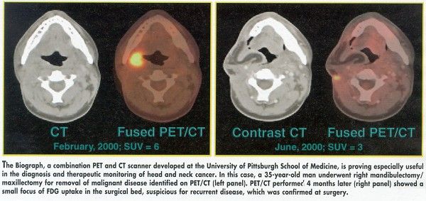
The combined scanner has also proved to be particularly useful in monitoring patients after treatment for the primary tumor. By itself, accurate CT or magnetic resonance imaging (MRI) of recurrent tumors is often especially difficult, since both surgery and radiation therapy can lead to disruption of normal spatial arrangement of neighboring tissues. Furthermore, the proximity of the brain, where normal physiologic activity is especially variable, complicates the interpretation of FDG-PET.
The images below show an example of monitoring with the combined scanner. In this patient, a primary tumor, itself detected by PET/CT, was surgically removed. Four months later, on a repeat scan, recurrence is evident in a small region of the original surgical bed.

Similar findings have proven the applicability of PET/CT in the assessment of recurrence in nine patients with thyroid cancer, and in seven of eight patients with skull base neoplasms. In the latter group, there was only one false negative, a tumor that was seen on MRI but not on PET/CT.
Thus, while still in its infancy, the combination of PET/CT offers clear advantages for the detection and monitoring of cancers in the head and neck, Dr. Meltzer said. It is to be hoped that this technology can be made widely available as a basic tool in the radiologist’s armamentarium, she added.