Effectiveness of Breast Cancer Screening: What Are the Metrics
As Smith et al[1] discuss, randomized controlled trials (RCTs) have shown that mammography reduces deaths due to breast cancer across ages 40 through 74 years, and within subsets by decade.[2]
As Smith et al[1] discuss, randomized controlled trials (RCTs) have shown that mammography reduces deaths due to breast cancer across ages 40 through 74 years, and within subsets by decade.[2] Even with these findings, screening mammography remains a topic of much discussion and debate.[3,4] Besides detecting cancers that need to be treated, screening can result in additional, unproductive testing and in treatment of tumors that would not have been life-threatening. These considerations are quite complex for mammography, and they are even more unresolved for newer technologies being developed or proposed for breast cancer screening. Useful discussions of these issues require careful definition of terms and metrics.
Let us first consider the definition of screening. The concept is to include asymptomatic women presenting for routine mammography. A screening mammogram should include both craniocaudal (CC) and mediolateral oblique (MLO) views. Failure to include the CC view was among the limitations of two of the RCTs of mammography.[5,6] For scheduling and billing, many women undergoing routine screening may be included in the “diagnostic” population at the discretion of the facility performing the screening, and this includes women with a personal history of breast cancer, women with implants, and even women with a benign breast biopsy in their history. A diagnostic mammogram begins with the standard views. What distinguishes diagnostic from screening mammography is supervision by a radiologist (at higher cost), and, if additional views or ultrasound are needed, these procedures are typically performed concurrently and the patient is given results of diagnostic mammography during the visit. Screening is typically performed as batch reading with results mailed. That said, some facilities perform “online” screening and review every examination while the patient waits. Online screening increases likelihood of additional testing (false positives, with additional billing)[7,8] without gain in sensitivity, and it is typically driven by patient demand for immediate results.[9] For all these reasons, tracking outcomes of “screening” in routine practice can be problematic.
Most women who die from breast cancer have not undergone regular screening mammography. Cady et al[10] reported in 2009 a series of 461 deaths due to breast cancer and found 116 (25%) of deaths were among asymptomatic women who had two or more screening mammograms at intervals of 2 years or less. Importantly, 345 deaths (75%) occurred in the 20% of women not screened by mammography in the prior 2 years (including 322 deaths among women never screened). Increasing participation rates in routine annual mammographic screening could further decrease mortality from breast cancer.
Mammography should result in fewer “advanced” breast cancers and more “early” breast cancers in a screened population. Again, there is a problem of definition. The National Cancer Institute defines “early stage breast cancer” as breast cancer that has not spread beyond the breast or axillary lymph nodes, that is stage IIIA or lower.[11] From the perspective of screening, however, the goal is to find breast cancer before it has spread to axillary lymph nodes and, ideally, when it is under 2 cm in size, that is, stage 0 or I. As seen in Table 2 of Smith et al,[1] mortality reduction in the RCTs of mammography closely parallels reduction in node-positive cancers. From this perspective, T2N0 (a subset of stage IIA) cancers should probably also be included among “early” breast cancers .
Among the harms of screening, Smith et al[1] discuss overdiagnosis. With rare exception, overdiagnosis more appropriately should be termed “overtreatment.” Imaging findings of cancer overlap with those of benign disease, and core needle biopsy is needed to distinguish these entities. From a core biopsy, we obtain the type of cancer, grade, and molecular markers. This information, possibly coupled with further gene testing of the malignancy,[12,13] can be used to guide and minimize harmful treatment when appropriate.
It is time to reach consensus about what we require from breast cancer screening to accept its effectiveness and measure its relative harms. Mammography is effective in women with fatty breasts, where the interval cancer rate (cancers detected clinically in the interval between screens) is 10% of cancers or less.[14] Inspired by poorer stage-distribution outcomes from mammographic screening in high-risk women[15-17] and those with dense breasts[14,18] additional types of breast cancer screening have been developed and are proven to increase detection of early breast cancer when added to mammography in such subsets of women.[19,20] Available technologies include magnetic resonance imaging (MRI) and ultrasound, with tomosynthesis and molecular breast imaging also currently undergoing validation. Is it reasonable to require RCTs with mortality as a primary endpoint to implement each such technology in practice?
Given the relatively low mortality from breast cancer, especially among women undergoing mammography, any RCT to examine an added technology would have to be extremely large and long-term. During the time of the trial, the new technology will likely improve, as will treatments. Given proven improvements in cancer detection, women are unlikely to accept randomization to the control group.
A more sensible and realistic approach would require cooperation of the entire multidisciplinary team involved in breast cancer detection and treatment. Full understanding of the screening history, breast density, risk factors, method of detection, tumor size, grade, receptor status, molecular markers, node status, treatment, and outcomes for every woman diagnosed with cancer-even as part of usual care-would facilitate analysis of the impact of new diagnostic and treatment methods. This would allow introduction of new technologies with simultaneous evidence collection. This approach has proven extremely useful with positron-emission tomography (PET) and the national PET registry, which provides Medicare coverage for PET studies if a patient is enrolled in a clinical trial or the registry.
I propose the following minimum criteria prior to implementing an additional imaging test for breast cancer screening where mammography is already available:
1) There should be a proven need for it, that is, the current practice of mammography is inadequate in a particular subgroup of women, as manifest by high interval cancer rate (> 10% of cancers diagnosed), higher mortality, and/or more advanced-stage distribution than women without that characteristic.
2) There should be a proven significant increase in breast cancer detection from adding such a test to mammography.
3) More than 75% of breast cancers identified with the technology should be node-negative.
4) Median size of invasive cancers identified should be 1 cm or smaller.
5) The test should be cost-effective. Absent mortality data, the cost to diagnose an invasive cancer should be comparable to that of mammography.
6) The test must be well tolerated by patients.
7) The false-positive rate should be as low as possible and should not exceed the 10% threshold recommended for mammography.
8) Radiation exposure should be as low as possible, ideally less than or comparable to that from an annual mammogram.
9) Direct biopsy guidance should exist for abnormalities seen only with use of the new screening method.
10) It should be possible to make the test widely available.
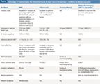
TABLE

Summary of Technologies for Potential Use in Breast Cancer Screening in Addition to Mammography
The table included in this commentary summarizes our current knowledge of potential supplemental screening modalities where data exist. Note that thermography has not been included, as it does not meet the above metrics for size of tumor or node status.[21]
In conclusion, this is a time of great opportunity and great challenge for breast cancer screening: opportunity, because we now have many possible tools to address the limitations of the past, and challenge, because we require systematic approaches to what is fundamentally a very personal choice with potential risks. As we tailor our approaches going forward, we must not lose sight of the important contribution that mammography makes to reducing mortality from breast cancer. At a minimum, we must require systematic data collection on the outcomes of screening as we introduce additional technologies.
Financial Disclosure:Dr. Berg has been a consultant and compensated speaker for SuperSonic Imagine; receives institutional research funding from Hologic, Inc.; has a pending research grant and equipment support to her institution from Gamma Medica, Inc., is a consultant to Naviscan, Inc., and serves on the medical advisory board of Philips, Inc.
References:
References
1. Smith RA, Duffy S, Tabár L. Breast Cancer Screening. Oncology. 2012;26:471-486.
2. Nelson HD, Tyne K, Naik A, et al. Screening for breast cancer: an update for the U.S. Preventive Services Task Force. Ann Intern Med. 2009;151:727-37.
3. Hendrick RE, Helvie MA. United States Preventive Services Task Force screening mammography recommendations: science ignored. AJR Am J Roentgenol. 2011;196:W112-16.
4. Jorgensen KJ, Keen JD, Gotzsche PC. Is mammographic screening justifiable considering its substantial overdiagnosis rate and minor effect on mortality? Radiology. 2011;260:621-7.
5. Kopans DB, Feig SA. The Canadian National Breast Screening Study: a critical review. AJR Am J Roentgenol. 1993;161:755-60.
6. Moss SM, Cuckle H, Evans A, et al. Effect of mammographic screening from age 40 years on breast cancer mortality at 10 years' follow-up: a randomised controlled trial. Lancet. 2006; 368:2053-60.
7. Burnside ES, Park JM, Fine JP, Sisney GA. The use of batch reading to improve the performance of screening mammography. AJR Am J Roentgenol. 2005;185:790-6.
8. Ghate SV, Soo MS, Baker JA, et al. Comparison of recall and cancer detection rates for immediate versus batch interpretation of screening mammograms. Radiology. 2005;235:31-5.
9. Lindfors KK, O'Connor J, Parker RA. False-positive screening mammograms: effect of immediate versus later work-up on patient stress. Radiology. 2001;218:247-53.
10. Cady B, Webb M, Webb M, et al. Death from breast cancer occurs predominantly in women not participating in mammographic screening. Presented at the American Society of Clinical Oncology (ASCO) 2009 Breast Cancer Symposium. San Francisco, CA. Abstract 24.
11. National Cancer Institute: Dictionary of Cancer Terms: Breast Cancer. Available at http://www.cancer.gov/dictionary?cdrid=446564. Accessed April 19, 2012.
12. Paik S, Shak S, Tang G, et al. A multigene assay to predict recurrence of tamoxifen-treated, node-negative breast cancer. N Engl J Med. 2004;351:2817-26.
13. van 't Veer LJ, Dai H, van de Vijver MJ, et al. Gene expression profiling predicts clinical outcome of breast cancer. Nature. 2002;415:530-6.
14. Mandelson MT, Oestreicher N, Porter PL, et al. Breast density as a predictor of mammographic detection: comparison of interval- and screen-detected cancers. J Natl Cancer Inst. 2000;92:1081-7.
15. Brekelmans CT, Seynaeve C, Bartels CC, et al. Effectiveness of breast cancer surveillance in BRCA1/2 gene mutation carriers and women with high familial risk. J Clin Oncol. 2001;19:924-30.
16. Tilanus-Linthorst M, Verhoog L, Obdeijn IM, et al. A BRCA1/2 mutation, high breast density and prominent pushing margins of a tumor independently contribute to a frequent false-negative mammography. Int J Cancer. 2002;102:91-5.
17. Komenaka IK, Ditkoff BA, Joseph KA, et al. The development of interval breast malignancies in patients with BRCA mutations. Cancer. 2004;100:2079-83.
18. Chiu SY, Duffy S, Yen AM, et al. Effect of baseline breast density on breast cancer incidence, stage, mortality, and screening parameters: 25-year follow-up of a Swedish mammographic screening. Cancer Epidemiol Biomarkers Prev. 2010;19:1219-28.
19. Berg WA. Tailored supplemental screening for breast cancer: what now and what next? AJR Am J Roentgenol. 2009;192:390-9.
20. Berg WA, Zhang Z, Lehrer D, et al. Detection of breast cancer with addition of annual screening ultrasound or a single screening MRI to mammography in women with elevated breast cancer risk. JAMA. 2012;307:1394-404.
21. Brenner RJ, Parisky Y. Alternative breast-imaging approaches. Radiol Clin North Am. 2007;45:907-23.
22. Kuhl C, Weigel S, Schrading S, et al. Prospective multicenter cohort study to refine management recommendations for women at elevated familial risk of breast cancer: the EVA trial. J Clin Oncol. 2010;28:1450-7.
23. Leach MO, Boggis CR, Dixon AK, et al. Screening with magnetic resonance imaging and mammography of a UK population at high familial risk of breast cancer: a prospective multicentre cohort study (MARIBS). Lancet. 2005;365:1769-78.
24. Sardanelli F, Podo F, Santoro F, et al. Multicenter surveillance of women at high genetic breast cancer risk using mammography, ultrasonography, and contrast-enhanced magnetic resonance imaging (the high breast cancer risk Italian 1 study): final results. Invest Radiol. 2011;46:94-105.
25. Warner E, Plewes DB, Hill KA, et al. Surveillance of BRCA1 and BRCA2 mutation carriers with magnetic resonance imaging, ultrasound, mammography, and clinical breast examination. JAMA. 2004;292:1317-25.
26. Berg WA. Supplemental screening sonography in dense breasts. Radiol Clin North Am. 2004;42:845-51.
27. Corsetti V, Houssami N, Ferrari A, et al. Breast screening with ultrasound in women with mammography-negative dense breasts: evidence on incremental cancer detection and false positives, and associated cost. Eur J Cancer. 2008;44:539-44.
28. Corsetti V, Houssami N, Ghirardi M, et al. Evidence of the effect of adjunct ultrasound screening in women with mammography-negative dense breasts: interval breast cancers at 1 year follow-up. Eur J Cancer. 2011;47:1021-6.
29. Kelly KM, Dean J, Comulada WS, et al. Breast cancer detection using automated whole breast ultrasound and mammography in radiographically dense breasts. Eur Radiol. 2010;20:734-42.
30. Skaane P, Gullien R, Eben EB, et al. Reading time of FFDM and tomosynthesis in a population-based screening program. MSVB31-07. Presented at the Radiological Society of North America (RSNA) 97th Scientific Assembly and Annual Meeting. Chicago, IL, Nov 27âDec 2, 2011.
31. Rhodes DJ, Hruska CB, Phillips SW, et al. Dedicated dual-head gamma imaging for breast cancer screening in women with mammographically dense breasts. Radiology. 2011;258:106-18.
32. Hagen AI, Kvistad KA, Maehle L, et al. Sensitivity of MRI versus conventional screening in the diagnosis of BRCA-associated breast cancer in a national prospective series. Breast. 2007;16:367-74.
33. Kriege M, Brekelmans CT, Boetes C, et al. Differences between first and subsequent rounds of the MRISC breast cancer screening program for women with a familial or genetic predisposition. Cancer. 2006;106:2318-26.
34. Kuhl CK, Schrading S, Leutner CC, et al. Mammography, breast ultrasound, and magnetic resonance imaging for surveillance of women at high familial risk for breast cancer. J Clin Oncol. 2005;23:8469-76.
35. Berg WA, Madsen KS, Schilling K, et al. Comparative effectiveness of positron emission mammography and MRI in the contralateral breast of women with newly diagnosed breast cancer. AJR Am J Roentgenol. 2012;198:219-32.
36. Plevritis SK, Kurian AW, Sigal BM, et al. Cost-effectiveness of screening BRCA 1/2 mutation carriers with breast magnetic resonance imaging. JAMA. 2006;295:2374-84.
37. Berg WA, Blume JD, Adams AM, et al. Reasons women at elevated risk of breast cancer refuse breast MR imaging screening: ACRIN 6666. Radiology. 2010;254:79-87.
38. Warner E. The role of magnetic resonance imaging in screening women at high risk of breast cancer. Top Magn Reson Imaging. 2008;19:163-9.
39. Hendrick RE. Radiation doses and cancer risks from breast imaging studies. Radiology. 2010;257:246-53.
40. Saslow D, Boetes C, Burke W, et al. American cancer society guidelines for breast screening with MRI as an adjunct to mammography. CA Cancer J Clin. 2007;57:75-89.
Gedatolisib Combo With/Without Palbociclib May Be New SOC in PIK3CA Wild-Type Breast Cancer
December 21st 2025“VIKTORIA-1 is the first study to demonstrate a statistically significant and clinically meaningful improvement in PFS with PAM inhibition in patients with PIK3CA wild-type disease, all of whom received prior CDK4/6 inhibition,” said Barbara Pistilli, MD.