Primary Cutaneous and Systemic Anaplastic Large Cell Lymphoma
Anaplastic large cell lymphoma (ALCL) is a biologic and clinically heterogenous subtype of T-cell lymphoma. Clinically, ALCL may present as localized (primary) cutaneous disease or widespread systemic disease. These two forms of ALCL are distinct entities with different clinical and biologic features. Both types share similar histology, however, with cohesive sheets of large lymphoid cells expressing the Ki-1 (CD30) molecule. Primary cutaneous ALCL (C-ALCL) is part of the spectrum of CD30+ lymphoproliferative diseases of the skin including lymphomatoid papulosis. Using conservative measures, 5-year disease-free survival rates are>90%. The systemic ALCL type is an aggressive lymphoma that may secondarily involve the skin, in addition to other extranodal sites. Further, systemic ALCL may be divided based on the expression of anaplastic lymphoma kinase (ALK) protein, which is activated most frequently through the nonrandom t(2;5) chromosome translocation, causing the fusion of the nucleophosmin (NPM) gene located at 5q35 to 2p23 encoding the receptor tyrosine kinase ALK. Systemic ALK+ ALCLs have improved prognosis compared with ALK-negative ALCL, although both subtypes warrant treatment with polychemotherapy. Allogeneic and, to a lesser extent, autologous stem cell transplantation play a role in relapsed disease, while the benefit of upfront transplant is not clearly defined. Treatment options for relapsed patients include agents such as pralatrexate (Folotyn) and vinblastine. In addition, a multitude of novel therapeutics are being studied, including anti-CD30 antibodies, histone deacetylase inhibitors, immunomodulatory drugs, proteasome inhibitors, and inhibitors of ALK and its downstream signaling pathways. Continued clinical trial involvement by oncologists and patients is imperative to improve the outcomes for this malignancy.
Anaplastic large cell lymphoma (ALCL) is a biologic and clinically heterogenous subtype of T-cell lymphoma. Clinically, ALCL may present as localized (primary) cutaneous disease or widespread systemic disease. These two forms of ALCL are distinct entities with different clinical and biologic features. Both types share similar histology, however, with cohesive sheets of large lymphoid cells expressing the Ki-1 (CD30) molecule. Primary cutaneous ALCL (C-ALCL) is part of the spectrum of CD30+ lymphoproliferative diseases of the skin including lymphomatoid papulosis. Using conservative measures, 5-year disease-free survival rates are > 90%. The systemic ALCL type is an aggressive lymphoma that may secondarily involve the skin, in addition to other extranodal sites. Further, systemic ALCL may be divided based on the expression of anaplastic lymphoma kinase (ALK) protein, which is activated most frequently through the nonrandom t(2;5) chromosome translocation, causing the fusion of the nucleophosmin (NPM) gene located at 5q35 to 2p23 encoding the receptor tyrosine kinase ALK. Systemic ALK+ ALCLs have improved prognosis compared with ALK-negative ALCL, although both subtypes warrant treatment with polychemotherapy. Allogeneic and, to a lesser extent, autologous stem cell transplantation play a role in relapsed disease, while the benefit of upfront transplant is not clearly defined. Treatment options for relapsed patients include agents such as pralatrexate (Folotyn) and vinblastine. In addition, a multitude of novel therapeutics are being studied, including anti-CD30 antibodies, histone deacetylase inhibitors, immunomodulatory drugs, proteasome inhibitors, and inhibitors of ALK and its downstream signaling pathways. Continued clinical trial involvement by oncologists and patients is imperative to improve the outcomes for this malignancy.
T-cell non-Hodgkin lymphomas (NHLs) represent a heterogeneous spectrum of lymphoid malignancies. While this disease is more common in Asia, there is significant geographic variation in its incidence, which has been attributed in part to risk factors such as human T-cell leukemia virus-1/2 (HTLV-1/2) and Epstein-Barr virus (EBV).[1] In a recent international clinicopathologic lymphoma study from 22 institutions in North America, Europe, and Asia, T-cell and natural killer (NK)-cell lymphomas accounted for approximately 12% of all NHLs.[2] The most common subtypes were peripheral T-cell lymphoma, not otherwise specified (PTCL-NOS; 26%), angioimmunoblastic T-cell lymphoma (AITL; 19%), NK/T-cell (10%), adult T-cell leukemia/lymphoma (ATLL; 10%), and anaplastic large cell lymphoma (ALCL, 12%: anaplastic lymphoma kinase [ALK]-positive 6.5%; ALCL, ALK-negative 5.5%).
ALCL was first described in 1985 based on the large pleomorphic cells expressing CD30, their propensity to invade sinusoids, and the cohesive appearance of the tumor.[3] ALCL occurs as two distinct entities-a widespread systemic disease or a localized (primary) cutaneous disease-with different clinical and biologic features.[4,5] However, both types share similar histology, with cohesive sheets of large lymphoid cells expressing the Ki-1 (CD30) molecule. In a subset of systemic ALCLs the t(2;5) chromosomal translocation occurs, which is known to cause the fusion of the nucleophosim (NPM) gene at 5q35 with the receptor tyrosine kinase ALK gene at 2p23 encoding the NPM-ALK fusion protein.[6-9] The updated World Health Organization (WHO) classification of hematopoietic tumors and lymphoid tissues now recognize this subclassification, dividing systemic ALCL into ALK-positive and ALK-negative disease.[4]
This article will provide a comprehensive review of the morphologic, immunologic, and molecular pathologic features of primary cutaneous-ALCL (C-ALCL) and systemic ALCL, including implications for prognosis. We will also describe standard and newer treatment approaches for patients with ALCL.
Clinical and Prognostic Features
Primary C-ALCL
Primary C-ALCL and lymphomatoid papulosis (LyP) represent a disease spectrum of CD30-positive lymphoproliferative disorders (CD30+ LPD) with many overlapping clinical, histologic, and immunophenotypical features as systemic ALCL.[4,10,11] Primary C-ALCL accounts for approximately 9% of all cutaneous T-cell lymphomas (CTCL) and affects older patients in the sixth decade with a median age of 61 years. It has rarely been described in the pediatric population.[12,13]
FIGURE 1
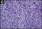
(A)
Cutaneous anaplastic large cell lymphoma (C-ALCL).
(B)
Pathology of C-ALCL.
(C)
Morphology.
LyP represents a benign, chronic recurrent, self-healing, papulonodular, and papulonecrotic CD30+ skin eruption.[13] Although 10% to 20% of patients with LyP may develop a lymphoid malignancy, the prognosis is otherwise excellent, showing a 100% 5-year survival. Most patients with primary C-ALCL present with solitary or localized skin tumors (see Figure 1A). Generalized or multifocal lesions are seen in about 20% of the patients.[11] Extracutaneous involvement is seen in 5% to 10% of patients at presentation and most commonly occurs with regional nodal involvement. Site of origin and ALK expression are important prognostic markers. Notably, C-ALCL lesions are known to regress spontaneously. C-ALCL patients have an excellent prognosis, with a 5-year disease-specific survival (DSS) exceeding 90%, as confirmed by several studies.[13,14] A recent report, however, describes a subset of C-ALCL patients with extensive limb disease (ELD) who have a worse treatment and survival outcome compared with patients who do not have ELD.[14] CD30/CD56 coexpression and increased fascin levels have also been associated with disease progression in selected cases.[15,16]
Systemic ALCL
ALCL, systemic type, accounts for approximately 2% to 3% of all NHLs.[17,18] Prior analyses showed that ALK+ ALCL was most frequently diagnosed in men prior to age 35 (male-to-female ratio: 3.0), while ALK-negative ALCL are usually older (median age, 61 years) with a male-to-female ratio of 0.9.[19-21] In a more recent series, Savage et al confirmed the younger median age for ALK+ vs ALK− ALCL (34 years vs 58 years, respectively, P < .001), while the male-to-female ratio was similar between ALK groups.[9] Approximately two-thirds of patients with systemic ALCL are known to have advanced-stage disease at presentation.[9,20,21] Systemic ALCL primarily involves lymph nodes, although extranodal sites may be involved. Based on ALK status, there is a similar distribution of nodal (ALK+, 54% vs ALK–, 49%) and extranodal disease (ALK+, 19% vs ALK–, 21%),[9] although other studies have shown more frequent extranodal disease in patients with ALK+ ALCL.[19-21] The most frequent extranodal sites in ALK– ALCL include bone, subcutaneous tissue, bone marrow, and spleen, whereas in ALK+ patients, the most common sites are skin, lung, liver, bone, and bone marrow.[9] There are rare reports of ALCL presenting as a leukemic disease, typically occurring in children and associated with a poor prognosis.[22] There has also been a recent association of ALCL occurring in women with breast implants. De Jong et al reported an odds ratio of 18.2 (95% confidence interval, 2.1–156.8) for ALCL associated with breast prostheses.[23]
The impact of NPM-ALK expression on patient outcome was first observed by Shiota in 1995.[24] In a series of 57 cases of adult ALCL, including T-cell and null cell phenotypes, 5-year overall survival (OS) was 67%. When stratified for expression of ALK, there was a significant favorable prognosis with ALK positivity, with a 5-year OS of 93% vs 37% (P < .00001) and 5-year FFS of 88% vs 37% (P < .0001).[25] The report by Savage et al confirmed the superior survival of systemic ALK+ ALCL compared with ALK-negative cases (5-year failure-free survival [FFS] 60% vs 36%; P = .015; and 5-year OS 70% vs 49%; P = .016); as previously discussed, however, ALK-positive patients were significantly younger than ALK-negative patients. Moreover, when they limited the survival comparison of ALK-negative and -positive cases to patients older or younger than 40 years of age, there were no differences in FFS or OS.[9]
When histologic subgroups within T-cell lymphoma are compared, most reports have shown that the survival of systemic ALCL is superior to survival of PTCL-NOS. Further, patients with systemic ALK-negative ALCL have a slightly improved survival compared with those who have PTCL-NOS,[26] although not all analyses support this.[21] Savage et al showed that, compared with patients who had PTCL-NOS, ALK-negative ALCL patients treated with curative intent had an improved 5-year FFS (39% vs 20%; P = .011) and 5-year OS (51% vs 32%; P = .028).[9]
TABLE 1
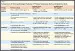
Comparison of Clinicopathologic Features of Primary Cutaneous ALCL and Systemic ALCL
The International Prognostic Index (IPI) is an independent prognostic marker that predicts survival in multivariate analysis across all peripheral T-cell lymphomas, including ALCL.[27] It identifies high-risk groups within both the ALK+ and ALK− ALCL subgroups (Table 1).[9] Another prognostic tool known as the ‘prognosis in T-cell lymphoma’ (PIT) scoring system was originally devised for PTCL-NOS patients; it incorporates age, performance status, LDH, and bone marrow involvement.[28] The PIT has also shown to be predictive of progression-free survival (PFS) and OS in ALCL.[9] In addition, expression of CD56, a neural cell adhesion molecule, was shown in a series of 143 patients with ALCL to be a predictor of poor survival (approximate 5-year OS: 28% vs 65%, P = .002).[20] The inferior outcome associated with CD56 was seen with ALK+ and ALK− patients.
Pathology
General Workup
Routine evaluation should include history and physical examination, complete blood count with differential, a metabolic panel including lactate dehydrogenase (LDH), lymph node excisional resection, skin biopsy of any suspicious cutaneous lesions for histology, bone marrow biopsy and aspirate, T-cell immunophenotyping, and gene rearrangement studies. Immunostaining with anti-ALK monoclonal antibodies and/or RT-PCR (reverse transcriptase polymerase chain reaction) should be performed to detect the upregulation of ALK for diagnostic and prognostic purposes as previously discussed. Full body imaging studies such as CT (computed tomography) and FDG-PET (fluorodeoxyglucose positron emission tomography) scans should be done to evaluate for the presence/extent of systemic disease. FDG-PET can be particularly helpful, especially in the detection of extranodal disease. Histopathologic and molecular results should be correlated with clinical findings and patients should be classified according to the WHO/European Organisation for Research and Treatment of Cancer (EORTC) consensus classification.[4]
Primary C-ALCL
• Morphology-Primary C-ALCL is characterized by cohesive sheets of large cells with an anaplastic, pleomorphic, or immunoblastic morphology (Figure 1B and 1C). However, there are no differences in prognosis or survival rate based on cytomorphology. According to the WHO/EORTC consensus classification, C-ALCL is defined by CD30 expression of at least 75% of the large lymphoid cells.[11] Unlike LyP, only a few neutrophils or eosinophils are present in C-ALCL, although neutrophil-rich (pyogenic) tumors have been seen.[29] Other variants, such as sarcomatoid and keratoacanthoma-like types, have also been described.[5,30] The atypical cells generally show an activated CD4+ T-helper cell phenotype with variable loss of T-cell markers and expression of cytotoxic proteins such as granzyme B, TIA-1, and perforin in about 50% of the cases.[31] Other activation markers such as CD25 (IL-2R) and HLA-DR are also found. CD8+ T-cell phenotype as well as a null CD4–CD8– phenotype and coexpression of CD56 and CD30 have rarely been reported.
• Immunophenotype-Unlike nodal ALCL, most C-ALCL express the cutaneous lymphocyte antigen (CLA) but lack epithelial membrane antigen (EMA) expression. The diagnostic and prognostic significance of tumor necrosis factor receptor–associated factor 1 (TRAF-1), multiple myeloma oncogene-1 (MUM-1), Bcl-2, and CD15 expression have not been found to reliably differentiate C-ALCL from other CD30+ LPD, secondary ALCL, or large-cell–transformation mycosis fungoides.[32]
CD30 signaling is known to have an effect of growth and survival of CD30+ lymphoproliferative diseases. Apoptosis rates and expression of apoptosis-related proteins such Bax, Bcl-x, c-kit, and Mcl-1 were analyzed in evolutional stages of LyP and C-ALCL, and CD30+ lymphoma cell lines.[33] While a higher apoptotic index was found in LyP than C-ALCL, the pro-apoptotic protein Bax was expressed at high levels in all evolutional stages of LyP and C-ALCL and may contribute to the spontaneous regression of cutaneous lesions. Expression of Bcl-2 appears to protect tumor cells from apoptosis in CD30+ LPD.[34] Stimulation of cell growth and apoptosis have been noted with activation of CD30 and therefore, CD30 ligand–mediated cytotoxicity may participate in the pathophysiology of clinical regression.[35] Interactions between Fas/APO-1 (CD95) and its ligand, FasL, have also been studied.[36] CD95 expression appears to be expressed at high levels in all cutaneous CD30+ LPD and suggest that CD95 activation may induce regression of CD30+ skin lesions.
• Molecular Characteristics-Molecular studies have shown that nearly all cases of C-ALCL are of clonal origin.[11] Array-based comparative genomic hybridization studies disclosed chromosomal imbalances in approximately 40% of the cases.[37] In contrast to primary nodal ALCL, patients with C-ALCL rarely carry the t(2;5) translocation and are usually ALK-negative.[38] More recently, C-ALCL was characterized by gains on chromosome 7q and 17q and losses on 6q and 13q.[39] In addition, C-ALCL showed higher expression of the skin-homing chemokine receptor genes CCR10 and CCR8, and aberrant expression of genes implicated in apoptosis (TNFRSF8/CD30, TRAF-1), and proliferation (IRF4/MUM-1, PRKCQ).[39]
FIGURE 2
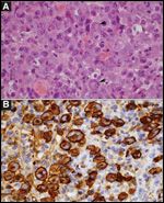
(A)
Anaplastic large cell lymphoma, ALK negative, H&E stain.
(B)
ALCL, ALK negative, CD30 Stain.FIGURE 3
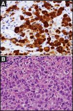
(A)
Anaplastic large cell lymphoma, common variant, ALK negative, H&E stain.
(B)
ALCL common variant, ALK Stain.
Systemic ALCL
• Morphology-Systemic ALCLs display a wide morphologic spectrum ranging from lymphomas rich in multinucleated giant cells to lymphomas with predominantly small lymphocytes.[40] The classic (common) variant (Figures 2A and 3A), small cell variant, and lymphohistiocytic variants are the most frequently encountered. The sarcomatoid, Hodgkin-like, neutrophil-rich, eosinophil-rich, and signet ring variants occur less frequently. All ALCL variants contain the characteristic hallmark cell that is large with a horseshoe-shaped nucleus and multiple visible nucleoli (Figures 2A and 3A).[40] However, the number of hallmark cells and the total number of tumor cells is variable in the different variants. For example, reactive histiocytes are much more numerous than lymphoma cells in the histiocyte-rich variant. Similarly, large lymphoma cells, including hallmark cells, are typically rare in the small-cell variant that contains predominantly small lymphocytes. The large lymphoma cells are characteristically located adjacent to blood vessels in the small-cell variant of ALCL.
• Immunophenotype-ALCL are T-cell NHLs, but it is not always possible to demonstrate T-cell lineage with immunohistochemical stains. Up to 75% are CD3-negative, and staining is variable with CD2, CD5, CD4, and CD8.[9,40] The 2008 study by Savage et al showed that ALK-negative ALCL tumors were more frequently CD2-positive (59% vs 23%, P < .001) and CD3-positive (45% vs 12%; P < .001) compared with ALK+ cases.[9] Analysis of T-cell–receptor genes is useful to establish T-cell lineage in cases in which immunohistochemistry is not informative.
Virtually all ALCL characteristically display strong, uniform, membranous staining for CD30 (Figure 2B) regardless of whether ALK is positive or negative.[5] The noteworthy exception to the CD30 staining pattern is the small-cell variant in which the less numerous large lymphoma cells are more consistently CD30-positive, while many of the small lymphoma cells show weak to negative staining. The different ALCL variants are positive for markers that are associated with activated, mature cytotoxic T cells including perforin, granzyme-B, and TIA-1.[41] Epithelial membrane antigen (EMA) is more frequently positive in the ALK+ cases compared with ALK– cases (EMA 83% vs 43%, respectively P < .001),[9] but EMA is also positive in many carcinomas and is therefore less specific than ALK.
Demonstration of positive staining for ALK is usually sufficient to establish the diagnosis in cases with typical morphology (Figure 3B). Approximately 40% of systemic ALCLs do not express ALK and are classified as systemic ALCL, ALK-negative subtype.[9] Although the small-cell variant is invariably ALK positive, morphologic features of the lymphoma cells are not sufficiently different to distinguish ALK-positive and ALK-negative cases based on hematoxylin and eosin stains.
TABLE 2

Characteristics of Fusion Proteins Associated With ALK-positive ALCL
• Molecular Characteristics-ALK positivity in ALCL indicates abnormal expression of the ALK protein and therefore rearrangement of the ALK gene located at chromosome 2p23, given that ALK protein is normally not expressed in lymphocytes.[42] By definition, the ALK gene is not rearranged in the ALK-negative cases. Several different gene fusion partners of the ALK gene have been described. The most common is the NPM gene located at chromosome 5q35, resulting in the (2;5)(p23;q35) translocation,[43] followed by the nonmuscle tropomyosin gene 3 located at chromosome 1q25, resulting in the (1;2)(q25;p23) translocation.[41] Inversion of the ALK gene, Inv(2)(p23q35), occurs less frequently. These three genetic alterations account for the vast majority (> 90%) of alterations in the ALK gene occurring in ALCL, but other rare fusion partners have been identified (Table 2).[41] Localization of ALK protein within the cell correlates with the different translocations. ALK staining localized to the nucleus and cytoplasm correlates with NPM-ALK chimeric fusion protein (Figure 3B), while cytoplasmic and/or membranous staining and absence of nuclear staining correlate with other types of fusion protein.
Genomic profiling has shown other genetic abnormalities in ALCL in addition to ALK gene rearrangement.[44] It is important to note, however, that other malignant diseases may rarely express the ALK gene, including neuroblastoma, rhabdomyosarcoma, inflammatory myofibroblastic tumors, and a subset of non–small-cell lung cancers.[8,45-48] In addition, there are reports of ALK detection in non-neoplastic and “normal” peripheral blood cells.[42] Other mechanisms of oncogenesis in ALCL include increased Bcl-2 expression,[21] hypermethylation,[44] and increased c-myc expression.[49] Furthermore, the NPM/ALK fusion protein constitutively activates phosphatidylinositol 3-kinase (PI3K)-AKT, JAK/STAT, and RAS/MEK/ERK, suggesting that these pathways may also be involved in the molecular pathogenesis of ALCL.[50,51]
Treatment
Primary C-ALCL
Primary C-ALCL is an indolent disease, therefore treatment measures should focus on noninvasive strategies. Localized radiation therapy or surgical excision for solitary or localized lesions is the preferred treatment for C-ALCL. Typically, radiation doses of 3000–3600 cGy are sufficient, and are associated with response rates greater than 90%. It is not known if radiation is warranted in addition to complete surgical excision, but if surgical margins are clear, it is appropriate to withhold radiation. Systemic therapy is indicated for patients refractory to local therapy, multifocal disease, and/or extracutaneous spread of disease. A well-tolerated option includes low-dose methotrexate. Kadin and colleagues used oral methotrexate at a dosage of 15–25 mg weekly for 12 months followed by maintenance therapy given at 2- to 4-week intervals (median total duration of methotrexate therapy for all patients, 39 months).[52,53] This was associated with an overall response rate (ORR) of 87%; approximately one-quarter of patients remained in durable remission after cessation of methotrexate (median duration, 10.6 years). Care should be taken to avoid long-term treatment with methotrexate, to avoid complications such as hepatic fibrosis. Other systemic options include doxorubicin-based chemotherapy (CHOP or CHOP-like), interferon-alpha (INF-α), or oral Bexarotene (LGD1069, Targretin).[11,13] Of note, the Dutch Cutaneous Lymphoma Group found that, compared with single-agent therapy,multiagent systemic chemotherapy did not result in a higher cure rate nor prevent future relapses in their experience.[13]
Systemic ALCL
• Chemotherapy-A standard therapeutic regimen does not exist for ALCL. Most adult ALCL treatment series have been small, retrospective, and/or were reported in an era in which patients may now be classified as having a different histology (eg, Hodgkin disease or diffuse large B-cell lymphoma). Nonetheless, regimens including anthracycline-based therapy, such as CHOP (cyclophosphamide, doxorubicin, vincristine, prednisone), have been most commonly studied. In 1997, Tilly et al treated a subset of systemic ALCL patients with combination chemotherapy regimens including ACVBP (doxorubicin [Adriamycin], cyclophosphamide, vindesine, bleomycin, prednisone) followed by a consolidation phase with high-dose methotrexate, ifosfamide, etoposide, asparaginase, and cytosine-arabinoside or m-BACOD (methotrexate, bleomycin, Adriamycin (doxorubicin) cyclophosphamide, vincristine, dexamethasone), VIMMM (VM26, ifosfamide, mitoxantrone, methyl-gag, methotrexate)/ACVBP, and CHOP.[54] The group of T-cell ALCL patients had a complete respone (CR) rate of 69%, with an associated 5-year OS of 63%. Of note, outcomes for ALCL patients were not stratified by ALK expression. In a retrospective report, Falini et al examined the outcomes according to ALK expression for ALCL patients treated with second- and third-generation polychemotherapy regimens.[19] They noted that ALK-negative patients had lower ORR and CR rates (84% and 56%, respectively) compared with ALK-positive patients (ORR 92% and CR 77%). Ten-year disease-free survival (DFS) rates from that analysis were 28% for ALK-negative vs 82% for ALK-positive patients.
Traditionally, in part as a result of the lack of clinical trial data and the heterogeneity in existing reports, chemotherapy for T-cell lymphomas have been extrapolated from data in aggressive B-cell lymphoma. Thus, CHOP has been the most commonly utilized regimen for the treatment of systemic ALCL. It is of interest that prior to the pre-rituximab (Rituxan) era, randomized clinical trials in aggressive NHL showed that intensified CHOP was associated with superior survival compared with classic CHOP-21. The NHL-B1 trial added etoposide (Toposar, VePesid) to CHOP and reduced the treatment interval from 3 to 2 weeks in young patients with aggressive NHL and good prognostic markers.[55] In this trial, 14% of patients had T-cell lymphoma (9.4% had systemic ALCL). The addition of etoposide improved the CR rate from 79% to 88% and increased 5-year EFS by 12%, while dose densification resulted in an increased OS by 10%. The subgroup of ALCL was too small to derive a statistical clinical benefit.
The NHL-B2 study had an identical trial design in 689 patients between the ages of 61–75 years.[56] Approximately 6% of patients had T-cell histology, with 3.5% ALCL. In a multivariate analysis, CHOP-14 was associated with improved EFS (relative risk 0.66, P = .003) and OS (relative risk 0.58, P < .001) compared with CHOP-21. These trials do not definitively support the use of intensified CHOP in systemic ALCL; a prospective controlled study in T-cell NHL would be needed. In a retrospective study evaluating clinical outcome in T-cell NHLs, intensive treatments such as hyper-CVAD/MA (fractionated cyclophosphamide, doxorubicin, vincristine, dexamethasone/methotrexate, and cytarabine) and/or early transplant produced no better results than CHOP.[57] The subset of T-cell NHL with ALCL in this study had a 3-year OS of 83%; most of these patients had received CHOP chemotherapy.
Transplantation
• Autologous stem cell transplant-Autologous stem cell transplant (ASCT) has been examined in relapsed ALCL.[58,59] Song et al compared 36 patients with relapsed/refractory T-NHL with 95 relapsed DLBCL patients and found comparable 3-year DFS.[60] On subgroup analysis, patients with ALCL had a superior outcome (3-year DFS 67%) compared with those who had other T-cell subtypes. The European Group for Blood and Marrow Transplantation (EBMT) reported data in 1999 among 64 patients with T-/null-cell ALCL.[61] Status at time of transplant significantly influenced outcomes; 12 of 33 patients in CR or PR relapsed compared with 14 of 16 with chemotherapy refractory disease at time of ASCT. A study from the GEL/TAMO (Spanish Cooperative Group for Bone Marrow Transplants in Lymphomas) of 123 patients with relapsed/refractory T-cell NHL, of which 25% cases were ALCL, showed that two to three adjusted IPI factors, > 1 extranodal site of disease, and elevated β2-microglobulin at time of transplant were associated with inferior survival.[62] At a median follow-up of 61 months, the 5-year OS and PFS rates were 45% and 34%, respectively. However, Smith et al reported modest outcomes with ASCT; among 32 relapsed/refractory T-cell NHL patients, of whom 21 had ALCL, 5-year relapse-free survival and OS were 18% and 34%, respectively.[63] Several small studies assessed the efficacy of ASCT for ALCL in first remission, with some reports showing 5-year survival > 80%.[64-66] These results are impressive, however the lack of a prospective control group makes the data difficult to interpret. Further, these patients were likely highly selected, and the expected outcomes according to IPI are not known. Other groups have reported on outcomes of T-cell NHL in first remission.[67-70] Patients with systemic ALCL have been included in these analyses, although they represent a minority of cases and it remains unclear if this strategy is superior to the conventional application of ASCT (ie, for relapsed disease).
When examining reports of ASCT in first remission, it is also important to analyze data as intent-to-treat, as many patients may never proceed to ASCT. Nickelsen et al recently compared outcomes of T-cell NHL (most common subtype: ALK-negative ALCL, 39%) vs aggressive B-cell lymphoma patients treated with four to six courses of dose-escalated CHOP plus etoposide, necessitating repeated ASCT.[71] Compared with 84% of B-cell NHL patients, 67% of T-cell NHL patients were able to receive all treatments. Three-year event-free survival (EFS) and OS were significantly inferior for T-NHL (26% and 45%, respectively) patients, compared with B-cell NHL patients (60% and 63%, respectively). Albeit not randomized, these data do not support the use of consolidative ASCT. Prospective randomized studies comparing conventional chemotherapy with ASCT are needed before ASCT may be considered standard therapy. Examination of high-risk patients by IPI and/or molecularly based prognostication may help identify patient groups that will garner benefit from consolidative ASCT.
• Allogeneic stem cell transplant-There are a handful of studies available describing the role of allogeneic stem cell transplant (alloSCT) in PTCL. An older French series of relapsed/refractory aggressive B-cell and T-cell patients treated with myeloablative conditioning showed comparable outcomes, with 5-year PFS and OS of 40% and 41%, respectively.[72] A more recent French retrospective report studied 77 T-cell lymphomas, of which 35% included ALCL.[73] Fifty-seven patients received myeloablative conditioning; 5-year toxicity-related mortality (TRM) was 33%. The 5-year EFS and OS for all patients was 53% and 57%, respectively, while the ALCL subtype was not significantly different, with 5-year EFS of 48% and OS of 55%. ALK status did not impact survival. Of note, chemotherapy-resistant patients also appeared to benefit from allo-SCT, with 5-year OS of 29%, and successful use of donor lymphocyte infusions suggested a graft-versus-lymphoma effect. Corradini et al reported on a small series of 17 patients (4 ALCL) with relapsed T-cell NHL who received salvage chemotherapy followed by allo-SCT with reduced-intensity conditioning and planned donor lymphocyte infusions.[74] All patients had sustained engraftment; the estimated 3-year PFS and OS rates were 64% and 81%, respectively. All four ALCL patients were event-free at 10, 12, 17, and 36 months. AlloSCT represents a treatment option for relapsed/refractory ALCL, especially for younger patients.
TABLE 3
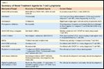
Summary of Novel Treatment Agents for T-cell Lymphoma
• Novel therapeutics-Several novel agents have been studied as single-agent therapy for relapsed/refractory patients and combined with chemotherapy for newly diagnosed T-cell NHL. Agents that have been combined with standard CHOP therapy for untreated T-cell NHL, including patients with systemic ALCL, include denileukin diftitox (Ontak), alemtuzumab (Campath), bevacizumab (Avastin), and bortezomib (Velcade) (see Table 3).[75-78] In a phase II multicenter trial, 24 patients with T-cell NHL (12% ALCL) received alemtuzumab with CHOP as first-line chemotherapy.[75] The ORR was 75% (CR 68%); all ALCL patients (3/3) had CRs that appeared durable. It will be difficult to discern the precise activity in ALCL in these trials because they are uncontrolled phase II trials and include many T-cell NHL subtypes, but the data are promising.
Several FDA-approved agents are clinically active as single-agents for relapsed or refractory ALCL. Pralatrexate (Folotyn) is a 10-deaza-aminopterin analog of methotrexate and a novel targeted antifolate that has shown higher affinity to the reduced folate carrier type 1 (RFC-1), increased accumulation and polyglutamylation in tumor cells compared with methotrexate. In a phase I/II study of relapsed/refractory lymphoma, the maximum tolerated dose of pralatrexate was 30 mg/m2 weekly for 6 weeks every 7 weeks.[79] Among 26 evaluable relapsed/refractory T-cell NHL patients, the ORR was 54% (47% by intent-to-treat). Two of the eight CRs seen were systemic ALCL patients (one ALK-negative and one ALK-positive) lasting 2 and > 22 months. Vinblastine is an old drug with recently demonstrated impressive clinical activity in ALCL. Brugieres et al reported a CR rate of 83% among 30 relapsed/refractory pediatric ALCL patients using weekly vinblastine.[80] Of 25 patients treated with vinblastine alone, 9 remained in a long-term CR (median, 7 years since the end of treatment). Interestingly, vinblastine was effective for treatment of subsequent patient relapses. CD25 has been shown to be expressed in pediatric[81] and adult patients[82] with systemic ALCL, although there are minimal data regarding the single-agent activity of denileukin diftitox in this disease.
As described previously, CD30 is uniformly expressed in cutaneous and systemic ALCL and thus represents a promising therapeutic target. Several anti-CD30 antibodies are under active development for the treatment of relapsed/refractory Hodgkin disease and systemic ALCL. MDX-060 (Medarex) is a fully human anti-CD30 IgG1k monoclonal antibody that has been shown to inhibit growth of CD-30 positive tumor cells in vitro and in tumor xenograft models.[83] It was developed in part to eliminate human antimouse antibody responses. In a phase I/II trial, MDX-060 was well tolerated, but clinical activity was modest, with an 8% response rate (6 of 72 patients) with 35% maintaining stable disease.[84] Six patients in the trial had ALCL, and among these patients one CR and one PR were noted. MDX-1401 is a second-generation antibody in which fucose is absent, thereby increasing the antibody’s antibody-dependent cellular cytotoxicity. In preclinical studies, MDX-1401 had improved antibody effector function over the fucose-containing parental anti-CD30 antibody (MDX-060).[85] A phase 1 trial of MDX-1401 is ongoing.
SGN-30 (Seattle Genetics) is a chimeric anti-CD30 monoclonal antibody with demonstrated activity in systemic ALCL. SGN-30 was evaluated in a phase II study of 38 patients with relapsed/refractory Hodgkin disease and 41 patients with systemic ALCL.[86]
In the ALCL group, two patients achieved CR and five additional patients achieved a PR (ORR 17%), with response durations ranging from 27 to ≥ 1,460 days. A phase II trial was recently reported using SGN-30 in heavily pretreated patients with C-ALCL, LyP, and transformed mycosis fungoides, which demonstrated promising results (ORR of 70%), with low toxicities.[87] Modifications to this antibody have also been made; SGN-35 is an antibody-drug conjugate, which was formed by coupling the anti CD-30 antibody, cAC10, to monomethyl auristatin E (MMAE), an antitubulin agent. In preclinical mouse xenograft models, SGN-35 also induced durable dose-dependent tumor regression compared with either untreated mice, or another control group receiving IgG-vcMMAE.[88] Reported in abstract form in two phase I studies, SGN-35 appears to have significant clinical activity in relapsed/refractory systemic ALCL.[89,90] Seven patients with systemic ALCL were treated on these relapsed/refractory CD30+ SGN-35 phase I trials (one q 3 week and one trial of weekly dosing); 86% of patients (6 of 7) achieved CR, which were durable. A pivotal phase II study in relapsed/refractory systemic ALCL is ongoing.[91]
Radioimmunoconjugates have potential therapeutic value in T-cell NHL. A radioimmunoconjugate in preclinical development is 131I anti-CD45 radioantibody.[92] This agent is particularly attractive in T-cell NHL since CD45 is a pan–T-cell antigen target. A radioimmunoconjugate consisting of the murine anti-CD30 monoclonal antibody Ki-4 labeled with iodine-131 showed clinical activity in patients with relapsed/refractory Hodgkin disease,[93] but has not been tested in T-cell HL. Other radioimmunoconjugates that could rationally be used in systemic ALCL include 131iodine-anti-CD25, 90yttrium-anti-CD25, and 90yttrium-anti-CD5.[94-96]
Histone deacetylases (HDACs) regulate cellular functions such as cell-cycle progression, proliferation, survival, transcription factors, and signal transduction, and are a target of interest in various lymphomas. The mechanism of HDAC inhibitors is related in part to chromatin relaxation with subsequent gene expression of tumor suppressors that allows tumor cell growth arrest. In 2006, vorinostat (Zolinza) was approved by the US Food and Drug Administration to treat cutaneous T-cell lymphoma, while in late 2009 romidepsin (Istodax) was approved for the treatment of CTCL patients who have received at least one prior systemic therapy. Clinical data using romidepsin for relapsed/refractory peripheral T-cell NHL have been encouraging.[97,98] In abstract form, At the 2008 meeting of the American Society of Hematology, Piekarz et al reported an ORR of 31% with single-agent romidepsin in 48 patients with relapsed/refractory T-cell NHL (4 CR and 11 PR).[98] The median duration of response was 9 months (range, 2 to ≥ 61 months).
Preclinical data support use of the proteasome inhibitor bortezomib in T-cell lymphoma cells.[99] Constitutive activation of the nuclear factor (NF)-kappa B p50 has been described in ALCL.[100] In relapsed/refractory cutaneous T-cell lymphoma, single-agent bortezomib was associated with an ORR of 67%.[101] Preliminary results were presented using combined bortezomib with gemcitabine (Gemzar) therapy in relapsed/refractory T-cell lymphoma.[102] In addition, preclinical data in T-cell and other NHL subtypes have shown that proteasome inhibitors combined with HDAC inhibitors are strongly synergistic.[103,104] A clinical trial examining the combination of vorinostat and bortezomib in relapsed/refractory T-cell NHL (including ALCL) is ongoing.[105]
Reference Guide
Therapeutic Agents
Mentioned in This Article
17-AAG
ACVBP (doxorubicin [Adriamycin],
cyclophosphamide, vindesine,
bleomycin, prednisone)
Alemtuzumab (Campath)
Asparaginase
Bevacizumab (Avastin)
Bexarotene (LGD1069, Targretin)
Bortezomib (Velcade)
CHOP (cyclophosphamide,
doxorubicin, vincristine,
prednisone)
Cyclophosphamide
Cytosine-arabinoside
Denileukin diftitox (Ontak)
Doxorubicin
Etoposide (Toposar, VePesid)
Flavopiridol (alvocidib, HMR 1275,
L-868275)
Gemcitabine (Gemzar)
Hyper-CVAD/MA (fractionated
cyclophosphamide, doxorubicin,
vincristine, dexamethasone/
methotrexate, and cytarabine)
Ifosfamide
IgG-vcMMAE
Interferon-alpha
131I anti-CD45
131I anti-CD25
131I Ki-4
Lenalidomide (Revlimid)
m-BACOD (methotrexate,
bleomycin, Adriamycin
[doxorubicin]), vincristine,
dexamethasone)
MDX-060 (Medarex)
MDX-1401
Methotrexate
Nutlin-3a
Pralatrexate (Folotyn)
Rituximab (Rituxan)
Romidepsin (Istodax)
SGN-30
SGN-35
Thalidomide (Thalomid)
VIMMM (VM26, ifosfamide,
mitoxantrone, methyl-gag,
methotrexate)
Vinblastine
Vorinostat (Zolinza)
90yttrium-anti-CD5
90yttrium-anti-CD25
Brand names are listed in parentheses only if a drug is not available generically and is marketed as no more than two trademarked or registered products. More familiar alternative generic designations may also be included parenthetically.
Immunomodulatory drugs (IMiDs) such as lenalidomide (Revlimid) have been investigated in a number of tumor types. IMiDs directly induce cell cycle arrest and possess potent antiangiogenic activity in vitro, which is likely to contribute to their antitumor effects. In addition, they help to minimize metastasis by reducing the expression of proangiogenic cytokines, such as vascular endothelial growth factor (VEGF), decreasing blood vessel cytokines, and affecting cell adhesion molecules. IMiDs also costimulate T cells and enhance antitumor immunity, which is mediated by T-helper-1 type cytokines. Clinical activity with thalidomide (Thalomid) in T-cell lymphoma has been reported anecdotally.[106] In preliminary analyses, single-agent lenalidomide had activity in relapsed/refractory T-cell NHL, including ALCL (ORR 30%).[107] Clinical trials examining lenalidomide in T-cell NHL are continuing.[108]
Currently no small-molecule inhibitors of ALK kinase are approved for clinical use. Several small molecules are being developed with the ability to inhibit ALK kinase activity, however.[109] This may be functionally achieved by directly inhibiting ALK kinase binding/activity,[110-112] by proteasomal-based degradation of ALK protein with agents such as 17-allylamino-17-demethoxygeldanamycin (17-AAG),[113] and by targeting pathways downstream of ALK.
For agents being developed to directly target ALK, it will be important to keep in mind the various mutants of ALK that continue to be indentified, some of which have been shown to be resistant to select small-molecule ALK inhibitors.[114] As discussed before, there are several signaling pathways downstream of NPM-ALK that lead to increased expression of cell-cycle regulators and transcription factors. SHP1 is a tyrosine phosphatase that functions in tumor suppression by inhibiting the JAK/STAT pathway. Its expression is decreased in ALK-positive ALCL cell lines as a result of gene hypomethylation. 5-aza-2'-deoxycytidine was shown to induce SHP1 expression in ALCL cell lines and subsequently downregulate pJAK3 and pSTAT3.[115] Bonvini et al reported that flavopiridol (alvocidib, HMR 1275, L-868275) has activity against ALCL cells by targeting cyclin-dependent kinases.[116] Furthermore, response to flavopiridol was increased when combined with ALK inihibitors. Drakos et al recently showed that nutlin-3a, a small molecule that targets Mdm2 (the critical negative regulator of p53), successfully activated p53 in ALK+ ALCL cells, resulting in cell-cycle arrest and induction of apoptosis.[117] Finally, several groups have undertaken proteomic approaches to identify the transcriptional signature for ALK signaling with therapeutic implications. 5-aminoimidazole-4-carboxamide ribonucleotide formyltransferase/inosine monophosphate cyclohydrolase (ATIC) is an enzyme that catalyzes purine synthesis. Boccalatte et al found that ALK-mediated ATIC phosphorylation enhanced its enzymatic activity, increasing its resistance to methotrexate.[118] Continued proteomics research is warranted to potentially predict the response of tumors to particular chemotherapy and/or targeted agents based on a transcriptional signature.
Conclusions
ALCL is a biologic and clinically heterogeneous subtype of T-cell lymphoma. Clinically, ALCL may present as localized (primary) cutaneous disease or widespread systemic disease. These two forms of ALCL are distinct entities with different clinical and biologic features. Both types share similar histology, with cohesive sheets of large lymphoid cells expressing the Ki-1 (CD30) molecule, however. Primary cutaneous ALCL (C-ALCL) is part of a spectrum of CD30+ lymphoproliferative diseases of the skin including lymphomatoidpapulosis. With conservative measures, 5-year disease-free survival rates exceed 90%. The systemic ALCL type is an aggressive lymphoma that may secondarily involve the skin, in addition to other extranodal sites. Furthermore, systemic ALCL is categorized based on expression of anaplastic lymphoma kinase (ALK) protein, which is activated most frequently through the nonrandom t(2;5) chromosome translocation causing the fusion of the nucleophosim (NPM) gene located at 5q35 to 2p23 and encoding the receptor tyrosine kinase ALK. Patients with systemic ALK+ ALCLs have improved prognosis compared with those who have ALK-negative ALCL, although both subtypes warrant treatment with polychemotherapy. Autologous and allogeneic stem cell transplantation have a role in relapsed disease, although the utility of upfront transplant remains undefined. There are several emerging treatment options for relapsed disease including pralatrexate therapy. In addition, a multitude of novel therapeutics are being studied, including anti-CD30 antibodies, HDAC inhibitors, IMiDs, proteasome inhibitors, and inhibitors of ALK and its downstream signaling pathways. Continued clinical trial involvement by oncologists and patients is critically important to improve the outcomes for this disease.
Financial Disclosure:Dr. Evens serves on the advisory boards of Seattle Genetics and Millennium. Dr. Rosen serves on the speakers bureau of Allos.
References:
References
1. The Non-Hodgkin’s Lymphoma Classification Project. A clinical evaluation of the International Lymphoma Study Group classification of non-Hodgkin’s lymphoma. Blood 89:3909-3918, 1997.
2. The International T-Cell Lymphoma Project. International peripheral T-cell and natural killer/T-cell lymphoma study: Pathology findings and clinical outcomes. J Clin Oncol 26:4124-4130, 2008.
3. Stein H, Mason DY, Gerdes J, et al. The expression of the Hodgkin’s disease associated antigen Ki-1 in reactive and neoplastic lymphoid tissue: Evidence that Reed-Sternberg cells and histiocytic malignancies are derived from activated lymphoid cells. Blood 66:848-858, 1985.
4. Swerdlow S, Campo E, Harris N, et al: WHO Classification of Tumours of Haematopoietic and Lymphoid Tissues, 4th ed. Lyon, France, IARC Press, 2008.
5. Stein H, Foss HD, Durkop H, et al: CD30(+) anaplastic large cell lymphoma: A review of its histopathologic, genetic, and clinical features. Blood 96:3681-3695, 2000.
6. Fischer P, Nacheva E, Mason DY, et al: A Ki-1 (CD30)-positive human cell line (Karpas 299) established from a high-grade non-Hodgkin’s lymphoma, showing a 2;5 translocation and rearrangement of the T-cell receptor beta-chain gene. Blood 72:234-240, 1988.
7. Mason DY, Bastard C, Rimokh R, et al: CD30-positive large cell lymphomas (‘Ki-1 lymphoma’) are associated with a chromosomal translocation involving 5q35. Br J Haematol 74:161-168, 1990.
8. Morris SW, Kirstein MN, Valentine MB, et al: Fusion of a kinase gene, ALK, to a nucleolar protein gene, NPM, in non-Hodgkin’s lymphoma. Science 263:1281-1284, 1994.
9. Savage KJ, Harris NL, Vose JM, et al: ALK- anaplastic large-cell lymphoma is clinically and immunophenotypically different from both ALK+ ALCL and peripheral T-cell lymphoma, not otherwise specified: Report from the International Peripheral T-Cell Lymphoma Project. Blood 111:5496-5504, 2008.
10. Querfeld C, Kuzel TM, Guitart J, et al: Primary cutaneous CD30+ lymphoproliferative disorders: New insights into biology and therapy. Oncology (Williston Park) 21:689-696; discussion 699-700, 2007.
11. Willemze R, Jaffe ES, Burg G, et al: WHO-EORTC classification for cutaneous lymphomas. Blood 105:3768-3785, 2005.
12. Kumar S, Pittaluga S, Raffeld M, et al: Primary cutaneous CD30-positive anaplastic large cell lymphoma in childhood: Report of 4 cases and review of the literature. Pediatr Dev Pathol 8:52-60, 2005.
13. Bekkenk MW, Geelen FA, van Voorst Vader PC, et al: Primary and secondary cutaneous CD30(+) lymphoproliferative disorders: A report from the Dutch Cutaneous Lymphoma Group on the long-term follow-up data of 219 patients and guidelines for diagnosis and treatment. Blood 95:3653-3661, 2000.
14. Woo DK, Jones CR, Vanoli-Storz MN, et al: Prognostic factors in primary cutaneous anaplastic large cell lymphoma: Characterization of clinical subset with worse outcome. Arch Dermatol 145:667-674, 2009.
15. Natkunam Y, Warnke RA, Haghighi B, et al: Co-expression of CD56 and CD30 in lymphomas with primary presentation in the skin: Clinicopathologic, immunohistochemical and molecular analyses of seven cases. J Cutan Pathol 27:392-399, 2000.
16. Kempf W, Levi E, Kamarashev J, et al: Fascin expression in CD30-positive cutaneous lymphoproliferative disorders. J Cutan Pathol 29:295-300, 2002.
17. Melnyk A, Rodriguez A, Pugh WC, et al: Evaluation of the Revised European-American Lymphoma classification confirms the clinical relevance of immunophenotype in 560 cases of aggressive non-Hodgkin’s lymphoma. Blood 89:4514-4520, 1997.
18. Pellatt J, Sweetenham J, Pickering RM, et al: A single-centre study of treatment outcomes and survival in 120 patients with peripheral T-cell non-Hodgkin’s lymphoma. Ann Hematol 81:267-272, 2002.
19. Falini B, Pileri S, Zinzani PL, et al: ALK+ lymphoma: Clinico-pathological findings and outcome. Blood 93:2697-2706, 1999.
20. Suzuki R, Kagami Y, Takeuchi K, et al: Prognostic significance of CD56 expression for ALK-positive and ALK-negative anaplastic large-cell lymphoma of T/null cell phenotype. Blood 96:2993-3000, 2000.
21. ten Berge RL, de Bruin PC, Oudejans JJ, et al: ALK-negative anaplastic large-cell lymphoma demonstrates similar poor prognosis to peripheral T-cell lymphoma, unspecified. Histopathology 43:462-469, 2003.
22. Takahashi D, Nagatoshi Y, Nagayama J, et al: Anaplastic large cell lymphoma in leukemic presentation: A case report and a review of the literature. J Pediatr Hematol Oncol 30:696-700, 2008.
23. de Jong D, Vasmel WL, de Boer JP, et al: Anaplastic large-cell lymphoma in women with breast implants. JAMA 300:2030-2035, 2008.
24. Shiota M, Nakamura S, Ichinohasama R, et al: Anaplastic large cell lymphomas expressing the novel chimeric protein p80NPM/ALK: A distinct clinicopathologic entity. Blood 86:1954-1960, 1995.
25. Gascoyne RD, Aoun P, Wu D, et al: Prognostic significance of anaplastic lymphoma kinase (ALK) protein expression in adults with anaplastic large cell lymphoma. Blood 93:3913-3921, 1999.
26. Gisselbrecht C, Gaulard P, Lepage E, et al: Prognostic significance of T-cell phenotype in aggressive non-Hodgkin’s lymphomas. Groupe d’Etudes des Lymphomes de l’Adulte (GELA). Blood 92:76-82, 1998.
27. Sonnen R, Schmidt WP, Muller-Hermelink HK, et al: The International Prognostic Index determines the outcome of patients with nodal mature T-cell lymphomas. Br J Haematol 129:366-372, 2005.
28. Gallamini A, Stelitano C, Calvi R, et al: Peripheral T-cell lymphoma unspecified (PTCL-U): A new prognostic model from a retrospective multicentric clinical study. Blood 103:2474-2479, 2004.
29. Boudova L, Kazakov DV, Jindra P, et al: Primary cutaneous histiocyte and neutrophil-rich CD30+ and CD56+ anaplastic large-cell lymphoma with prominent angioinvasion and nerve involvement in the forehead and scalp of an immunocompetent woman. J Cutan Pathol 33:584-589, 2006.
30. Lin JH, Lee JY: Primary cutaneous CD30 anaplastic large cell lymphoma with keratoacanthoma-like pseudocarcinomatous hyperplasia and marked eosinophilia and neutrophilia. J Cutan Pathol 31:458-461, 2004.
31. Kummer JA, Vermeer MH, Dukers D, et al: Most primary cutaneous CD30-positive lymphoproliferative disorders have a CD4-positive cytotoxic T-cell phenotype. J Invest Dermatol 109:636-640, 1997.
32. Benner MF, Jansen PM, Meijer CJ, et al: Diagnostic and prognostic evaluation of phenotypic markers TRAF1, MUM1, BCL2 and CD15 in cutaneous CD30-positive lymphoproliferative disorders. Br J Dermatol 161:121-127, 2009.
33. Greisser J, Doebbeling U, Roos M, et al: Apoptosis in CD30-positive lymphoproliferative disorders of the skin. Exp Dermatol 14:380-385, 2005.
34. Paulli M, Berti E, Boveri E, et al: Cutaneous CD30+ lymphoproliferative disorders: Expression of bcl-2 and proteins of the tumor necrosis factor receptor superfamily. Hum Pathol 29:1223-1230, 1998.
35. Wahl AF, Klussman K, Thompson JD, et al: The anti-CD30 monoclonal antibody SGN-30 promotes growth arrest and DNA fragmentation in vitro and affects antitumor activity in models of Hodgkin’s disease. Cancer Res 62:3736-3742, 2002.
36. Mori M, Manuelli C, Pimpinelli N, et al: CD30-CD30 ligand interaction in primary cutaneous CD30(+) T-cell lymphomas: A clue to the pathophysiology of clinical regression. Blood 94:3077-3083, 1999.
37. Mao X, Orchard G, Lillington DM, et al: Genetic alterations in primary cutaneous CD30+ anaplastic large cell lymphoma. Genes Chromosomes Cancer 37:176-185, 2003.
38. Herbst H, Sander C, Tronnier M, et al: Absence of anaplastic lymphoma kinase (ALK) and Epstein-Barr virus gene products in primary cutaneous anaplastic large cell lymphoma and lymphomatoid papulosis. Br J Dermatol 137:680-686, 1997.
39. van Kester MS, Tensen CP, Vermeer MH, et al: Cutaneous anaplastic large cell lymphoma and peripheral T-cell lymphoma NOS show distinct chromosomal alterations and differential expression of chemokine receptors and apoptosis regulators. J Invest Dermatol 130:563-575, 2010.
40. Benharroch D, Meguerian-Bedoyan Z, Lamant L, et al: ALK-positive lymphoma: A single disease with a broad spectrum of morphology. Blood 91:2076-2084, 1998.
41. Delsol G, Falini B, Muller-Hermelink HK et al: Anaplastic large cell lymphoma (ALCL), ALK-positive, in: WHO Classification of Tumors of Haematopoietic and Lymphoid Tissues 4th ed. Lyon, WHO Press, 2008.
42. Pulford K, Lamant L, Morris SW, et al:Detection of anaplastic lymphoma kinase (ALK) and nucleolar protein nucleophosmin (NPM)-ALK proteins in normal and neoplastic cells with the monoclonal antibody ALK1. Blood 89:1394-1404, 1997.
43. Morris SW, Kirstein MN, Valentine MB, et al: Fusion of a kinase gene, ALK, to a nucleolar protein gene, NPM, in non-Hodgkin’s lymphoma. Science 267:316-317, 1995.
44. Salaverria I, Bea S, Lopez-Guillermo A, et al:. Genomic profiling reveals different genetic aberrations in systemic ALK-positive and ALK-negative anaplastic large cell lymphomas. Br J Haematol 140:516-526, 2008.
45. Falini B, Bigerna B, Fizzotti M, et al: ALK expression defines a distinct group of T/null lymphomas (“ALK lymphomas”) with a wide morphological spectrum. Am J Pathol 153:875-886, 1998.
46. Griffin CA, Hawkins AL, Dvorak C, et al: Recurrent involvement of 2p23 in inflammatory myofibroblastic tumors. Cancer Res 59:2776-2780, 1999.
47. Lamant L, Pulford K, Bischof D, et al: Expression of the ALK tyrosine kinase gene in neuroblastoma. Am J Pathol 156:1711-1721, 2000.
48. Soda M, Choi YL Enomoto M, et al: Identification of the transforming EML4-ALK fusion gene in non-small-cell lung cancer. Nature 448:561-566, 2007.
49. Inghirami G, Macri L, Cesarman E, et al: Molecular characterization of CD30+ anaplastic large-cell lymphoma: High frequency of c-myc proto-oncogene activation. Blood 83:3581-3590, 1994.
50. Bai RY, Ouyang T, Miething C, et al: Nucleophosmin-anaplastic lymphoma kinase associated with anaplastic large-cell lymphoma activates the phosphatidylinositol 3-kinase/Akt antiapoptotic signaling pathway. Blood 96:4319-4327, 2000.
51. Chiarle R, Voena C, Ambrogio C, et al: The anaplastic lymphoma kinase in the pathogenesis of cancer. Nat Rev Cancer 8:11-23, 2008.
52. Kadin ME, Carpenter C: Systemic and primary cutaneous anaplastic large cell lymphomas. Semin Hematol 40:244-256, 2003.
53. Vonderheid EC, Sajjadian A, Kadin ME: Methotrexate is effective therapy for lymphomatoid papulosis and other primary cutaneous CD30-positive lymphoproliferative disorders. J Am Acad Dermatol 34:470-481, 1996.
54. Tilly H, Gaulard P, Lepage E, et al: Primary anaplastic large-cell lymphoma in adults: clinical presentation, immunophenotype, and outcome. Blood 90:3727-3734, 1997.
55. Pfreundschuh M, Trumper L, Kloess M, et al: Two-weekly or 3-weekly CHOP chemotherapy with or without etoposide for the treatment of young patients with good-prognosis (normal LDH) aggressive lymphomas: Results of the NHL-B1 trial of the DSHNHL. Blood 104:626-633, 2004.
56. Pfreundschuh M, Trumper L, Kloess M, et al: Two-weekly or 3-weekly CHOP chemotherapy with or without etoposide for the treatment of elderly patients with aggressive lymphomas: Results of the NHL-B2 trial of the DSHNHL. Blood 104:634-641, 2004.
57. Escalon MP, Liu NS, Yang Y, et al: Prognostic factors and treatment of patients with T-cell non-Hodgkin lymphoma: The M. D. Anderson Cancer Center experience. Cancer 103:2091-2098, 2005.
58. Jagasia M, Morgan D, Goodman S, et al: Histology impacts the outcome of peripheral T-cell lymphomas after high dose chemotherapy and stem cell transplant. Leuk Lymphoma 45:2261-2267, 2004.
59. Jantunen E, Wiklund T, Juvonen E, et al: Autologous stem cell transplantation in adult patients with peripheral T-cell lymphoma: A nation-wide survey. Bone Marrow Transplant 33:405-410, 2004.
60. Song KW, Mollee P, Keating A, et al: Autologous stem cell transplant for relapsed and refractory peripheral T-cell lymphoma: Variable outcome according to pathological subtype. Br J Haematol 120:978-985, 2003.
61. Fanin R, Ruiz de Elvira MC, Sperotto A, et al: Autologous stem cell transplantation for T and null cell CD30-positive anaplastic large cell lymphoma: analysis of 64 adult and paediatric cases reported to the European Group for Blood and Marrow Transplantation (EBMT). Bone Marrow Transplant 23:437-442, 1999.
62. Rodriguez J, Conde E, Gutierrez A, et al: The adjusted International Prognostic Index and beta-2-microglobulin predict the outcome after autologous stem cell transplantation in relapsing/refractory peripheral T-cell lymphoma. Haematologica 92:1067-1074, 2007.
63. Smith SD, Bolwell BJ, Rybicki LA, et al: Autologous hematopoietic stem cell transplantation in peripheral T-cell lymphoma using a uniform high-dose regimen. Bone Marrow Transplant 40:239-243, 2007.
64. Deconinck E, Lamy T, Foussard C, et al: Autologous stem cell transplantation for anaplastic large-cell lymphomas: Results of a prospective trial. Br J Haematol 109:736-742, 2000.
65. Fanin R, Silvestri F, Geromin A, et al: Primary systemic CD30 (Ki-1)-positive anaplastic large cell lymphoma of the adult: Sequential intensive treatment with the F-MACHOP regimen (+/- radiotherapy) and autologous bone marrow transplantation. Blood 87:1243-1248, 1996.
66. Fanin R, Sperotto A, Silvestri F, et al: The therapy of primary adult systemic CD30-positive anaplastic large cell lymphoma: Results of 40 cases treated in a single center. Leuk Lymphoma 35:159-169, 1999.
67. Corradini P, Tarella C, Zallio F, et al: Long-term follow-up of patients with peripheral T-cell lymphomas treated up-front with high-dose chemotherapy followed by autologous stem cell transplantation. Leukemia 20:1533-1538, 2006.
68. Reimer P, Rudiger T, Geissinger E, et al: Autologous stem-cell transplantation as first-line therapy in peripheral T-cell lymphomas: Results of a prospective multicenter study. J Clin Oncol 27:106-113, 2009.
69. Rodriguez J, Conde E, Gutierrez A, et al: Frontline autologous stem cell transplantation in high-risk peripheral T-cell lymphoma: A prospective study from the GEL-TAMO Study Group. Eur J Haematol 79:32-38, 2007.
70. Mercadal S, Briones J, Xicoy B, et al: Intensive chemotherapy (high-dose CHOP/ESHAP regimen) followed by autologous stem-cell transplantation in previously untreated patients with peripheral T-cell lymphoma. Ann Oncol 19:958-963, 2008.
71. Nickelsen M, Ziepert M, Zeynalova S, et al: High-dose CHOP plus etoposide (MegaCHOEP) in T-cell lymphoma: A comparative analysis of patients treated within trials of the German High-Grade Non-Hodgkin Lymphoma Study Group (DSHNHL). Ann Oncol 20:1977-1984, 2009.
72. Dhedin N, Giraudier S, Gaulard P, et al: Allogeneic bone marrow transplantation in aggressive non-Hodgkin’s lymphoma (excluding Burkitt and lymphoblastic lymphoma): A series of 73 patients from the SFGM database. Societ Francaise de Greffe de Moelle. Br J Haematol 107:154-161, 1999.
73. Le Gouill S, Milpied N, Buzyn A, et al: Graft-versus-lymphoma effect for aggressive T-cell lymphomas in adults: A study by the Societe Francaise de Greffe de Moelle et de Therapie Cellulaire. J Clin Oncol 26:2264-2271, 2008.
74. Corradini P, Dodero A, Zallio F, et al: Graft-versus-lymphoma effect in relapsed peripheral T-cell non-Hodgkin’s lymphomas after reduced-intensity conditioning followed by allogeneic transplantation of hematopoietic cells. J Clin Oncol 22:2172-2176, 2004.
75. Gallamini A, Zaja F, Patti C, et al: Alemtuzumab (Campath-1H) and CHOP chemotherapy as first-line treatment of peripheral T-cell lymphoma: Results of a GITIL (Gruppo Italiano Terapie Innovative nei Linfomi) prospective multicenter trial. Blood 110:2316-2323, 2007.
76. National Institutes of Health: A Pilot Study to Determine the Safety of the Combination of Ontak in Combination With CHOP in Peripheral T-Cell Lymphoma. Last updated June 5, 2009. Available at: http://clinicaltrials.gov/ct2/show/NCT00337987. Accessed May 19, 2010.
77. National Cancer Institute: Bevacizumab and Combination Chemotherapy in Treating Patients With Peripheral T-Cell Lymphoma or Natural Killer Cell Neoplasms. Last updated April 22, 2009. Available at: http://clinicaltrials.gov/ct2/show/NCT00217425. Accessed May 19, 2010.
78. Lee J, Suh C, Kang HJ, et al: Phase I study of proteasome inhibitor bortezomib plus CHOP in patients with advanced, aggressive T-cell or NK/T-cell lymphoma. Ann Oncol 19:2079-2083, 2008.
79. O’Connor OA, Horwitz S, Hamlin P, et al: Phase II-I-II study of two different doses and schedules of pralatrexate, a high-affinity substrate for the reduced folate carrier, in patients with relapsed or refractory lymphoma reveals marked activity in T-cell malignancies. J Clin Oncol 27:4357-4364, 2009.
80. Brugieres L, Pacquement H, Le Deley MC, et al: Single-drug vinblastine as salvage treatment for refractory or relapsed anaplastic large-cell lymphoma: A report from the French Society of Pediatric Oncology. J Clin Oncol 27:5056-5061, 2009.
81. Gualco G, Chioato L, Weiss LM, et al: Analysis of human T-cell lymphotropic virus in CD25+ anaplastic large cell lymphoma in children. Am J Clin Pathol 132:28-33, 2009.
82. Janik JE, Morris JC, Pittaluga S, et al: Elevated serum-soluble interleukin-2 receptor levels in patients with anaplastic large cell lymphoma. Blood 104:3355-3357, 2004.
83. Borchmann P, Treml JF, Hansen H, et al: The human anti-CD30 antibody 5F11 shows in vitro and in vivo activity against malignant lymphoma. Blood 102:3737-3742, 2003.
84. Ansell SM, Horwitz SM, Engert A, et al:Phase I/II study of an anti-CD30 monoclonal antibody (MDX-060) in Hodgkin’s lymphoma and anaplastic large-cell lymphoma. J Clin Oncol 25:2764-2769, 2007.
85. Cardarelli PM, Moldovan-Loomis MC, Preston B, et al: In vitro and in vivo characterization of MDX-1401 for therapy of malignant lymphoma. Clin Cancer Res 15:3376-3383, 2009.
86. Forero-Torres A, Leonard JP, Younes A, et al: A phase II study of SGN-30 (anti-CD30 mAb) in Hodgkin lymphoma or systemic anaplastic large cell lymphoma. Br J Haematol 146:171-179, 2009.
87. Duvic M, Reddy SA, Pinter-Brown L, et al: A phase II study of SGN-30 in cutaneous anaplastic large cell lymphoma and related lymphoproliferative disorders. Clin Cancer Res 15:6217-6224, 2009.
88. Francisco JA, Cerveny CG, Meyer DL, et al: cAC10-vcMMAE, an anti-CD30-monomethyl auristatin E conjugate with potent and selective antitumor activity. Blood 102:1458-1465, 2003.
89. Younes A F-TA, Bartlett NL: Multiple complete responses in a phase I dose-escalation study of the antibody-drug conjugate SGN-35 in patients with relapsed or refractoryCD-30 positive lymphomas (abstract 1006). Presented at the American Society of Hematology 50th annual meeting, San Francisco, CA, December 6–9, 2008. Available at http://ash.confex.com/ash/2008/webprogram/Paper8532.html. Accessed May 21, 2010.
90. Bartlett NL F-TA, Rosenblatt JD: Complete remissions with weekly dosing of SGN-35, a novel antibody-drug conjugate (ADC) targeting CD30, in a phase I dose-escalation study in patients with relapsed or refractory Hodgkin Lymphoma (HL) or systemic anaplastic large cell lymphoma (abstract 8500). Presented at the 2009 annual meeting of the American Society of Clinical Oncology, Orlando, FL, May 29–June 2, 2009. Available at http://www.asco.org/ASCOv2/Meetings/Abstracts?&vmview=abst_detail_view&confID=65&abstractID=33524. Accessed May 21, 2010.
91. National Institutes of Health: A Phase 2 Open Label Trial of SGN-35 for Systemic Anaplastic Large Cell Lymphoma. Available at http://clinicaltrials.gov/ct2/show/study/NCT00866047?show_locs=Y#locn. Accessed May 21, 2010.
92. Gopal AK, Pagel JM, Fromm JR, et al: 131I anti-CD45 radioimmunotherapy effectively targets and treats T-cell non-Hodgkin lymphoma. Blood 113:5905-5910, 2009.
93. Schnell R, Dietlein M, Staak JO, et al: Treatment of refractory Hodgkin’s lymphoma patients with an iodine-131-labeled murine anti-CD30 monoclonal antibody. J Clin Oncol 23:4669-4678, 2005
94. Dancey G, Violet J, Malaroda A, et al: A phase I clinical trial of CHT-25, a 131I-labeled chimeric anti-CD25 antibody showing efficacy in patients with refractory lymphoma. Clin Cancer Res 15:7701-7710, 2009.
95. Foss FM, Raubitscheck A, Mulshine JL, et al: Phase I study of the pharmacokinetics of a radioimmunoconjugate, 90Y-T101, in patients with CD5-expressing leukemia and lymphoma. Clin Cancer Res 4:2691-2700, 1998.
96. Waldmann TA, White JD, Carrasquillo JA, et al: Radioimmunotherapy of interleukin-2R alpha-expressing adult T-cell leukemia with yttrium-90-labeled anti-Tac. Blood 86:4063-4075, 1995.
97. Woo S, Gardner ER, Chen X, et al: Population pharmacokinetics of romidepsin in patients with cutaneous T-cell lymphoma and relapsed peripheral T-cell lymphoma. Clin Cancer Res 15:1496-1503, 2009.
98. Piekarz R, Wright J, Frye R, et al: Results of a phase 2 NCI multicenter study of romidepsin in patients with relapsed peripheral T-cell lymphoma (PTCL) (abstract 1657). Blood 114:661, 2009.
99. Zhao WL, Liu YY, Zhang QL, et al: PRDM1 is involved in chemoresistance of T-cell lymphoma and down-regulated by the proteasome inhibitor. Blood 111:3867-3871, 2008.
100. Mathas S, Johrens K, Joos S, et al: Elevated NF-kappaB p50 complex formation and Bcl-3 expression in classical Hodgkin, anaplastic large-cell, and other peripheral T-cell lymphomas. Blood 106:4287-4293, 2005.
101. Zinzani PL, Musuraca G, Tani M, et al: Phase II trial of proteasome inhibitor bortezomib in patients with relapsed or refractory cutaneous T-cell lymphoma. J Clin Oncol 25:4293-4297, 2007.
102. Evens AM GL, Gordon LI, Patton D, et al: Phase I results of combination gemcitabine and bortezomib (Velcade®) for relapsed/refractory nodal T-cell non-Hodgkin lymphoma (T-NHL) and aggressive B-cell NHL (abstract 2005). Presented at the American Society of Hematology 50th annual meeting, San Francisco, CA, December 6–9, 2008. Available at http://ash.confex.com/ash/2008/webprogram/Paper11233.html. Accessed May 21, 2010.
103. Heider U, Rademacher J, Lamottke B, et al: Synergistic interaction of the histone deacetylase inhibitor SAHA with the proteasome inhibitor bortezomib in cutaneous T cell lymphoma. Eur J Haematol 82:440-449, 2009.
104. Zhang QL, Wang L, Zhang YW, et al: The proteasome inhibitor bortezomib interacts synergistically with the histone deacetylase inhibitor suberoylanilide hydroxamic acid to induce T-leukemia/lymphoma cells apoptosis. Leukemia 23:1507-1514, 2009.
105. National Institutes of Health: Combination of Vorinostat and Bortezomib in Relapsed or Refractory T-Cell Non-Hodgkin’s Lymphoma. Available at: http://www.clinicaltrial.gov/ct2/show/NCT00810576. Accessed May 21, 2010.
106. Damaj G, Bouabdallah R, Vey N, et al: Single-agent thalidomide induces response in T-cell lymphoma. Eur J Haematol 74:169-171, 2005.
107. Dueck GS, Chua N, Prasad A, et al: Activity of lenalidomide in a phase II trial for T-cell lymphoma: Report on the first 24 cases (abstract 8524). J Clin Oncol 27(suppl):15s, 2009. Available at http://www.asco.org/ASCOv2/Meetings/Abstracts?&vmview=abst_detail_view&confID=65&abstractID=30959. Accessed May 21, 2010.
108. National Institutes of Health: A Phase II Clinical Trial of Lenalidomide for T-cell Non-Hodgkin’s Lymphoma. Available at http://clinicaltrials.gov/ct2/show/NCT00322985. Accessed May 21, 2010.
109. Li R, Morris SW: Development of anaplastic lymphoma kinase (ALK) small-molecule inhibitors for cancer therapy. Med Res Rev 28:372-412, 2008.
110. McDermott U, Iafrate AJ, Gray NS, et al: Genomic alterations of anaplastic lymphoma kinase may sensitize tumors to anaplastic lymphoma kinase inhibitors. Cancer Res 68:3389-3395, 2008.
111. Galkin AV, Melnick JS, Kim S, et al: Identification of NVP-TAE684, a potent, selective, and efficacious inhibitor of NPM-ALK. Proc Natl Acad Sci U S A 104:270-275, 2007.
112. Christensen JG, Zou HY, Arango ME, et al: Cytoreductive antitumor activity of PF-2341066, a novel inhibitor of anaplastic lymphoma kinase and c-Met, in experimental models of anaplastic large-cell lymphoma. Mol Cancer Ther 6:3314-3322, 2007.
113. Bonvini P, Dalla Rosa H, Vignes N, et al: Ubiquitination and proteasomal degradation of nucleophosmin-anaplastic lymphoma kinase induced by 17-allylamino-demethoxygeldanamycin: Role of the co-chaperone carboxyl heat shock protein 70-interacting protein. Cancer Res 64:3256-3264, 2004.
114. Lu L, Ghose AK, Quail MR, et al: ALK mutants in the kinase domain exhibit altered kinase activity and differential sensitivity to small molecule ALK inhibitors. Biochemistry 48:3600-3609, 2009.
115. Han Y, Amin HM, Frantz C, et al: Restoration of SHP1 expression by 5-AZA-2'-deoxycytidine is associated with downregulation of JAK3/STAT3 signaling in ALK-positive anaplastic large cell lymphoma. Leukemia 20:1602-1609, 2006.
116. Bonvini P, Zorzi E, Mussolin L, et al: The effect of the cyclin-dependent kinase inhibitor flavopiridol on anaplastic large cell lymphoma cells and relationship with NPM-ALK kinase expression and activity. Haematologica 94:944-955, 2009.
117. Drakos E, Atsaves V, Schlette E, et al: The therapeutic potential of p53 reactivation by nutlin-3a in ALK+ anaplastic large cell lymphoma with wild-type or mutated p53. Leukemia 23:2290-2299, 2009.
118. Boccalatte FE, Voena C, Riganti C, et al: The enzymatic activity of 5-aminoimidazole-4-carboxamide ribonucleotide formyltransferase/IMP cyclohydrolase is enhanced by NPM-ALK: New insights in ALK-mediated pathogenesis and the treatment of ALCL. Blood 113:2776-2790, 2009.