Single-Tube Assay for CLL Optimizes Detection of Minimal Residual Disease
A single tube 10-color assay for the detection of residual disease in chronic lymphocytic leukemia provides at least equivalent results to the international standardized flow cytometry approach to detection and requires fewer cells, uses fewer reagents, and allows for simpler analysis, according to a new study.
A single tube 10-color assay for the detection of residual disease in chronic lymphocytic leukemia (CLL) provides at least equivalent results to the international standardized flow cytometry approach (ISA) to detection and requires fewer cells, uses fewer reagents, and allows for simpler analysis, according to Mary M. Sartor, BSc, MSc, of the flow cytometry unit, Institute of Clinical Pathology and Medical Research, Westmead Hospital, Sydney, Australia.

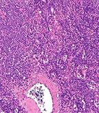
B-cell chronic lymphocytic leukemia/small cell lymphoma; intermediate magnification micrograph, H&E stain; source: Nephron, Wikimedia Commons
By directly removing contaminating events, the assay improves the accuracy of residual disease detection in CLL “and may reclassify the status” of some patients following chemotherapy, said Sartor. “The result has the potential to stop some patients previously thought to have very low level residual disease from being exposed to unnecessary therapy.” She and David J. Gottlieb, a professor at the University of Sydney, Sydney, Australia, reported findings of a recent study of the assay in the journal Clinical Cytometry.
“Recent advances in chemoimmunotherapy make the use of overall survival problematic for timely analysis of trials involving new therapies,” noted the investigators. “As a result, although the level of minimal residual disease (MRD) achieved is not included in the standard International Workshop on Chronic Lymphocytic Leukemia (IWCLL) definition of clinical remission, it is increasingly used as a secondary or even primary endpoint in clinical trials.” As treatments for many hematological malignancies improve, there is a growing trend toward the measurement of very low level disease (minimal residual disease or MRD) to identify patients who might appear to have had excellent responses to treatment but who are at high risk of future relapse and might benefit from further therapy. In CLL, allele-specific oligonucleotide PCR (ASOPCR) of the immunoglobulin gene of the B-CLL clone, the current “gold standard” for measuring MRD in CLL, “is labor intensive and can only be performed on patients that have a pretreatment sample available.” Results are not generally available rapidly enough for “real time” decision-making.
Multiparameter flow cytometry has the advantages of rapid analysis without requirement for patient-specific reagents and can give results available within a few hours. Co-expression of CD19+ and CD5+ with demonstration of clonality by immunoglobulin light chain restriction, the most commonly used flow cytometry method to evaluate MRD in CLL, “lacks sensitivity and is limited by the inclusion of normal hemopoietic cells and inability to demonstrate light chain restriction at very low B cell numbers,” said the investigators. Other approaches that have looked at the differential expression of various cell surface markers and combinations have been complicated by treatment effects and phenotypic overlap with normal cells.
The present study was designed in conjunction with an Australasian Lymphoma and Leukemia Group trial of lenalidomide consolidation therapy in patients with detectable MRD after completion of chemoimmunotherapy for CLL. “We developed a 10-color single tube flow cytometry assay based on the ISA methodology to optimize the accuracy and sensitivity of MRD detection,” said Sartor. “Placing all of the informative markers in one tube reduces the total number of lymphocytes required for analysis in patients with lymphopenia following chemotherapy and facilitates direct exclusion of contaminating T cells during cell analysis.” She emphasized that the assay was developed solely for use by a reference laboratory experienced in multiparameter flow cytometry for CLL in a multi-institutional clinical trial setting and was not appropriate for general purpose flow laboratories not experienced in MRD analysis.
The flow cytometry study population included 58 patients with CLL (median age 61 years, range 41–78 years). Treatment consisted of fludarabine alone or in combination with cyclophosphamide and/or rituximab (n = 52); or reduced intensity allogeneic stem cell transplantation (n = 6). As in previous studies of CLL, residual disease levels of less than 0.01%, 0.01% to 1%, and greater than 1% were taken to indicate levels of residual disease with significantly different prognostic outcomes. Normal blood and bone marrow were obtained from transplant donors. Sensitivity assays were performed using dilution with normal peripheral blood (n = 94) and bone marrow (n = 35). A total of 129 samples were analyzed at various times post-treatment.
A single tube incorporated all monoclonal antibodies: CD81FITC, CD22PE, CD3ECD, CD5PercP5.5, CD20PECY7, CD79bAPC, CD38A700, CD43APC Alexa750, CD19eFluor 450, and CD45KO. A “modified ISA gating strategy” was employed to remove contaminating events. Clinical samples were compared using the four-tube ISA and the single tube 10-color assay. In 80 samples analyzed with both assays, there was an “excellent correlation between the two methods, said Sartor. Removal of contaminating events in the single tube assay led to a significant reduction in residual disease values (P = .0014).
The investigators acknowledge that despite its advantages, the single tube assay is not yet “a realistic widespread alternative” to the current ISA method for assessing CLL MRD. “Validation of the assay across multiple sites would be essential for more generalized introduction of this technology.” Nevertheless they concluded that, “in its current form in a centralized and experienced flow cytometry laboratory, the 10-color single tube assay permits the most accurate flow based CLL MRD detection currently available.”