Sjögren Syndrome Induced by Immune Checkpoint Inhibitors in a Patient with Advanced Renal Cell Carcinoma
This clinical quandary details a Mexican man, aged 77 years, who presented to the oncology clinic with a sternal mass. Based on the results, the patient fulfilled the 2016 American College of Rheumatology/European League Against Rheumatism classification criteria for Sjögren syndrome, thus the diagnosis triggered by immune checkpoint inhibitors was definitively established.
Oncology (Williston Park). 2021;35(8):486-490.
DOI: 10.46883/ONC.2021.3508.0486
Conde-Flores is a medical oncology fellow at Médica Sur in Mexico City, Mexico.
Remolina-Bonilla is a clinical researcher at Instituto Nacional de Ciencias Médicas y Nutrición Salvador Zubirán in Mexico City.

Castro-Alonso is an associate professor in the Department of Internal Medicine at Hospital Regional de Alta Especialidad de Oaxaca in Oaxaca, Mexico.
Martínez-Ibarra is a clinical researcher at Instituto Nacional de Ciencias Médicas y Nutrición Salvador Zubirán in Mexico City.

Hernández-Molina is an associate professor in the Department of Immunology and Rheumatology at Instituto Nacional de Ciencias Médicas y Nutrición Salvador y Zubirán in Mexico City.
Chapa-Ibargüengoitia is an associate professor in the Department of Imaging and Radiology at Instituto Nacional de Ciencias Médicas y Nutrición Salvador y Zubirán.

Gamboa-Domínguez is an associate professor in the Department of Anatomic Pathology at Instituto Nacional de Ciencias Médicas y Nutrición Salvador y Zubirán in Mexico City.

Bourlon is an associate professor in the Department of Hematology and Medical Oncology and head of
the Urologic Oncology Clinica at Instituto Nacional
de Ciencias Médicas y Nutrición Salvador Zubirán in Mexico City. She is also on the IDEA Award Committee and the Program Scientific Committee of the American Society of Clinical Oncology.

A Mexican man, aged 77 years, presented to the oncology clinic with a sternal mass. His past medical history was relevant for tobacco use for a 50 pack-year index, hypertension, and atrial fibrillation. Laboratories reported hemoglobin 10.8 g/dL with no other relevant results. A bone scintigraphy revealed multiple axial and extra-axial metastases; a CT scan showed a left kidney mass, osteolytic lesions with soft tissue component, and pancreatic metastases. An incisional biopsy of the sternal mass reported metastatic clear renal cell carcinoma. Diagnosis of intermediate-risk clear cell renal carcinoma was made. He underwent a left radical nephrectomy confirming the diagnosis.
The patient started first-line systemic treatment with nivolumab (Opdivo) 3 mg/kg plus ipilimumab (Yervoy) 1 mg/kg every 3 weeks. After the third cycle, he complained about dryness in his mouth and eyes. Thus, we initially established the diagnosis of grade 2 sicca syndrome (sicS) secondary to immune checkpoint inhibitors (ICIs; according to Common Terminology Criteria for Adverse Events [CTCAE], version 5.0) and started a diagnostic approach to sicS. Immunotherapy was delayed and the patient began supportive measures with artificial saliva, nystatin/almagate oral solutions, and hyaluronate/chondroitin ophthalmic droplets.
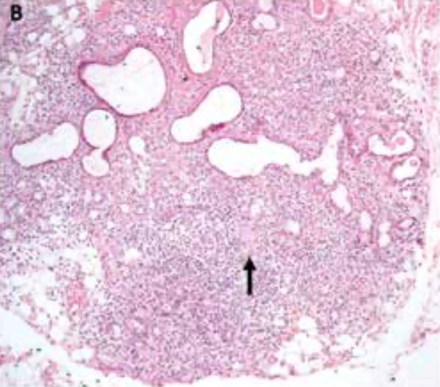
Direct interrogation was negative for other rheumatologic symptoms (fever, weight loss, skin lesions, arthralgia, lymphadenopathies, and swollen salivary glands). Physical exam showed decreased tear pool in the lower ophthalmic conjunctiva and a dry tongue with absent sublingual salivary pool (Figure 1A). Schirmer I test revealed wetting of less than 5 mm in both eyes; unstimulated whole salivary flow rate was 0.1 ml/min. Complement C3/C4 proteins, anti–Sjögren-syndrome-related antigen A and B autoantibodies (anti-Ro/SSA; anti-La/SSB), rheumatoid factor (RF), antinuclear antibodies (ANA), serum amylase, HIV, and hepatitis C virus antibodies were negative. Salivary gland ultrasonography showed normal parotid gland echotexture and echogenicity. Minor salivary gland biopsy reported focal lymphocytic sialadenitis with fibrosis, with a Focus score of 3 (Figure 1B). Based on the results, the patient fulfilled the 2016 American College of Rheumatology/European League Against Rheumatism (EULAR) classification criteria for Sjögren syndrome (Sjös)1, thus the diagnosis of Sjögren syndrome triggered by ICIs was definitively established.
FIGURE 1. Dry Mouth Before Systemic Steroids Course (A) Clinical examination
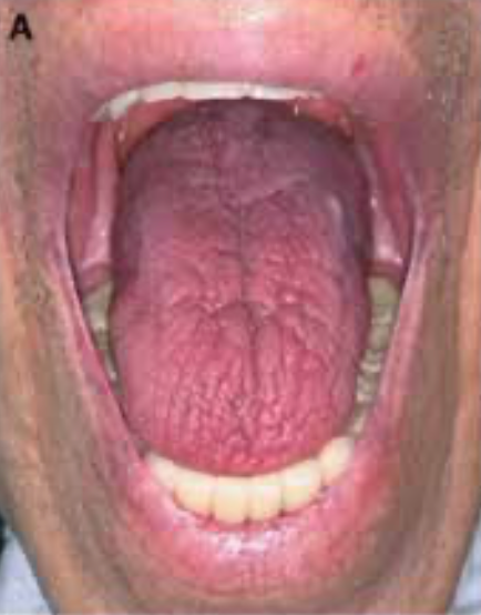
FIGURE 1. Dry Mouth Before Systemic Steroids Course (B) Histopathological examination revealed minor salivary gland with atrophy of acinar cells and ductal dilatation. Acinar structures are infiltrated by lymphocytes, plasma cells, and scattered giant cells (arrow). Collagen fibers surround with duct ectasia and inspissated luminal acidophilic clusters (HE X40). Immunohistochemical reactions in sequential sections of salivary gland
Discussion
Blockade of immunologic checkpoints has reshaped cancer therapeutics over the last decade. Immune checkpoint inhibitors (ICIs) restore endogenous T-cell antitumor activity, achieving long-term tumor responses.2 This immune system enhancement sometimes can be excessive, resulting in a plethora of immune-related adverse events (irAEs).3 IrAEs can be toxicities affecting a single organ or multiple organs, and they are generally dependent on the class of drug: 18% to 37% of patients treated with ipilimumab (an anti–CTLA-4
antibody) experience grade 3 or greater events, compared with about 10% of patients treated with anti–PD-1 or anti–PD-L1 antibodies as monotherapy, and 46% to 55% of patients treated with the combination of anti–PD-1 plus anti–CTLA-4 are affected by irAEs.2,4-7
irAEs usually involve the skin or the gastrointestinal, endocrine, or respiratory systems, but involvement in any organ and tissue can occur.8,9 Rheumatologic irAEs, according to the available data, are reported in 1.5% to 22% of patients who use immunotherapy; the wide range is explained by the different, inconsistent definitions used in studies.8,10-12 The largest cohorts of patients with rheumatologic irAEs come from single-center studies; for instance, a Mayo Clinic retrospective database reported 43 cases among 1293 patients treated with ICIs (74% with single-agent anti–PD-[L]1 agents), resulting in a prevalence of 3.3%.13 A French prospective cohort with 524 patients reported 6.6% (n = 35) of patients experiencing rheumatologic irAEs, where 77% were treated with anti–PD-(L)1 drugs.14 The reason for the scarcity in reported rheumatologic irAEs is multifactorial; explanations include the inconsistency in coding musculoskeletal symptoms, fewer deaths or hospital admissions due to rheumatologic irAEs, underreporting in clinical trials, and late-onset events (occurring up to 2 years after patients have started ICI therapy) not reported in studies.12,15 When rheumatologic disorders preexist, disease flares or irAEs have been reported at rates ranging from 38% to 67%.16
The most commonly reported rheumatologic irAEs are arthralgia and myalgia, with frequencies of 1% to 43% and 2% to 21%, respectively. Arthritis was reported in 1% to 7% of patients; seronegative inflammatory arthritis is described in only 0.7% of patients treated with anti–PD-1 antibodies and in 2.5% of those treated with combination ICIs. Additionally, myalgia was seen in 2% to 18% of participants in trials of nivolumab and ipilimumab, while muscle weakness was reported in 1%.17
ICIs can induce the activation and proliferation of autoreactive immune cells that may lead to SicS, which resembles primary Sjös.18 The first 4 cases of SicS triggered by cancer immunotherapy were reported by Laura C. Cappelli, MD, MHS, MS, and colleagues in 2016; half of those patients presented with additional nonrheumatologic irAEs.19 More recent series describe ICI-induced sicca syndrome as a clinical entity, with an incidence rate ranging in clinical trials from 1.2% to 24.2%, depending on the criteria used in their definitions.8,20 In the REISAMIC registry, a French multicenter irAEs database, Sebastian Le Burel, MD, reported a 0.3% prevalence of CTCAE grade 2 or higher SicS in patients treated with anti–PD-(L)1 monotherapy; this frequency increased to 2.5% among patients treated with a combination of anti–CTLA-4 plus anti–PD-(L)1 agents.8
SicS is rarely reported in clinical trials. This might be because its symptoms are common in the population, with a prevalence reaching up to 30% in persons 65 years or older, and their occurrence may not be attributed to ICIs, leading to underreporting.15 A recent case series identified 20 patients who developed SicS that was associated with ICI therapy and exhaustively described the clinical presentation. The median age of the affected patients was 57 years (range, 26-78); 70% were male, 20% had a history of autoimmune diseases, and 85% were treated with anti–PD-(L)1 monotherapy. Half of these affected patients had concomitant nonrheumatologic irAEs. Dry-mouth symptoms were present in all, appearing at a median of 70 days (range, 30-206) following ICI treatment initiation. Dry mouth was usually abrupt in onset; it was often worse with physical exertion or at night, with patients awakening at night with their tongue stuck to their teeth or hard palate and needing to drink water. Almost all (95%) had salivary hypofunction (unstimulated whole salivary flow <1.5 mL/15 minutes); 45% (n = 9) patients reported dry eyes, and 55% (n = 5) had aqueous tear deficiency (Schirmer’s test 5 mm wetting/5 minutes in at least 1 eye).21,22 In another multinational registry, among 26 patients with SicS, clinical features were as follows: 96% dry mouth, 65% dry eyes, 62% abnormal ocular tests, and 86% abnormal oral tests.22
Nevertheless, among the SicS cases, few have been attributed to Sjös. Sjös is a chronic inflammatory autoimmune disease that involves mainly exocrine glands (eg, salivary and lachrymal glands); it can be conceptualized as an autoimmune epithelitis. Among patients with Sjös, dry mouth is the most frequent clinical manifestation (80%), followed by dry eyes in 50% and arthralgia in 20%.23 For instance, in a multicenter register of 26 patients with sicca syndrome triggered by PD-(L)1 ICIs, 10 of them fulfilled the current Sjös criteria.22
When prescribing ICIs, high suspicion of SicS is warranted; if symptoms occur, a careful history and complete physical evaluation, including oral and ocular diagnostic tests of dryness, should be conducted. Mucositis should be excluded as an alternative irAE with similar symptoms. Serum autoantibodies, viral screening, and minor salivary gland biopsy should be performed to differentiate between Sjös and other etiologies.24 Prompt referral to a rheumatologist is recommended to prevent delay in diagnosis; according to one study, only one-third of the patients with rheumatologic irAEs were ever seen by a rheumatologist.25
On the other hand, several features distinguish ICI-induced Sjös from primary Sjös: The former is characterized by older age, male predominance, and abrupt onset, whereas in ICI-induced Sjös, fewer patients have dry eyes or abnormal ocular tests.22,26 These and other differences are depicted in the Table.25-27
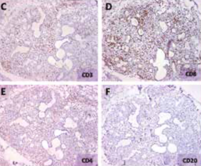
Regarding autoantibodies, most patients with ICI-induced Sjös have a seronegative immunological pattern that is not present in primary Sjös, consistent with reports for other ICI-induced irAEs that have a primary autoimmune counterpart. In the International ImmunoCancer Registry, ANA and anti-Ro/SSA were the most prevalent antibodies with 52% and 20%, respectively; RF and anti-La/SSB were present in 9% and 8% of patients.22 Other immunological markers included low C3/C4 (6%) and positive cryoglobulins (10%).22,26 Minor salivary gland biopsy reveals differences in histopathological findings as well. The typical focal lymphocytic sialadenitis of primary Sjös is present in roughly half of the ICI-induced Sjös cases, with many patients exhibiting mild chronic sialadenitis. The type of lymphocyte infiltrate is also dissimilar: In ICI-triggered patients, it is a predominant CD3+ T-cell infiltrate (with a slight predominance of CD4+ over CD8+ T cells), and CD20+ B cells are virtually absent. This pattern was similar to the one observed in our patient, except we found a predominance of CD8+ over CD4+ T cells (Figures 1C-1F). This phenotype contrasts with primary Sjös, in which CD20+ B cells represent 20% to 62% of the lymphocyte infiltrate.22,26 All these differences are summarized in the Table.
Specific treatment recommendations for inflammatory arthritis, myositis, and polymyalgia-like syndrome, but not for SicS/Sjös, have been proposed by the American Society for Clinical Oncology guidelines.27 Recent recommendations for the management of SicS/Sjös following ICIs have been issued by EULAR, based mostly on expert opinion and the small case series published.11 Experts agree that all patients with ICI-induced SicS/Sjös require topical treatments aimed at alleviating their ocular and oral dryness, preventing dental decay, and managing oral candidiasis. Mild symptoms (CTCAE grade 1) can be managed with just the aforementioned local therapies, as well as oral stimulators of salivary gland function, like pilocarpine or cevilemine.11,24 For symptoms of CTCAE grade 2 or greater, additional treatments should be prescribed. Therefore, answer A is incorrect, because the patient has grade 2 SicS/Sjös and local therapies by themselves are insufficient treatments.
In the case of severe symptoms (CTCAE grade 2 or 3), ICI therapy should be at least temporally withheld. Unlike patients with primary Sjös, many patients with ICI-induced SicS/Sjös may receive systemic steroids, and most of them experience improvement. Doses varied greatly among patients in the cases reported, but experts suggest about 40 mg of prednisone equivalent (range, 10-80 mg), followed by subsequent tapering.24,25 Upon improvement of SicS, ICIs could be resumed, ideally after an interval of 3 months.16,21,27 Permanent ICI discontinuation should be considered in patients with recurrence of intractable, severe symptomatology. Therefore, answers B and C are incorrect: Neither withholding immunotherapy nor oral steroids should be the sole treatment recommended. Thus, D is the correct answer.
Functional prognosis for ICI-induced SicS/Sjös is better than for primary Sjös, as previously stated. Almost 75% of patients were prescribed systemic steroids, of whom nearly 80% reported subjective improvement; however, fewer patients (30%) experienced a return to normal salivary flow rates, and none reported complete resolution of their symptoms.21 Many of these patients will experience long-term salivary hypofunction, with increased risk of dental sequelae (eg, caries, recurrent candidiasis infections) and reduced quality of life. Continuous surveillance (eg, every 3-4 months) with dentists or oral medicine specialists is recommended to avoid those complications16 .
Finally, in relation to oncological outcomes, one single-center prospective cohort study with 524 patients who received treatment with ICIs (of whom 35 were referred to a rheumatology department for irAEs), found that patients with rheumatologic irAEs had greater tumor response rates compared with patients who did not present them: 85.7% vs 35.3% (odds ratio, 8.8; 95% CI, 3.2-29.8; P <.0001). However, no statistically significant differences in tumor response, between patients with rheumatologic vs nonrheumatologic irAEs (85.7% vs 75.1%; P = .18).14
TABLE. Differences Between Checkpoint-induced Sjögren Syndrome and Classical Sjögren Syndromea
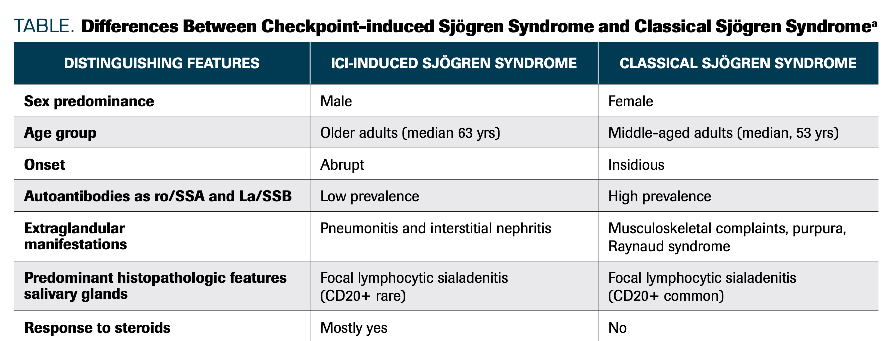
FIGURE 1. Dry Mouth Before Systemic Steroids Course
(C-F), all X100: A CD3/CD8-positive T-cell population dominates the inflammatory infiltrate (C and D). A few CD4 positive lymphocytes (E) and scattered B (CD20) cells (F) were recognized in the background.
Outcome of This Case
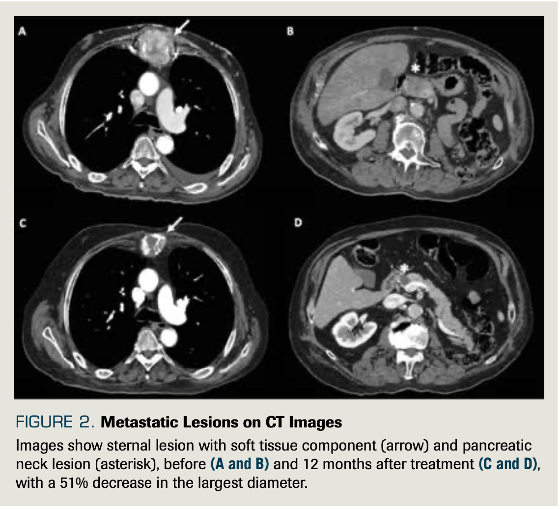
Systemic steroids were started at 0.5 mg/kg methylprednisolone equivalent (50 mg orally administered prednisone daily). The patient was followed up weekly; after 4 weeks of prednisone 50 mg daily, he noted partial improvement in saliva and tear production (Figure 1G). We decided to taper prednisone and restart nivolumab monotherapy. Symptoms were not aggravated upon resuming immunotherapy. A CT scan after 12 months of immunotherapy treatment revealed partial response with a 51% reduction of target lesions (Figure 2).
FIGURE 1. Dry Mouth Before Systemic Steroids Course Improvement in saliva production after systemic steroid course (G).
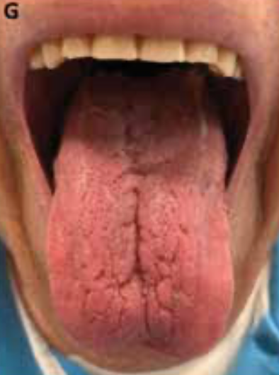
FIGURE 2. Metastatic Lesions on CT Images
Images show sternal lesion with soft tissue component (arrow) and pancreatic neck lesion (asterisk), before (A and B) and 12 months after treatment (C and D), with a 51% decrease in the largest diameter.
Financial Disclosure: The authors have no relationship with any product or provider of any service mentioned in this article.
About the SERIES EDITORS: Maria T. Bourlon, MD is associate professor, Head Urologic Oncology Clinic; national researcher, Instituto Nacional de Ciencias Médicas y Nutrición Salvador Zubirán, Mexico City, Mexico. She is also a member of ASCO’s IDEA Working Group.
E. David Crawford, MD, is chairman, Prostate Conditions Education Council; editor in chief, Grand Rounds in Urology; and professor of Urology, University of California San Diego, La Jolla, CA.
References
1. Shiboski CH, Shiboski SC, Seror R, et al; International Sjögren’s Syndrome Criteria Working Group. 2016 American College of Rheumatology/European League Against Rheumatism classification criteria for primary Sjögren’s syndrome: a consensus and data‐driven methodology involving three international patient cohorts. Arthritis Rheumatol. 2017;69(1):35-45. doi:10.1002/art.39859
2. Bertrand A, Kostine M, Barnetche T, Truchetet M-E, Schaeverbeke T. Immune related adverse events associated with anti-CTLA-4 antibodies: systematic review and meta-analysis. BMC Med. 2015;13:211. doi:10.1186/s12916-015-0455-8
3. Day D, Hansen AR. Immune-related adverse events associated with immune checkpoint inhibitors. BioDrugs.2016;30(6):571-584. doi:10.1007/s40259-016-0204-3
4. Wang Y, Zhou S, Yang F, et al. Treatment-related adverse events of PD-1 and PD-L1 inhibitors in clinical trials: a systematic review and meta-analysis. JAMA Oncol. 2019;5(7):1008-1019. doi:10.1001/jamaoncol.2019.0393
5. Martins F, Sofiya L, Sykiotis GP, et al. Adverse effects of immune-checkpoint inhibitors: epidemiology, management and surveillance. Nat Rev Clin Oncol. 2019;16(9):563-580. doi:10.1038/s41571-019-0218-0
6. Xu H, Tan P, Ai J, et al. Antitumor activity and treatment-related toxicity associated with nivolumab plus ipilimumab in advanced malignancies: a systematic review and meta-analysis. Front Pharmacol. 2019; 10:1300. doi:10.3389/fphar.2019.01300
7. Haanen JBAG, Carbonnel F, Robert C, et al; ESMO Guidelines Committee. Management of toxicities from immunotherapy: ESMO Clinical Practice Guidelines for diagnosis, treatment and follow-up. Ann Oncol. 2017;28(suppl 4): iv119-iv142. doi: 10.1093/annonc/mdx225
8. Le Burel S, Champiat S, Mateus C, et al. Prevalence of immune-related systemic adverse events in patients treated with anti-programmed cell death 1/anti-programmed cell death-ligand 1 agents: a single-centre pharmacovigilance database analysis. Eur J Cancer. 2017;82:34-44. doi:10.1016/j.ejca.2017.05.032
9. Kumar V, Chaudhary N, Garg M, Floudas CS, Soni P, Chandra AB. Current diagnosis and management of immune related adverse events (irAEs) induced by immune checkpoint inhibitor therapy. Front Pharmacol. 2017;8:49. doi:10.3389/fphar.2017.00049
10. Lidar M, Giat E, Garelick D, et al. Rheumatic manifestations among cancer patients treated with immune checkpoint inhibitors. Autoimmun Rev. 2018;17(3):284-289. doi:10.1016/j.autrev.2018.01.003
11. Kostine M, Finckh A, Bingham CO, et al. EULAR points to consider for the diagnosis and management of rheumatic immune-related adverse events due to cancer immunotherapy with checkpoint inhibitors. Ann Rheum Dis. 2021;80(1):36-48. doi:10.1136/annrheumdis-2020-217139
12. Calabrese LH, Calabrese C, Cappelli LC. Rheumatic immune-related adverse events from cancer immunotherapy. Nat Rev Rheumatol. 2018;14(10):569-579. doi:10.1038/s41584-018-0074-9
13. Richter MD, Crowson C, Kottschade LA, Finnes HD, Markovic SN, Thanarajasingam U. Rheumatic syndromes associated with immune checkpoint inhibitors: a single-center cohort of sixty-one patients. Arthritis Rheumatol. 2019;71(3):468-475. doi:10.1002/art.40745
14. Kostine M, Rouxel L, Barnetche T, et al; FHU ACRONIM. Rheumatic disorders associated with immune checkpoint inhibitors in patients with cancer - clinical aspects and relationship with tumour response: a single-centre prospective cohort study. Ann Rheum Dis. 2018;77(3):393-398. doi:10.1136/annrheumdis-2017-212257
15. Abdel-Wahab N, Suarez-Almazor ME. Frequency and distribution of various rheumatic disorders associated with checkpoint inhibitor therapy. Rheumatology (Oxford). 2019;58(Suppl 7):vii40-vii48. doi:10.1093/rheumatology/kez297
16. Steven NM, Fisher BA. Management of rheumatic complications of immune checkpoint inhibitor therapy – an oncological perspective. Rheumatology (Oxford). 2019;58(Suppl 7):vii29-vii39. doi:10.1093/rheumatology/kez536
17. Cappelli LC, Gutierrez AK, Bingham CO III, Shah AA. Rheumatic and musculoskeletal immune-related adverse events due to immune checkpoint inhibitors: a systematic review of the literature. Arthritis Care Res (Hoboken). 2017;69(11):1751-1763. doi:10.1002/acr.23177
18. Vivino FB. Sjogren’s syndrome: clinical aspects. Clin Immunol. 2017;182:48-54. doi:10.1016/j.clim.2017.04.005
19. Cappelli LC, Gutierrez AK, Baer AN, et al. Inflammatory arthritis and sicca syndrome induced by nivolumab and ipilimumab. Ann Rheum Dis. 2017;76(1):43-50. doi:10.1136/annrheumdis-2016-209595
20. Melissaropoulos K, Klavdianou K, Filippopoulou A, Kalofonou F, Kalofonos H, Daoussis D. Rheumatic manifestations in patients treated with immune checkpoint inhibitors. Int J Mol Sci. 2020;21(9):3389. doi:10.3390/ijms21093389
21. Warner BM, Baer AN, Lipson EJ, et al. Sicca syndrome associated with immune checkpoint inhibitor therapy. Oncologist. 2019;24(9):1259-1269. doi:10.1634/theoncologist.2018-0823
22. Ramos-Casals M, Maria A, Suárez-Almazor ME, et al; ICIR. Sicca/Sjögren’s syndrome triggered by PD-1/PD-L1 checkpoint inhibitors. data from the International ImmunoCancer Registry (ICIR). Clin Exp Rheumatol. 2019;37(Suppl 118;3):114-122.
23. Kostine M, Truchetet ME, Schaeverbeke T. Clinical characteristics of rheumatic syndromes associated with checkpoint inhibitors therapy. Rheumatology (Oxford). 2019;58(Suppl 7):vii68-vii74. doi:10.1093/rheumatology/kez295
24. Mavragani CP, Moutsopoulos HM. Sicca syndrome following immune checkpoint inhibition. Clin Immunol. 2020;217:108497. doi:10.1016/j.clim.2020.108497
25. Liew DFL, Leung JLY, Liu B, Cebon J, Frauman AG, Buchanan RRC. Association of good oncological response to therapy with the development of rheumatic immune-related adverse events following PD-1 inhibitor therapy. Int J Rheum Dis. 2019;22(2):297-302. doi:10.1111/1756-185X.13444
26. Christodoulou MI, Kapsogeorgou EK, Moutsopoulos HM. Characteristics of the minor salivary gland infiltrates in Sjögren’s syndrome. J Autoimmun. 2010;34(4):400-407. doi:10.1016/j.jaut.2009.10.004
27. Leipe J, Mariette X. Management of rheumatic complications of ICI therapy: a rheumatology viewpoint. Rheumatology (Oxford). 2019;58(Suppl 7):vii49-vii58. doi:10.1093/rheumatology/kez360
