In-Transit Melanoma: An Individualized Approach
The management of in-transit metastases is challenging, since the treatments and extent of disease vary greatly based on the number, depth, location, and distribution of lesions, and on their biological behavior.
The majority of locoregional recurrences in melanoma occur in the form of intradermal or subcutaneous local or in-transit metastasis. In-transit melanoma represents contamination of the lymphatic space that, if treated, can result in long-term cure in a subset of patients. The management of in-transit metastases is challenging, since the treatments and extent of disease vary greatly based on the number, depth, location, and distribution of lesions, and on their biological behavior. A number of different treatment options exist, but there is no level 1 evidence to guide clinical decision-making. Herein we present our institutional treatment algorithm, which allows for individualization based on the patient's presentation.
Introduction
The simultaneous push for standardized treatment guidelines and "individualized medicine" may appear contradictory. If we develop evidence-based guidelines to follow as best practice for all patients with a disease, then how can we at the same time be individualizing care? However, personalized medicine does not prohibit the use of guidelines, and as we become more sophisticated, gene expression patterns-as opposed to the current crude gross clinical findings utilized today-will play a more central role in governing the treatment options we pursue. We are beginning to realize that most cancers, including melanoma, do not represent a single disease but rather are a phenotypic representation of a diverse array of underlying genetic changes that likely can be broken down into a few subtypes. Given that there are no randomized trials comparing the different approaches to in-transit melanoma, we have developed guidelines for patients who present with this pattern of disease, and we outline here our institutional preferred treatment decisions based on the extent and type of disease. Clearly we remain at the beginning stages of understanding this complex disease, and we look forward to modifying our algorithm as knowledge in this field advances.
Incidence and Pathophysiology
In-transit metastases develop from regional contamination of the intralymphatic system and occur shortly (mean, 16 months) after definitive treatment of the primary melanoma in 10% of patients.[1] The network of lymphatics draining from the primary tumor to the regional basin is quite robust. The clinical manifestation of an in-transit metastasis occurs when malignant cells present in the lymphatic system multiply to the point that they become palpable or visible at or just below the skin. These isolated lesions may appear indolent and easily treatable. However, such lesions are frequently just the tip of the iceberg, and an individual lesion often represents the most advanced presentation of many subclinical in-transit metastases. This is the reason localized treatment often fails and recurrence remains the rule. In-transit melanoma is associated with aggressive clinical and pathological factors such as increasing age, greater Breslow thickness, ulceration, high tumor mitotic rate, presence of angiolymphatic invasion, positive sentinel lymph nodes (SLNs), and extremity location.[1,2] While SLN metastases are associated with a 24% incidence of subsequent in-transit metastasis, the SLN procedure itself does not increase the incidence of in-transit disease.[2]
These in-transit metastases must be distinguished from a true local recurrence, which often involves the epidermis, representing regrowth of residual primary disease from incomplete resection. Given the recommendations of contemporary guidelines for wide local excision, true local recurrences should be an uncommon event, except in certain settings, such as lentigo maligna of the face. In-transit metastases are associated with a significantly worse prognosis compared with true local recurrence and therefore are classified as stage IIIB or IIIC disease, depending on the status of the regional lymph nodes as defined below (B = negative; C = positive).[3]
Because the definitions used in the literature vary, we want to clarify that we are using the term "in-transit metastasis" to include satellites, microsatellites, and in-transit metastasis. Microsatellites are defined as "any discontinuous nest of intralymphatic metastatic cells > 0.05 mm in diameter that is clearly separated by normal dermis (not fibrosis or inflammation) from the main invasive component of melanoma by a distance of at least 0.3 mm."[3] Satellite and in-transit metastases are cutaneous and/or subcutaneous metastases that occur between the primary melanoma and the first echelon regional lymph nodes, which have arbitrarily been distinguished on the basis of whether they are located within or more than 2 cm from the primary tumor. There does not appear to be a survival difference between patients with satellites and patients with in-transit metastases; the pathogenesis is identical, as is treatment-and the differences in terminology are chiefly of historical interest, since the 2-cm proximity rule has no anatomic or physiologic basis.[4] Prognosis after in-transit recurrence is largely the result of pathological features of the primary melanoma and the presence or absence of synchronous lymph node metastasis, as well as of the disease-free interval prior to recurrence.[5,6] Satellites and in-transit metastases are both considered intralymphatic metastases and are classified as N2c, with 5- and 10-year survivals of 69% and 52%, respectively, which are somewhat better than the 5- and 10-year survivals (59% and 43%, respectively) for stage IIIB patients overall, but still similar enough to make this classification the "closest fit."[3] Patients with both satellites/in-transit metastases and nodal metastases have a worse prognosis than patients who have either satellites/in-transit metastases or nodal metastases alone.[4,7] Consequently, the presence of a satellite or in-transit metastasis in addition to one or more positive lymph nodes is staged as N3.[3,8]
Algorithm
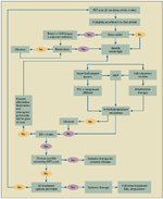
FIGURE 1

Algorithm for the Treatment of In-Transit Melanoma
It is understood that patients with in-transit disease are at high risk for further locoregional and distant recurrence. Many potential treatment options exist for this disease, with no accepted "best practice." Patients with this pattern of disease vary greatly in terms of the number, depth, location, and distribution of lesions. With an acknowledgement of these complexities and limitations, we have reviewed the literature and have developed a clinical algorithm (Figure 1) to facilitate the individualization of treatment and to improve patient outcomes. This algorithm will evolve as our understanding of the biology and immunology of melanoma disease progression matures.
Intralymphatic spread is not a single disease entity but rather represents diverse and complex patterns of failure, with patients frequently experiencing multiple treatments; this makes comparing outcomes challenging. For each treatment there is a select group of patients who will have a complete response and long-term survival. Although there is a lack of level I evidence to guide management, there are some consistent principles in the literature that we have used as the basis for our treatment algorithm.
Surgical Resection
Patients who develop locoregional recurrence should be adequately restaged with positron-emission tomography (PET)–computed tomography (CT), since 10% to 20% of these patients may have simultaneous distant relapse necessitating a change in management.[2,9] However, if the disease is indeed localized, then the simplest technique should be employed first-ie, complete excision and primary closure when possible. There are no evidence-based data to support a particular width of peripheral margins of excision for in-transit metastases, and the wide local excision guidelines for a primary lesion do not apply. A single cutaneous metastasis can be narrowly resected, and we typically utilize a 5-mm gross margin. These individual lesions are, in general, well circumscribed, and negative microscopic margins should be obtained if excision is performed for therapeutic rather than diagnostic purposes. Unfortunately, surgical excision is not always feasible, and in patients with too many lesions to resect, recurrence within a short time period following resection, or numerous recurrences, alternative treatment approaches should be considered as outlined below. A multifocal metastasis within a circumscribed area may be resected en-bloc. Primary closure is preferred; however, lack of redundant skin and lymphedema from previous interventions may necessitate rotation flaps or skin grafting. Skin grafting has the advantage of not impairing lymphatic flow. Rarely, uncontrollable pain, extensive locoregional tumor progression, or ulcerating and fungating lesions exhaust other treatment measures, and palliative amputation may remain the only option. Initial reports of amputation for advanced melanoma were associated with poor outcomes.[10] Recent reports of salvage amputation still report a 36% incidence of stump recurrence, with approximately 20% of patients surviving more than 5 years.[11,12] We employ limb-preserving therapies whenever possible, reserving amputations for cases in which all other measures have been exhausted.
In the absence of prior regional nodal dissection, patients with first-time in-transit recurrences should undergo nodal restaging with SLN biopsy, since up to 50% of patients have synchronous nodal metastasis.[13-16] Prior wide local excision does not appear to adversely impact the ability to detect lymphatic metastases, and SLN biopsy has been found to be accurate in the setting of recurrent melanoma.[14,15] SLN biopsy and subsequent lymphadenectomy for nodal positivity are prognostic, identifying patients with a higher risk of relapse (median disease-free survival of 16 months for node-positive status compared with 36 months when SLNs were negative); the procedure also offers excellent regional control.[14]
Isolated Limb Perfusion and Isolated Limb Infusion
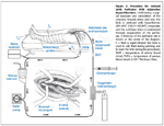
First described by Creech in 1958, isolated limb perfusion (ILP) involves exclusion of the extremity from the systemic circulation, thereby allowing regional delivery of chemotherapy at doses 10 to 25 times higher than can safely be tolerated with systemic administration. This requires cannulation of a major blood vessel of the limb; typically, an iliac or femoral approach is used for the lower limb and an axillary approach for the arm (Figure 2). Perfusion of the extremity proceeds with high flow (500 to 1000 mL/min), with a membrane oxygenator used to maintain oxygenation and an appropriate acid-base balance. The limb is perfused for 60 to 90 minutes with chemotherapy. Systemic leak is minimized and circulating chemotherapy is washed out prior to recirculation, resulting in few to no systemic side effects. The perfused extremity inevitably experiences some local toxicity, ranging from mild erythema to epidermolysis; approximately one third of patients develop a Common Terminology Criteria for Adverse Events (CTAE) grade 3 or higher toxicity, although fascial compartment syndrome or deep tissue damage necessitating amputation results only rarely (in < 3% of cases).[17,18] Retrospective studies have shown that approximately 85% of patients experience an objective response after ILP, and 50% have a complete response.[19,20] It may take 3 to 6 months to see the maximal benefit; however, in patients who experience a complete response, the response is often long-lived, with a 32-month median duration.[17] The overall 5-year survival rate after ILP in a large series was 32%, and improved overall survival was closely associated with achievement of complete response.[21]
FIGURE 2

Procedure for Isolated Limb Perfusion With Adjunctive Hyperthermia
Melphalan (L-phenylalanine mustard) is the standard chemotherapy for perfusion. Melphalan is an alkylating agent with melanoma tumor selectivity, since phenylalanine is an essential component of melanin synthesis. Other agents, including interleukin (IL)-2, interferon (INF)-α, cisplatin, temozolomide (Temodar), and ADH-1 (Exherin), have been used alone or in combination with melphalan without significant improvements in response compared with melphalan alone.[22-24] Hyperthermia is typically used as an adjunct to improve response rates.[25] Hyperthermia induces cutaneous vasodilatation and improves drug uptake and cytotoxicity when temperatures exceed 38.5°C (101.3°F).[26] Temperatures over 42°C (107.6°F) are directly cytotoxic to malignant cells but may exacerbate locoregional toxicity.[27] The addition of tumor necrosis factor (TNF)-α to the perfusate results in tumor vasculature destruction, increased cytotoxicity, and modulation of the immune response. TNF-α also augments melphalan uptake into melanoma cells, and promising 4- to 6-fold improved response rates have been reported, especially for bulky or treatment-resistant disease.[28,29] However, TNF-α is associated with significantly increased toxicity and was shown to be no better than melphalan alone in a randomized prospective trial; as a result, it is no longer available off protocol in the United States.[30]
Isolated limb infusion (ILI) is a simpler and less invasive technique with less regional toxicity; it was first described in 1994 by Thompson et al of the Sydney Melanoma Unit.[31] Melphalan is infused at lower concentrations, with lower flow (55 to 75 mL/min), and for a shorter duration (30 minutes) through a percutaneous approach. A membrane oxygenator is not used in the circuit, resulting in a hypoxic and acidotic environment, which may increase cytotoxic activity. However, the same degree of hyperthermia is not obtained, which may contribute to the reduced complete response rate of 30% and the shorter median duration of response (24 months) compared with ILP.[17,18,32] CTAE grade 3 toxicities are only reported in 20% of patients undergoing ILI, and no treatment-related amputations have been reported.[17] The shorter duration of treatment and the significantly less invasive nature of the approach make this technique ideal for the elderly and those with significant comorbidities.[32,33] Another advantage of ILI is the avoidance of long-term morbidity associated with a lymph node dissection. However, occult nodal micrometastasis may be left behind in nearly half of patients[13,14]; this is demonstrated in patterns of relapse, with 75% (21 of 28) of in-field progressions occurring in the undissected regional nodal basin after an ILI.[34]
Both procedures can be repeated and still retain reasonable response rates.[35,36] Therefore, repeat perfusion should be considered in patients who had a prior response but later experienced progression in the extremity. ILI clearly has a distinct advantage in ease of repeating the procedure and decreased morbidity. However, the difference in response rates between the two types of regional therapy when repeated are even more pronounced than the difference between them when used as initial treatment-favoring ILP over ILI.[17,37] Regional chemotherapy in the form of ILP or ILI has the theoretical advantage of sterilizing the entire lymphatic system that is perfused, resulting in treatment of both clinical and subclinical disease. However, the techniques are limited to patients with disease isolated to an extremity. Patients with disease of the head, neck, or trunk are not candidates. Also, because the perfusion catheters are advanced distally from the point of entry into the vessel, the most proximal portion of the extremity will remain outside the treatment field. Therefore we recommend hyperthermic ILP (HILP) for healthy patients with unresectable in-transit melanoma of the extremities. We currently do not offer ILI but consider referring patients with significant comorbidities or advanced age for this less toxic but less effective alternative.
Intralesional Therapy
Intralesional therapy has the distinct advantage of delivering therapy directly into the malignant lesion. Intradermal and subcutaneous lesions lend themselves to this approach since they are easily accessible. The number of agents that can be injected into a lesion and result in cellular death is considerable. However, patients with intralymphatic disease often have multiple lesions, and in-field recurrence following treatment of an individual focus whether by resection or injection of a toxin is the rule. Therefore, the real goal of intralesional therapy is not only to destroy the target lesion but also to stimulate regression of other, untreated lesions. This can be accomplished when the injectant induces a regional and systemic immunization to melanoma antigens.
Bacille Calmette-Gurin (BCG) was the first intralesional treatment described, and it demonstrated local control in 90% of injected intradermal metastases as well as evidence of inducing systemic immune response to melanoma antigens. As early as 1974, Morton et al described a 17% response rate in distant lesions for patients undergoing intralesional injection with BCG.[38]
Intralesional IL-2 allows intratumoral concentrations that are much higher than can be delivered systemically, without significant toxicity. A clinical trial using this approach has demonstrated a complete response in 97% of injected lesions. Unfortunately, despite a significant increase in INF-γ–positive, tumor-specific CD8-positive T cells, there were no objective responses in distant untreated metastasis.[39]
Intralesional injection of granulocyte macrophage colony–stimulating factor (GM-CSF) has demonstrated a high response rate, as well as offering hope of regional immune stimulation and regression of noninjected lesions. A novel extension of this approach was the development of an oncolytic herpes simplex virus type 1 (HSV-1) encoded with the genetic sequence to produce GM-CSF (OncoVEXGM-CSF). A phase I clinical trial demonstrated a 26% objective response rate in both injected and noninjected lesions, highlighting the dual mechanism of action that includes both a direct oncolytic effect in injected tumors and a secondary immune-mediated antitumor effect in noninjected tumors.[40] Despite 80% of the treated patients in this study having stage IV disease, a 58% 1-year survival was observed, which is significantly better than the historical 1-year survival rates of 25% for stage IV disease.[41] Based on these preliminary results, an international prospective, randomized phase III clinical trial in patients with unresectable stage IIIB or IIIC or stage IV melanoma (OPTIM) is currently underway.[42]
Pulsed Dye Laser Therapy
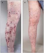
Another approach for the treatment of intradermal in-transit melanoma metastases is the use of pulsed dye laser therapy (PDL) alone or in conjunction with topical imiquimod (Aldara). The rationale for the use of PDL is that it photocoagulates tumor-associated vasculature and thus induces an inflammatory response in addition to direct tumor photothermolysis. The PDL laser settings used have generally been a 9- to 15-J/cm2 fluence, a 3- to 40-millisecond pulse duration, and a 7-mm spot size.[43-45] Imiquimod, a toll-like receptor agonist, is often used as an adjunct in order to augment the local inflammatory response produced with PDL treatment, potentially creating a systemic antitumor immunity that may induce responses at other untreated sites of disease. Naylor et al reported a complete response in one patient who had disease involving the skin, lung, and regional lymph nodes within 7 months of completing PDL therapy to the in-transit metastasis only.[44] The unique features of PDL that make it attractive include its ease of delivery and tolerability, and its minimal toxicity. Although the results have been mixed, overall patients have tolerated treatment well, with the main side effects being localized pain and swelling, and with most patients reporting an overall improvement in quality of life.[43-45] PDL also has the ability to treat small (< 3 cm) metastases that are inaccessible to ILP (ie, located on the head/neck or trunk).[46] Because of its limited depth of penetration, this technique is only appropriate for superficial intradermal lesions (Figure 3).
Radiotherapy
FIGURE 3

Dramatic Response to Pulsed Dye Laser (PDL) Treatment
Melanoma has long been considered a radioresistant tumor; however, more contemporary data suggest that the radiosensitivity of melanoma is more heterogeneous.[47,48] External beam radiation is rarely used as primary treatment for locoregional recurrent melanoma; however, it can be useful for unresectable in-transit melanoma. A single institutional review of 120 patients with advanced melanoma, including 33 patients with in-transit disease, reported a 3-month response rate in stage III patients of 77%, including 44% with a complete response. Complete response was closely associated with improved survival. The median survival for patients specifically with in-transit melanoma was 19 months, and the 1- and 5-year overall survivals were 69% and 32%, respectively.[49] Radiation was well tolerated, and 95% of patients were able to complete treatment; patients who received a radiation dose of less than 40 Gy had a significantly worse overall survival than those receiving 40 to 50 Gy. Radiation doses of greater than 50 Gy did not seem to improve overall survival.[49] As in other tumors, radiotherapy is often most effective in treating microscopic residual disease, as opposed to gross disease, serving as an adjuvant therapy that can be utilized following local R0 resections of in-transit metastasis confined to a limited field. Because it obliterates the lymphatic network, radiation therapy is also associated with lymphedema, and circumferential treatment would thus be a relative contraindication. External beam radiation has also been demonstrated to be very effective in treating in-transit melanoma of the head and neck, and given the lack of other effective treatment modalities and the cosmetic concerns about this location, we routinely utilize primary or adjuvant radiation in patients with in-transit melanoma of the head and neck.[49]
REFERENCE GUIDE
Therapeutic Agents
Mentioned in This Article
ADH-1 (Exherin)
Bacille Calmette Gurin
Bevacizumab (Avastin)
Bleomycin
Carboplatin
Cisplatin
Dacarbazine
Granulocyte macrophage colony– stimulating factor (GM-CSF)
Imiquimod (Aldara)
Interferon-α
Interleukin-2 (IL-2)
Ipilimumab (Yervoy)
Melphalan
Oncolytic herpes simplex virus type 1 encoding GM-CSF (Onco
VEXGM-CSF
)
Paclitaxel
Sorafenib (Nexavar)
Temozolomide (Temodar)
Tumor necrosis factor (TNF)-α
Vemurafenib (Zelboraf))
Brand names are listed in parentheses only if a drug is not available generically and is marketed as no more than two trademarked or registered products. More familiar alternative generic designations may also be included parenthetically.
Systemic Therapy
As noted, in-transit disease represents advanced stage III disease that is potentially curable but often unresectable. In a recent retrospective review, 56% of patients with in-transit disease as the sole site of first recurrence subsequently developed distant disease. Large in-transit metastasis with a diameter ≥ 2 cm was the only independent predictor of distant metastasis (odds ratio [OR], 9.69).[2] For patients who fail to respond to locoregional therapy or who develop synchronous or metachronous distant metastasis, systemic therapy is indicated.
Until recently, dacarbazine (DTIC) and IL-2 were the only FDA-approved systemic agents for metastatic melanoma. High-dose IL-2 has demonstrated only a 16% response rate, and the drug’s approval was based on the rare but potentially curative 6% durable complete response rate.[50] Nonvisceral disease is consistently associated with a higher rate of complete response to high-dose IL-2-up to 50% in the setting of M1a disease in some studies-and this therapy therefore may be considered for those with in-transit disease.[50-52] The obvious limitation remains its significant toxicity profile and low overall complete response rate.
Recently there has been dramatic improvement in the systemic treatment of patients with disseminated melanoma. Ipilimumab (Yervoy), a fully humanized IgG1 monoclonal antibody against cytotoxic T lymphocyte–associated antigen 4 (CTLA-4), was recently approved by the FDA as immunotherapy for the treatment of metastatic melanoma. Ipilimumab in combination with DTIC significantly improved duration of response and overall survival compared with DTIC alone in a large randomized controlled trial.[53] However, this study consisted primarily of patients with stage IV disease, with only 2% to 3% of patients having unresectable stage III disease. Ipilimumab following HILP with melphalan is currently being investigated for patients with in-transit melanoma, and the results of this study may lead to further utilization of this agent for locoregionally advanced melanoma (protocol ID 10-101, NCT01323517).
BRAF is a serine threonine protein kinase that is a component of the mitogen-activated kinase pathway. About half of human melanomas have been shown to have an activating mutation in BRAF due to a substitution of glutamic acid for valine at amino acid 600 (V600E).[54,55] Sorafenib (Nexavar) is a nonspecific, multikinase inhibitor that has been demonstrated to enhance the cytotoxic effects of melphalan in melanoma cell lines as well as in an animal model [56]; unfortunately, in the clinical setting it did not add any benefit to carboplatin and paclitaxel in the treatment of stage III and IV melanoma.[57] Vemurafenib (PLX4032; Zelboraf) is a potent inhibitor of mutated BRAF that was recently shown to improve overall survival in patients with stage IIIC or stage IV melanoma that tested positive for the BRAF V600E mutation [58]. Subsequently vemurafenib was approved by the FDA for unresectable melanoma. Currently there are no studies evaluating vemurafenib specifically in locoregionally advanced melanoma.
There is also great interest in the inhibition of vascular endothelial growth factor (VEGF) in melanoma. Bevacizumab (Avastin) is a monoclonal antibody against VEGF that is used for the treatment of metastatic colorectal, brain, non–small-cell lung, and breast cancers. In a phase II trial of carboplatin and paclitaxel with bevacizumab in stage IV melanoma, there was a median progression-free survival of 6 months and an overall survival of 12 months.[59] A phase III trial of carboplatin and paclitaxel with or without bevacizumab in patients with stage IV melanoma demonstrated a clinically but not statistically significant difference in progression-free survival (5.6 months vs 4.2 months) and in overall survival (12.3 months vs 9.2 months).[60] However, only patients with stage IV melanoma were included in these trials, not those with in-transit disease. Bevacizumab in combination with melphalan has demonstrated a marked synergistic antitumor effect in a xenograft model, suggesting VEGF inhibition may alter vascular permeability, leading to increased melphalan delivery during HILP for in-transit melanoma.[61]
Electrochemotherapy
Electrochemotherapy (ECT) consists of chemotherapy with subsequent application of electrical fields to increase tumor cell membrane permeability. When applied to in-transit and cutaneous metastases, electroporation has the potential to enhance penetration of intralesional and intravenous chemotherapy. Cisplatin and bleomycin have demonstrated high efficacy in this setting. Although both agents are given in subtherapeutic doses, the electroporation enhances cytotoxicity 10- to 700-fold.[62] Objective response rates of up to 96% have been reported, with 50% to 78% complete responses.[63,64] Unfortunately, ECT fails to induce a regional and systemic immunization to melanoma antigens, and most patients experience progression.[63] Although this approach is promising, we do not currently offer ECT, and therefore it is not included in our algorithm.
Conclusion
Unfortunately, despite aggressive treatment, relapse is the rule rather than the exception in patients with in-transit melanoma. Survival is mainly dictated by the biological behavior of the disease, and most patients can expect to experience multiple treatment regimens. In-transit melanoma is not a single disease entity; instead, it represents diverse and complex patterns of failure. For each treatment, there is a select group of patients who will have a complete response and long-term survival. Future studies will need to identify which patient and tumor characteristics respond to each approach. Since these tumors are easily accessible, they offer the unique opportunity for pre- and post-treatment tissue analysis to identify genetic signatures. A better understanding of chemotherapy resistance pathways, biologic targets, gene expression profiles, and markers predictive of response to each therapy will allow rational preoperative determination of individualized treatment as opposed to the broad generalized clinically based guidelines we have outlined.
Financial Disclosure:The authors have no significant financial interest or other relationship with the manufacturers of any products or providers of any service mentioned in this article.
References:
References
1. Stucky CC, Gray RJ, Dueck AC, et al. Risk factors associated with local and in-transit recurrence of cutaneous melanoma. Am J Surg. 2010;200:770-4; discussion 74-5.
2. Pawlik TM, Ross MI, Johnson MM, et al. Predictors and natural history of in-transit melanoma after sentinel lymphadenectomy. Ann Surg Oncol. 2005;12:587-96.
3. Edge SB, Compton CC. The American Joint Committee on Cancer: the 7th edition of the AJCC cancer staging manual and the future of TNM. Ann Surg Oncol. 2010;17:1471-4.
4. Buzaid AC, Ross MI, Balch CM, et al. Critical analysis of the current American Joint Committee on Cancer staging system for cutaneous melanoma and proposal of a new staging system. J Clin Oncol. 1997;15:1039-51.
5. Francken AB, Accortt NA, Shaw HM, et al. Prognosis and determinants of outcome following locoregional or distant recurrence in patients with cutaneous melanoma. Ann Surg Oncol. 2008;15:1476-84.
6. Karakousis CP, Balch CM, Urist MM, et al. Local recurrence in malignant melanoma: long-term results of the multiinstitutional randomized surgical trial. Annals of Surgical Oncology. 1996;3:446-52.
7. Balch CM, Buzaid AC, Soong SJ, et al. Final version of the American Joint Committee on Cancer staging system for cutaneous melanoma. J Clin Oncol. 2001;19:3635-48.
8. Balch CM, Soong SJ, Gershenwald JE, et al. Prognostic factors analysis of 17,600 melanoma patients: validation of the American Joint Committee on Cancer melanoma staging system. J Clin Oncol. 2001;19:3622-34.
9. Soong SJ, Harrison RA, McCarthy WH, et al. Factors affecting survival following local, regional, or distant recurrence from localized melanoma. J Surg Oncol. 1998;67:228-33.
10. Jaques DP, Coit DG Brennan MF. Major amputation for advanced malignant melanoma. Surg Gynecol Obstet. 1989;169:1-6.
11. Kroon HM, Lin DY, Kam PC, Thompson JF. Major amputation for irresectable extremity melanoma after failure of isolated limb infusion. Ann Surg Oncol. 2009;16:1543-7.
12. Kapma MR, Vrouenraets BC, Nieweg OE, et al. Major amputation for intractable extremity melanoma after failure of isolated limb perfusion. Eur J Surg Oncol. 2005;31:95-9.
13. Reintgen M, Nobo C, Giuliano R, et al. Regional therapy for recurrent metastatic melanoma confined to the extremity: hyperthermic isolated limb perfusion vs isolated limb infusion. Cancers. 2010;2:43-50.
14. Yao KA, Hsueh EC, Essner R, et al. Is sentinel lymph node mapping indicated for isolated local and in-transit recurrent melanoma? Ann Surg. 2003;238:743-7.
15. Coventry BJ, Chatterton B, Whitehead F, et al. Sentinel lymph node dissection and lymphatic mapping for local subcutaneous recurrence in melanoma treatment: longer-term follow-up results. Ann Surg Oncol. 2004;11:203S-7S.
16. Aloia TA, Grubbs E, Onaitis M, et al. Predictors of outcome after hyperthermic isolated limb perfusion: role of tumor response. Arch Surg. 2005;140:1115-20.
17. Raymond AK, Beasley GM, Broadwater G, et al. Current trends in regional therapy for melanoma: lessons learned from 225 regional chemotherapy treatments between 1995 and 2010 at a single institution. J Am Coll Surg. 2011;213:306-16.
18. Beasley GM, Petersen RP, Yoo J, et al. Isolated limb infusion for in-transit malignant melanoma of the extremity: a well-tolerated but less effective alternative to hyperthermic isolated limb perfusion. Ann Surg Oncol. 2008;15:2195-205.
19. Grunhagen DJ, Brunstein F, Graveland WJ, et al. One hundred consecutive isolated limb perfusions with TNF-alpha and melphalan in melanoma patients with multiple in-transit metastases. Ann Surg. 2004;240:939-47; discussion 47-8.
20. Sanki A, Kam PC, Thompson JF. Long-term results of hyperthermic, isolated limb perfusion for melanoma: a reflection of tumor biology. Ann Surg. 2007;245:591-6.
21. Olofsson R, Lindner P. Long-term follow-up of all isolated limb perfusions for in-transit metastasis of malignant melanoma in Sweden during 25 years. Society of Surgical Oncology 64th Annual Cancer Symposium. San Antonio, TX. Mar 2-5, 2011.
22. Daryanani D, de Vries EG, Guchelaar HJ, et al. Hyperthermic isolated regional perfusion of the limb with carboplatin. Eur J Surg Oncol. 2000;26:792-7.
23. Ariyan S, Poo WJ, Bolognia J. Regional isolated perfusion of extremities for melanoma: a 20-year experience with drugs other than L-phenylalanine mustard. Plast Reconstr Surg. 1997;99:1023-9.
24. Beasley GM, Riboh JC, Augustine CK, et al. Prospective multicenter phase II trial of systemic ADH-1 in combination with melphalan via isolated limb infusion in patients with advanced extremity melanoma. J Clin Oncol. 29:1210-5.
25. Barbour AP, Thomas J, Suffolk J, et al. Isolated limb infusion for malignant melanoma: predictors of response and outcome. Ann Surg Oncol. 2009;
16:3463-72.
26. Stehlin JS, Giovanella BC, de Ipolyi PD, et al. Results of hyperthermic perfusion for melanoma of the extremities. Surg Gynecol Obstet. 1975;140:339-48.
27. Klaase JM, Kroon BB, van Geel BN, et al. Patient- and treatment-related factors associated with acute regional toxicity after isolated perfusion for melanoma of the extremities. Am J Surg. 1994;167:618-20.
28. Lienard D, Ewalenko P, Delmotte JJ, et al. High-dose recombinant tumor necrosis factor alpha in combination with interferon gamma and melphalan in isolation perfusion of the limbs for melanoma and sarcoma. J Clin Oncol. 1992;10:52-60.
29. Fraker DL, Alexander HR, Andrich M, Rosenberg SA. Treatment of patients with melanoma of the extremity using hyperthermic isolated limb perfusion with melphalan, tumor necrosis factor, and interferon gamma: results of a tumor necrosis factor dose-escalation study. J Clin Oncol. 1996;14:479-89.
30. Cornett WR, McCall LM, Petersen RP, et al. Randomized multicenter trial of hyperthermic isolated limb perfusion with melphalan alone compared with melphalan plus tumor necrosis factor: American College of Surgeons Oncology Group Trial Z0020. J Clin Oncol. 2006;24:4196-201.
31. Thompson JF, Saw RPM, Kam PC. Isolated limb infusion with melphalan for recurrent melanoma: a simple alternative to isolated limb perfusion. Reg Cancer Treat. 1994;7:188-92.
32. Beasley GM, Caudle A, Petersen RP, et al. A multi-institutional experience of isolated limb infusion: defining response and toxicity in the US. J Am Coll Surg. 2009;208:706-15; discussion 15-7.
33. Kroon HM, Lin DY, Kam PC, Thompson JF. Safety and efficacy of isolated limb infusion with cytotoxic drugs in elderly patients with advanced locoregional melanoma. Ann Surg. 2009;249:1008-13.
34. Beasley GM, Riboh JC, Augustine CK, et al. Prospective multicenter phase II trial of systemic ADH-1 in combination with melphalan via isolated limb infusion in patients with advanced extremity melanoma. J Clin Oncol. 2011;29:1210-5.
35. Kroon HM, Lin DY, Kam PC, Thompson JF. Efficacy of repeat isolated limb infusion with melphalan and actinomycin D for recurrent melanoma. Cancer. 2009;115:1932-40.
36. Grunhagen DJ, van Etten B, Brunstein F, et al. Efficacy of repeat isolated limb perfusions with tumor necrosis factor alpha and melphalan for multiple in-transit metastases in patients with prior isolated limb perfusion failure. Ann Surg Oncol. 2005;12:609-15.
37. Chai CY, Beasley GM, Marzban SS, et al. A multi-institutional experience of repeat limb regional perfusion therapies. Society of Surgical Oncology 64th Annual Cancer Symposium. San Antonio, TX. Mar 2-5, 2011.
38. Morton DL, Eilber FR, Holmes EC, et al. BCG immunotherapy of malignant melanoma: summary of a seven-year experience. Ann Surg. 1974;180:635-43.
39. Weide B, Derhovanessian E, Pflugfelder A, et al. High response rate after intratumoral treatment with interleukin-2: results from a phase 2 study in 51 patients with metastasized melanoma. Cancer. 2010;116:4139-46.
40. Senzer NN, Kaufman HL, Amatruda T, et al. Phase II clinical trial of a granulocyte-macrophage colony-stimulating factor-encoding, second-generation oncolytic herpesvirus in patients with unresectable metastatic melanoma. J Clin Oncol. 2009;27:5763-71.
41. Korn EL, Liu PY, Lee SJ, et al. Meta-analysis of phase II cooperative group trials in metastatic stage IV melanoma to determine progression-free and overall survival benchmarks for future phase II trials. J Clin Oncol. 2008;26:527-34.
42. Kaufman HL, Bines SD. OPTIM trial: a phase III trial of an oncolytic herpes virus encoding GM-CSF for unresectable stage III or IV melanoma. Future Oncol. 2010;6:941-9.
43. Kottschade LA, Weenig RH, Otley CC, et al. The use of pulsed dye laser in the treatment of melanoma metastatic to the skin: a Mayo Clinic case series. J Am Acad Dermatol. 2010;62:e22-5.
44. Naylor MF, Chen WR, Teague TK, et al. In situ photoimmunotherapy: a tumour-directed treatment for melanoma. Br J Dermatol. 2006;155:1287-92.
45. Zeitouni NC, Dawson K, Cheney RT. Treatment of cutaneous metastatic melanoma with imiquimod 5% cream and the pulsed-dye laser. Br J Dermatol. 2005;152:376-7.
46. Kozlov AP, Moskalik KG. Pulsed laser radiation therapy of skin tumors. Cancer. 1980;46:2172-8.
47. Rofstad EK. Local tumor control following single dose irradiation of human melanoma xenografts: relationship to cellular radiosensitivity and influence of an immune response by the athymic mouse. Cancer Res. 1989;49:3163-7.
48. Rofstad EK. Radiation biology of malignant melanoma. Acta Radiol Oncol. 1986;25:1-10.
49. Seegenschmiedt MH, Keilholz L, Altendorf-Hofmann A, et al. Palliative radiotherapy for recurrent and metastatic malignant melanoma: prognostic factors for tumor response and long-term outcome: a 20-year experience. Int J Radiat Oncol Biol Phys. 1999;44:607-18.
50. Atkins MB, Lotze MT, Dutcher JP, et al. High-dose recombinant interleukin 2 therapy for patients with metastatic melanoma: analysis of 270 patients treated between 1985 and 1993. J Clin Oncol. 1999;17:2105-16.
51. Tarhini AA, Kirkwood JM, Gooding WE, et al. Durable complete responses with high-dose bolus interleukin-2 in patients with metastatic melanoma who have experienced progression after biochemotherapy. J Clin Oncol. 2007;25:3802-7.
52. Phan GQ, Attia P, Steinberg SM, et al. Factors associated with response to high-dose interleukin-2 in patients with metastatic melanoma. J Clin Oncol. 2001;19:3477-82.
53. Robert C, Thomas L, Bondarenko I, et al. Ipilimumab plus dacarbazine for previously untreated metastatic melanoma. N Engl J Med. 2011;364:2517-26.
54. Curtin JA, Fridlyand J, Kageshita T, et al. Distinct sets of genetic alterations in melanoma. N Engl J Med. 2005;353:2135-47.
55. Davies H, Bignell GR, Cox C, et al. Mutations of the BRAF gene in human cancer. Nature. 2002;417:949-54.
56. Augustine CK, Toshimitsu H, Jung SH, et al. Sorafenib, a multikinase inhibitor, enhances the response of melanoma to regional chemotherapy. Mol Cancer Ther. 2010;9:2090-101.
57. Hauschild A, Agarwala SS, Trefzer U, et al. Results of a phase III, randomized, placebo-controlled study of sorafenib in combination with carboplatin and paclitaxel as second-line treatment in patients with unresectable stage III or stage IV melanoma. J Clin Oncol. 2009;27:2823-30.
58. Chapman PB, Hauschild A, Robert C, et al. Improved survival with vemurafenib in melanoma with BRAF V600E mutation. N Engl J Med. 2011; 364:2507-16.
59. Perez DG, Suman VJ, Fitch TR, et al. Phase 2 trial of carboplatin, weekly paclitaxel, and biweekly bevacizumab in patients with unresectable stage IV melanoma: a North Central Cancer Treatment Group study, N047A. Cancer. 2009;115:119-27.
60. O'Day S, Kim K, Sosman J. BEAM: a randomized phase II study evaluating the activity of bevacizumab in combination with carboplatin plus paclitaxel in patients with previously untreated advanced melanoma. Eur J Cancer. 2009;7: abstract 13.
61. Padussis J, Augustine C, Tyler D. Bevacizumab augmentation of regional melphalan in a melanoma xenograft model. Society of Surgical Oncology 63rd Annual Cancer Symposium. St. Louis, MO. Mar 3-7, 2010.
62. Jaroszeski MJ, Dang V, Pottinger C, et al. Toxicity of anticancer agents mediated by electroporation in vitro. Anticancer Drugs. 2000;11:201-8.
63. Campana LG, Mocellin S, Basso M, et al. Bleomycin-based electrochemotherapy: clinical outcome from a single institution’s experience with 52 patients. Ann Surg Oncol. 2009;16:191-9.
64. Sersa G, Stabuc B, Cemazar M, et al. Electrochemotherapy with cisplatin: clinical experience in malignant melanoma patients. Clin Cancer Res. 2000;6:863-7.