3 Things You Should Know About Managing Advanced Melanoma With Oncolytic Viral Immunotherapies
Innovative oncolytic virus therapies transform advanced melanoma treatment, enhance patient outcomes, and overcome resistance to traditional immunotherapies.


The experts.

LEARNING OBJECTIVES
Upon successful completion of this activity, you should be better prepared to:
• Analyze clinical and safety evidence for second-line therapies, including oncolytic viruses, TILs, and combination immunotherapies, for patients with melanoma after progression on PD-1 inhibitors.
• Identify imaging and procedural strategies for effective lesion prioritization during oncolytic immunotherapy.
• Evaluate the current and evolving impact of oncolytic immunotherapy on interventional oncology practices in melanoma care.
• Summarize the mechanisms, administration techniques, and clinical benefits of oncolytic viruses and combination therapies for melanoma.
RELEASE DATE: July 1, 2025
EXPIRATION DATE: July 1, 2026
Accreditation/Credit Designation
Physicians’ Education Resource®, LLC, is accredited by the Accreditation Council for Continuing Medical Education (ACCME) to provide continuing medical education for physicians. Physicians’ Education Resource®, LLC, designates this enduring material for a maximum of 0.25 AMA PRA Category 1 Credits™. Physicians should claim only the credit commensurate with the extent of their participation in the activity.
Acknowledgment of commercial support
This activity is supported by an educational grant from Replimune Group, Inc.
Off-label Disclosure and Disclaimer
This activity may or may not discuss investigational, unapproved, or off-label use of drugs. Learners are advised to consult prescribing information for any products discussed. The information provided in this activity is for accredited continuing education purposes only and is not meant to substitute for the independent clinical judgment of a health care professional relative to diagnostic, treatment, or management options for a specific patient’s medical condition. The opinions expressed in the content are solely those of the individual faculty members, and do not reflect those of PER® or any company that provided commercial support for this activity.
INSTRUCTIONS FOR PARTICIPATION and HOW TO RECEIVE CREDIT
1. Read this activity in its entirety.
2. Go to https://gotoper.com/sir25melanomaoncolytic-postref to access and complete the posttest.
3. Answer the evaluation questions.
4. Request credit using the drop down menu.
YOU MAY IMMEDIATELY DOWNLOAD YOUR CERTIFICATE.
Melanoma represents the fifth most diagnosed cancer in the United States each year, with incidences continuing to rise. Over the past decade, targeted immunotherapies have been the mainstay of treatment for advanced melanoma cases. However, greater understanding of the unique tumor microenvironment in melanoma has led to new and emerging intratumoral agents (eg, genetically engineered oncolytic viruses) administered by interventional radiologists. These agents have revolutionized the treatment landscape for this challenging malignancy. Here are 3 things you should know about managing advanced melanoma with oncolytic viral immunotherapies.
1 Novel strategies aim to overcome resistance to PD-1 inhibitors in melanoma.
Significant advancements in advanced melanoma treatment have been made over the past decade after the advent of immune checkpoint inhibitors (ICIs). Prior to ICIs becoming available, metastatic melanoma was nearly always fatal, with a median overall survival (mOS) of 6 to 9 months.1 Approval of ipilimumab, a CTLA-4 inhibitor, in 2011 marked a turning point, followed shortly by the development of PD-1 inhibitors (nivolumab and pembrolizumab), which significantly improved mOS and progression-free survival (PFS).2,3 Despite these advancements, 40% to 65% of patients exhibit primary resistance, and 20% to 30% may develop acquired resistance, highlighting the need for effective second-line strategies.4,5
To address resistance, novel combination strategies have emerged. Dual checkpoint inhibition with ipilimumab and nivolumab, as demonstrated in the CheckMate 067 trial (NCT01844505), improved mOS to nearly 6 years (71.9 months).6 Meanwhile, PD-1/LAG-3 combinations, such as nivolumab/relatlimab, have shown an increase in PFS in treatment-naive patients (10.1 vs 4.6 months), with emerging data for fianlimab/cemiplimab supporting further exploration.7,8 Targeted therapies such as BRAF/MEK inhibitor combinations (eg, dabrafenib/trametinib) offer another route, particularly for the 50% of patients with BRAF-mutated melanoma, though concerns about acquired resistance remain.9
Moreover, adoptive cellular therapies, such as tumor-infiltrating lymphocytes (TILs), are also gaining traction, with lifileucel becoming the first FDA-approved TIL therapy in 2024, demonstrating objective response rates (ORRs) of 31.5% and durable responses in pretreated patients.10,11 While IL-2 toxicity poses challenges, TIL therapies are typically not associated with severe cytokine-related toxicities seen with chimeric antigen receptor T-cell approaches.12 Emerging IL-2–sparing agents, such as OBX-115, represent the next generation of promising cell therapies under investigation.13
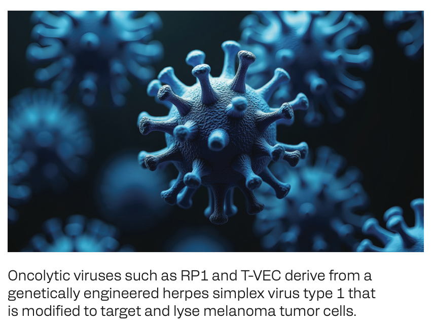
Lastly, intratumoral oncolytic viruses have introduced new therapeutic approaches that circumvent the challenges faced with therapeutic resistance. Talimogene laherparepvec (T-VEC) is an oncolytic virus built on a herpes simplex virus type 1 (HSV-1) backbone. T-VEC is the first FDA-approved oncolytic virus for advanced melanoma, producing durable response rates of 16.3% in the OPTiM trial (NCT00769704).14 RP1 (vusolimogene oderparepvec), another HSV-1–derived virus, has also shown promising results in the phase 2 IGNYTE trial (NCT03767348) when given in combination with nivolumab, regardless of prior PD-1 exposure or resistance. The study demonstrated an ORR of 33.6%, including a complete response (CR) rate of 15% and a generally mild toxicity profile.15 Additionally, the multicenter phase 3 IGNYTE-3 trial (NCT06264180) is actively underway, evaluating the safety and efficacy of RP1 combined with nivolumab vs treatment of physician’s choice in patients with advanced melanoma who progressed on anti–PD-1 and anti–CTLA-4 therapy.16


Oncolytic viruses such as RP1 and T-VEC derive from a genetically engineered herpes simplex virus type 1 that is modified to target and lyse melanoma tumor cells.
2 Practical coordination of care is criticalin advanced melanoma.
Given the expanded complexity of the therapeutic arsenal used to treat advanced melanoma, reliance on interdisciplinary collaboration outside of the multidisciplinary tumor board is paramount. By integrating the unique perspectives from interventional radiologists (IRs), medical oncologists, and surgeons alike, the optimal therapy can be better tailored to meet the unique needs of each patient.The emergence of intratumoral immunotherapies—particularly oncolytic viruses such as T-VEC and RP1—has ultimately redefined the IR’s role in treating patients with advanced melanoma. Oncolytic viruses and vaccine-based immunotherapies have become the leading focus of clinical research in advanced melanoma, outnumbering trials for all other therapeutic classes.17 Intratumoral injections offer a promising alternative and/or adjunct to systemic immunotherapies, bypassing issues with poor drug penetration and resistance, while also mounting systemic immune responses against tumors elsewhere in the body.15,17,18
Eligibility for intratumoral injection depends on several lesion-specific factors, with accessibility and safety being paramount. Ideal lesions are generally greater than or equal to 1 cm for solid tumors or greater than or equal to 1.5 cm for lymph nodes, with larger lesions often exhibiting higher metabolic activity on PET imaging as well. Pre- and intraprocedural imaging, such as diffusion-weighted and contrast-enhanced studies, are indispensable for evaluating lesion architecture and tumor microenvironment, thereby guiding the safest and most effective treatment plan.17 Imaging modalities such as CT and MRI aid in lesion mapping, while ultrasound and CT fluoroscopy provide real-time procedural guidance for needle placement and identifying areas of tumor necrosis.17
Compared with biopsies, intratumoral injections are straightforward, using smaller-gauge needles (21- to 23-gauge) for deeper targets and those as small as 30-gauge for superficial skin lesions.17 While familiar to IRs, the injection technique must account for tumor density and internal pressure, as these influence drug dispersion. In general, a commonly employed injection technique uses a single, end-hole needle and a fanning motion.17 Furthermore, tumor characteristics such as stromal density and proximity to vital structures may present challenges. For dense or fibrous tumors, the peripheral edge is often targeted for injection to ensure uniform drug distribution.


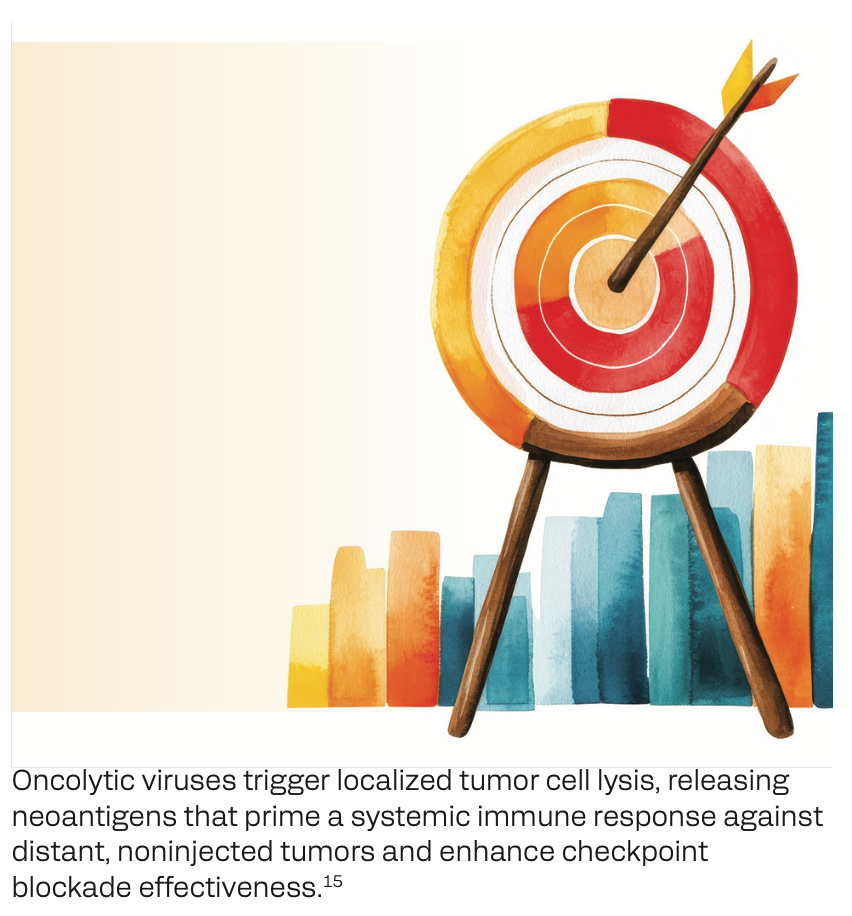
Oncolytic viruses trigger localized tumor cell lysis, releasing neoantigens that prime a systemic immune response against distant, noninjected tumors and enhance checkpoint blockade effectiveness.15
Procedure documentation is also critical for ensuring smooth continuity of care. Having a standardized reporting protocol enables subsequent IRs to identify the optimal lesions for injection in future treatment sessions.17 Postinjection follow-up typically includes imaging surveillance and monitoring for adverse events. Most oncolytic viral therapies, including RP1, are well tolerated, with minimal systemic toxicity. Localized inflammation or mild injection-site reactions may occur but are typically self-limiting.17
3 Oncolytic virus-based therapeutic approaches for melanoma continue to expand.
Oncolytic viruses have emerged as a transformative strategy in the treatment of advanced melanoma, especially for patients who have progressed on ICIs.Agents such as T-VEC and RP1 exemplify the practicality of administering intratumoral therapies to stimulate systemic immune responses.14,15 Looking closer at RP1, it is engineered to express granulocyte-macrophage colony-stimulating factor (GM-CSF) and a fusogenic protein, promoting immunogenic cell death and enhancing lymphocyte infiltration of the tumor microenvironment (TME). Combining oncolytic viruses with ICIs capitalizes on their complementary mechanisms—oncolytic viruses prime the immune system through the release of tumor neoantigens, while ICIs remove the brakes on the immune system, enhancing cytotoxic T-cell activity.15
Regarding the transmissibility of herpetic infections after RP1 injections, a study by Bommareddy and colleagues showed this to be safe for patients, caregivers at home, and medical staff administering treatments. Swab samples that tested positive for the presence of a virus showed no evidence of RP1 DNA, alleviating concerns about viral shedding and caregiver transmission.19 Furthermore, oncolytic viral injections may not be considered safe in select patients, such as those with immunocompromising conditions such as active herpetic infections, hematological malignancies, HIV, or an active pregnancy.20,21


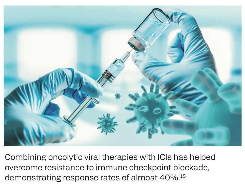
Combining oncolytic viral therapies with ICIs has helped overcome resistance to immune checkpoint blockade, demonstrating response rates of almost 40%.15
Looking ahead, several emerging therapies are demonstrating encouraging results in early clinical trials for advanced melanoma. In the neoadjuvant setting, the ongoing phase 2 HCC 22-138 trial (NCT06216938) is evaluating the ability of RP1 to stimulate immune responses in sentinel lymph nodes, potentially preventing melanoma recurrence following surgery.22 Meanwhile, RP2 (a derivative of RP1) incorporates an anti–CTLA-4–like molecule, further amplifying the immune response with intratumoral injection. RP2 is currently under investigation alone and in combination with nivolumab in a study evaluating patients with advanced uveal melanoma.23
Beyond oncolytic viruses, T-cell receptor–based therapies, such as IMA203, are expanding therapeutic possibilities. Targeting preferentially expressed antigens in melanoma (PRAME), a melanoma- associated antigen, IMA203 demonstrated an ORR of 67% in a phase 1 trial involving heavily pretreated patients with PRAME- positive tumors.24 Together, these innovative therapies are reshaping the treatment landscape of advanced melanoma and expanding the clinical toolkit to overcome resistance and further improve clinical outcomes for these patients.
Key References
6. Wolchok JD, Chiarion-Sileni V, Rutkowski P, et al. Final, 10-year outcomes with nivolumab plus ipilimumab in advanced melanoma.N Engl J Med. 2025;392(1):11-22. doi:10.1056/NEJMoa2407417
15. Wong M, Sacco J, Robert C, et al. Efficacy and safety of RP1 combined with nivolumab in patients with anti–PD-1–failed melanoma from the IGNYTE clinical trial. J Clin Oncol. 2024;42 (16 suppl):9517. doi:10.1200/JCO.2024.42.16_suppl.9517
17. Sheth RA, Wehrenberg-Klee E, Patel SP, Brock KK, Fotiadis N, de Baère T. Intratumoral injection of immunotherapeutics: state of the art and future directions. Radiology. 2024;312(1):e232654. doi:10.1148/radiol.232654
For FULL reference list, visit https://www.gotoper.com/sir25melanomaoncolytic-postref
CME Posttest Questions
1 In the IGNYTE study, which evaluated the combination of the oncolytic virus RP1 and nivolumab in patients with melanoma and prior progression on anti–PD-1–based therapy, which of the following was shown?
A. Favorable objective response rate (ORR) (33.6%), with no complete responses (CRs)
B. Favorable ORR (33.6%), with nearly half of these being CRs
C. Low ORR (12.3%), but a high disease control rate (80.5%)
D. Most patients had progressive disease as a best response.
2 Your 59-year-old patient with unresectable, metastatic cutaneous melanoma is being considered for intratumoral immunotherapy. He has 6 lesions that measure from 1.3 to 2.1 cm in diameter, with superficial tumors located on his upper back. He is not on anticoagulant therapy, and his platelet count is 50 × 103/µL. Which of the following parameters confirms this patient’s candidacy when assessing eligibility for intratumoral injection?
A. Anatomic location of tumor on his back
B. Anticoagulation status and bleeding risk
C. Platelet count
D. Size and quantity of accessible tumors at least 1 cm in diameter
3 According to recent data, which of the following classes of agents has the greatest number of planned or active early-phase clinical trials for intratumoral immunotherapy?
A. Immune checkpoint inhibitors
B. Nucleic acids
C. Oncolytic viruses/vaccines
D. Pattern recognition receptor agonists
Claim Your CME Credit at https://www.gotoper.com/sir25melanomaoncolytic-postref
To learn more about this topic, including information on multidisciplinary management strategies for patients with advanced melanoma, go to https://www.gotoper.com/sir25melanomaoncolytic-activity
CME Provider Contact information
Physicians’ Education Resource®, LLC
2 Commerce Drive, Suite 110, Cranbury, NJ 08512
Toll-Free: 888-949-0045
Local: 609-378-3701
Fax: 609-257-0705
info@gotoper.com


![A third of patients had a response [to lifileucel], and of the patients who have a response, half of them were alive at the 4-year follow-up.](/_next/image?url=https%3A%2F%2Fcdn.sanity.io%2Fimages%2F0vv8moc6%2Fcancernetwork%2F6b7c9a3270c71a70749ba86000cfc78a29d74309-2988x1702.png%3Ffit%3Dcrop%26auto%3Dformat&w=3840&q=30 "A third of patients had a response [to lifileucel], and of the patients who have a response, half of them were alive at the 4-year follow-up.")


