Automated System Improves Accuracy of HER-2 Scoring
SAN ANTONIO-By using a system combining color-based imaging and automated microscopy, pathologists were able to significantly improve their accuracy in evaluating HER-2 protein expression in breast cancer tissue, said Kenneth Bloom, MD, of Rush-Presbyterian-St. Luke’s Medical Center, Chicago.
SAN ANTONIOBy using a system combining color-based imaging and automated microscopy, pathologists were able to significantly improve their accuracy in evaluating HER-2 protein expression in breast cancer tissue, said Kenneth Bloom, MD, of Rush-Presbyterian-St. Luke’s Medical Center, Chicago.
In a study presented at the 23rd Annual San Antonio Breast Cancer Symposium, 10 pathologists evaluated slides from 129 patients with invasive breast cancer for HER-2 expression using manual immunohistochemistry (IHC) and fluorescence in situ hybridization (FISH). The IHC analysis was then repeated using ChromaVision Medical System’s automated cellular imaging system (ACIS). More than 1,250 staining intensity scores were evaluated.
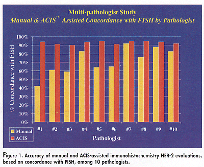
The pathologists’ previous experience in evaluating HER-2 status ranged from 2 to 3 analyses a week to more than 50 analyses a day. This range of experience was reflected in their accuracy in manual IHC scoring, which ranged from 42% to 92%, based on concordance with FISH (Figure 1). Scoring accuracy improved to a range of 91% to 95% using ACIS-assisted IHC, Dr. Bloom reported.
With use of ACIS, the pathologist who was the least accurate manually (42%) improved to an accuracy equal to that of the most accurate manual reader (92%) (see Figure 1). "Everybody becomes as good as the expert with ACIS," he said.

Using ACIS benefits the pathologist by establishing consistency and uniformity. "The ACIS system can virtually eliminate interlaboratory variability and allow for consistent and reliable interpretation of IHC results among pathologists and laboratories," Dr. Bloom said. Agreement among the 10 pathologists was 72% using manual IHC, which improved to 95% with ACIS-assisted IHC. "Any pathologist evaluating the same slide will get the same result," he said.
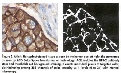
With an IHC-based test, like HercepTest (DAKO, Carpinteria, California), which was used in the study, the pathologist must analyze the amount and intensity of the color stain. However, the human eye is limited in its ability to distinguish various hues of the same color. ACIS, by comparison, is designed to identify minute differences between cells. It can distinguish up to 256 levels of intensity of a single color, compared to 4 levels discernible by the human eye, according to ChromaVision (Figure 2).

The ACIS system scans an area of a slide selected by the pathologist and shows that area at high power on a monitor. The system uses color criteria and pattern recognition software to identify the cells that overexpress HER-2, and then it reports the score to the pathologist.
Breast cancer patients whose tumors overexpress HER-2 are candidates for trastuzumab (Herceptin), a monoclonal antibody that binds to HER-2 receptors and slows tumor growth.
The HercepTest is scored manually from 0 to 3+ (0 to 1+, negative; 2+, weak positive; and 3+ strong positive). The cut-off point for determining trastuzumab treatment in the United States is 2+. Thus, accurate readings of slides for HER-2 expression are important because they affect the course of therapy for the patient, Dr. Bloom said.
While FISH also produces a quantitative measurement, it measures copies of the HER-2/neu gene rather than the expression of HER-2/neu protein. It is also more costly and complex to use than ACIS, according to ChromaVision.
The ACIS software can detect one abnormal cell among more than 100 million normal cells, ChromaVision said. The technology had its origins in the US military’s Star Wars missile defense program, where it was used to distinguish between active nuclear warheads and decoys. When the military spun off the technology, ChromaVision (San Juan Capistrano, Calif) acquired the patent rights.
ACIS, approved by the FDA in 1999, has been available for HER-2 testing for about 1 year. The technology is also being used to measure estrogen and progesterone receptors, Ki-67, and p53, and to evaluate cancer micrometastases in bone marrow, lymph nodes, and other tissue.
ChromaVision supplies the technology to labs on a fee-per-use basis. Clients receive the entire computerized microscopy system, but ChromaVision retains ownership of the system itself.