Company Offers Computer-Aided Detection of Breast Cancer
WESTLAKE VILLAGE, Calif-The first computer-aided detection (CAD) system for mammography (Image-Checker from R2 Technology) received FDA approval only 2 years ago, and, to date, only a handful of institutions and mammography centers offer the service on site.
WESTLAKE VILLAGE, CalifThe first computer-aided detection (CAD) system for mammography (Image-Checker from R2 Technology) received FDA approval only 2 years ago, and, to date, only a handful of institutions and mammography centers offer the service on site.
Larry Chespak, MD, a California radiologist who early on saw the technologys potential, established a new company, iMammogram.com Inc., last year to provide CAD directly to women seeking a CloserLook, the companys trademarked name for its service.
CAD is not a diagnostic service. Its simply a tool, a spell-checker, to help radiologists detect up to 20% more cancers during routine screening mammography, Dr. Chespak said in an interview with ONI. Theres no magic here for doctors. CAD wont turn a bad mammog-rapher into a good one, but it will turn a good mammographer into a better one, he said.
Partnerships With Centers
Women can order the service directly from the companys website (found at www.iMammogram.com), but the companys goal is to establish partnerships with mammography centers. In an ongoing pilot program with several centers, radiologists at the centers offer CAD to all women receiving screening mammo-grams. The radiologists read the mam-mograms with the CAD printouts in front of them, he said.
Since CAD is not reimbursable by Medicare or private insurers, the patient pays iMammogram.com for the service and may also be charged a small handling fee by the mammography center.
The centers are getting no money from us; we feel that would be unethical, Dr. Chespak said. He also told ONI that the company would provide the service free of charge to low-income women, as determined by the imaging center.

We would like to be able to do every screening mammogram that comes through the center, he said. He added that high volume would allow radiologists at the centers to become familiar with CAD quickly, reducing the length of the learning curve.
When a woman requests CloserLook, the center sends her most recent screening mammogram study (four images) to iMammogram.com where the films are digitalized and scanned by the computer for suspicious areas.
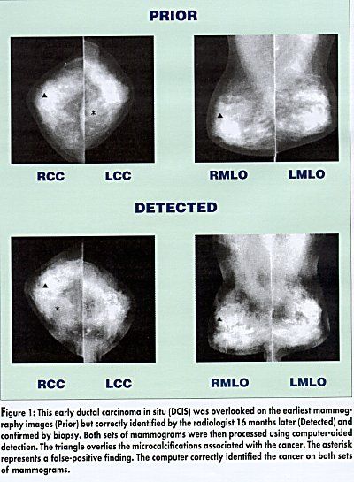
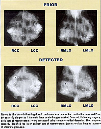
The ImageChecker looks at every pixel and does a billion calculations for each film, he said. It searches for clusters of bright spots suggestive of microcalcifica-tions, which it marks with a triangle, and for patterns of dense regions with spiculation suggestive of masses, which it marks with an asterisk (see Figures 1 and 2). The marks appear only on the CAD printout, not the original mammography film. The CAD printout is the patients actual breast, not a diagram of the breast, Dr. Chespak noted.
Sensitivity
In the studies of ImageChecker that led to its approval, he said, the sensitivity of the machine was 99% for microcalcifications (400 of 406 detected) and 75% for masses (506 of 677 detected). The latest algorithm, which all of our machines have (ImageChecker Version 2.2 Software Release), will pick up close to 90% of masses, he said.
Dr. Chespak calls his service especially timely in todays highly regulated medical environment, where radiologists must read films at a fast, cost-effective pace. Many imaging centers are losing money on mammograms, and there are places that are no longer doing them for that reason, he said.
Although at some academic institutions, radiology departments do double reads, its hard to rationalize having a second radiologist read every study, Dr. Chespak said. Statistically, CAD is essentially equal to a second read in improving detection rates. It saves time and money for the radiology center and, of course, benefits the patient.
Dr. Chespak said that the technology is gaining acceptance. Women are asking for it. Radiologists are showing it to their colleagues.

He expects that the cost of the service will come down as volume increases. His company currently has several Image-Checker machines (costing about $200,000 each) and plans to add several more by the end of the year, depending on volume. The actual scanning only takes about 6 minutes per case, but the technician must also spend some time entering the patients data.
Dr. Chespak stressed that iMammogram.com has taken several measures to ensure the security of patient data. Medical privacy is guaranteed. The images are identified with a bar code and stored on disks, away from disks containing the patient information, so the two cant be put together without the access codes.
Teleradiology
Dr. Chespak said that the company has signed a letter of intent with a leading PACS/ASP provider to develop a teleradiology system to allow us to transmit images electronically and also store images more efficiently. His company already offers digital archiving under the trademark ImageStore. Electronic transmission is currently being pilot tested, and he expects to be able to offer it to all centers within the next 6 months.
With this system, mammography films are scanned (digitized) at the radiology center and then electronically transmitted to iMammogram.com for CAD analysis. With this system, we will be able to take the turn around time from 2 days to 10 minutes, he said.
Film Archiving
Over the next 10 years, as more radiology centers acquire CAD technology, Dr. Chespak expects that iMammograms main role may lie in film archiving.
When diagnosing a patient, I sometimes look back at 10 years of mam-mograms. These things are heavy. Theyre cumbersome and expensive to store, taking up about a third of the space at most imaging centers. Theyre also susceptible to loss. I know of one hospital in Los Angeles that lost 25% of their images due to flooding, he said.
He said that his company could store all of a centers films on just a few optical disks, but that few centers are willing to do this because of the large initial investmentup to $5 or $6 millionfor conversion of back files.
Perhaps more important than saving storage space, film archiving has the potential to allow electronic transmission of mammography films between centers, and this is a long-term goal of iMam-mogram.com.
He explained that the low-resolution CAD readouts can be sent easily over the Internet and read on standard computer monitors, but that actual mammography films must be sent at a high resolution, requiring a very large band width for transmission and high-resolution monitors for viewing electronically.
Monitors a Problem
The band width will be there in a year, we think. No one is worried about that. Its the display that is the problem, Dr. Chespak said, adding that high-resolution monitors suitable for viewing mammography films cost upwards of $20,000. We know the technology eventually will be there to allow this, he said.
When radiologists can transmit actual x-ray films easily over the Internet and read them in their offices, he said, the need for film archiving services, such as that offered by iMammogram.com, will be even greater.