Compound Ultrasound Improves Assessment of Nodules
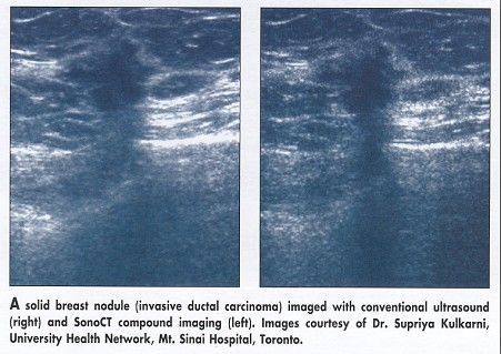
SEATTLE, Washington-Compound ultrasound imaging improves the evaluation of solid breast nodules and the retroareolar region of the breast, according to two studies from University Health Network, Mt. Sinai Hospital, Toronto, Canada. The studies were reported at the 101st Annual Meeting of the American Roentgen Ray Society.
SEATTLE, WashingtonCompound ultrasound imaging improves the evaluation of solid breast nodules and the retroareolar region of the breast, according to two studies from University Health Network, Mt. Sinai Hospital, Toronto, Canada. The studies were reported at the 101st Annual Meeting of the American Roentgen Ray Society.
Images From Various Angles
"In conventional ultrasound, each scan line hits the target object in a single scan plane," said Supriya Kulkarni, MD, a fellow at Mt. Sinai Hospital. "Real-time compound imaging utilizes multiple coplanar tomographic images obtained from various angles to produce a single compound image." The net result is a marked reduction of acoustic artifact and noise, she said.
In the first study (abstract 16), compound imaging with the SonoCT scanner system (ATL Ultrasound, Bothwell, Washington) was compared with conventional (fundamental) ultrasound imaging for the evaluation of solid breast nodules.
The study included 163 image pairs from 51 consecutive patients referred for routine breast sonography; each pair consisted of a conventional image and a compound image obtained with identical projection and compression, Dr. Kulkarni said.

Two blinded reviewers with no previous experience with compound imaging graded the features of each image individually and classified the nodules as benign, malignant, or indeterminate; these classifications were subsequently correlated with pathologic findings. Each image pair was also assessed side by side in a preference study.
"Compound imaging significantly reduced posterior artifact, both shadowing and enhancement, and improved visualization of retrolesional tissue," Dr. Kulkarni said. Shape, echodensity, echotexture, and homogeneity did not change with compound imaging, she added. In 58% of cases, the reviewers felt that the compound image permitted better overall evaluation of the nodules.
Overall, 22% of nodules were reclassified with compound imaging; 12% changed from indeterminate to benign, 4% from benign to indeterminate, and 6% from malignant to indeterminate.
"After reviewing the images, we found that loss of posterior artifact and improved margin resolution led to the changes in classification," Dr. Kulkarni said.
Conventional imaging still has a role in the evaluation of solid breast nodules, Dr. Kulkarni emphasized. "Preservation of posterior artifact is imperative for the accurate characterization of solid breast nodules. In fact, the loss of this artifact can be used to change the classification. Hence, we suggest that solid breast nodules are most optimally assessed using a combination of compound and fundamental imaging," she said.
Evaluating Retroareolar Tissue
In the second study (abstract 17), SonoCT compound imaging was compared with conventional ultrasound for the evaluation of the retroareolar tissue. This area of the breast is notoriously difficult to evaluate with current imaging modalities, including conventional ultrasound, said Derek Muradali, MD, a staff radiologist at Mt. Sinai Hospital.
"On ultrasound, the retroareolar region is difficult to assess because of shadowing from the nipple and the periareolar tissue," he noted.
The study included 126 image pairs of the retroareolar region from 40 consecutive patients referred for routine breast sonography. As in the first study, two reviewers graded features of each image individually and then compared the compound and conventional ultrasound images in a side-by-side preference study.
With compound imaging, the depth of shadowing improved in 73.8% of images, density of shadowing improved in 65.7% of images, duct wall morphology in 59.5%, lumen morphology in 48.4%, and morphology of parenchymal tissue and fat lobules in 69.8%, Dr. Muradali said.
The reviewers judged the overall anatomic detail to be better with compound imaging for at least 75% of the image pairs.
"Compound imaging improves visualization of the retroareolar region by decreasing tissue shadowing and increasing anatomic resolution," he said.
Future Research
Dr. Kulkarni concluded: "The future research now is to define the artifacts and explain their physical basis. This will permit imagers to become more familiar with the appearances of these artifacts and improve the interpretation of compound images."