Dynamic RODEO Images Show Laser Ablation of a Small Breast Tumor
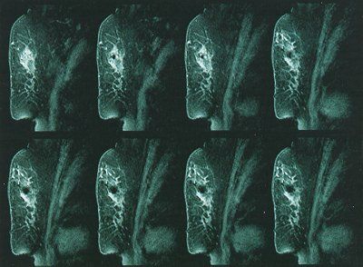
Serial dynamic RODEO (Rotating Delivery of Excitation Off-resonance) magnetic resonance images (top left to bottom right) generated at approximately 1- to 2-minute intervals during the course of interstitial laser photocoagulation treatment of a small breast cancer. Prior to laser treatment, pre- and post-gadolinium contrast RODEO images were generated to allow accurate placement of a needle tip into the lesion. A bare-tip laser fiber was then inserted into the needle and connected to the laser. The laser tip was pre-charred; then 3 watts of continuous power were applied for 10 minutes. The top left image is before initiation of laser treatment. The next image (to the right of the top left image) depicts the zone of pre-charring. As the laser ablation progresses, the hypointense zone increases in size. The final dynamic image on the bottom right shows an approximately 1.4 cm zone of ablation. Images courtesy of Dr. Steven E. Harms, professor of radiology, University of Arkansas for Medical Sciences.


Serial dynamic RODEO (Rotating Delivery of Excitation Off-resonance) magnetic resonance images (top left to bottom right) generated at approximately 1- to 2-minute intervals during the course of interstitial laser photocoagulation treatment of a small breast cancer. Prior to laser treatment, pre- and post-gadolinium contrast RODEO images were generated to allow accurate placement of a needle tip into the lesion. A bare-tip laser fiber was then inserted into the needle and connected to the laser. The laser tip was pre-charred; then 3 watts of continuous power were applied for 10 minutes. The top left image is before initiation of laser treatment. The next image (to the right of the top left image) depicts the zone of pre-charring. As the laser ablation progresses, the hypointense zone increases in size. The final dynamic image on the bottom right shows an approximately 1.4 cm zone of ablation. Images courtesy of Dr. Steven E. Harms, professor of radiology, University of Arkansas for Medical Sciences.


