Gated 4D PET/CT: Improving Radiation Treatment Planning for Patients With Lung Cancer
This special supplement to Oncology News International comprises expertcommentary and selected reports from the 2004 meetings of RSNA andASTRO about new imaging techniques, with a focus on state-of-the-art magneticresonance imaging, positron emission tomography, computed tomography,and complementary modalities for improving the diagnosis, staging, andtreatment of a variety of cancers. Evident in these reports is the increasingcollaboration between the specialties of radiation oncology and diagnosticradiology as imaging technology continues to evolve.
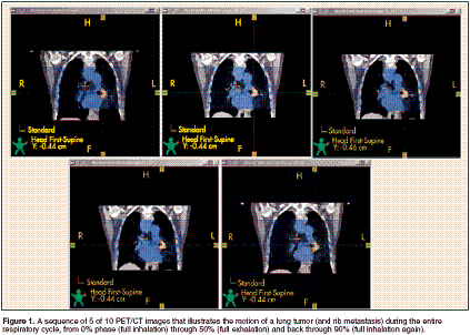
TEANECK, New Jersey-Respiratorygating during positron emissiontomography (PET) and computedtomography (CT) can reduceuncertainties about tumor location,thereby improving radiation treatmentplanning for patients with lung cancer,according to a pilot study presentedat the 46th Annual Meeting of theAmerican Society for Therapeutic Radiologyand Oncology (abstract 1056)."We have been using PET/CT atour facility for about 3 years now, sowe are very comfortable using functionalimaging with PET in treatmentplanning for radiation oncology," saidlead author Allan J. Caggiano, MS, amedical physicist at Holy Name Hospital,Teaneck, New Jersey. "We decidedto try to extend this technologyto include imaging the patients whilethey are breathing, trying to eliminatesome of the errors associated withmoving tumors under respiration," hetold ONI in an interview.Two-Day ProtocolEighteen patients with lung cancerhave participated in the 2-day protocol.On the first day, an immobilizationdevice was made, patients weretrained in regular breathing, and baselinemeasures were obtained; on thesecond day, ungated and gated (4D)PET/CT images were obtained, thelatter during coached breathing.Treatment plans were generatedfrom both ungated and gated PET/CTs. Tumor motion on gated PET/CTs was assessed during 10 phases ofrespiration, ranging from one full inhalationto the next (see Figure 1), andwas classified as low (< 1.0 cm), medium(1.0 to 2.0 cm), or high (> 2.0 cm).

Overall, 15 (83%) of the 18 patientscompleted the rigorous imagingprotocol. The gated PET and CTimages were successfully fused for eachof the 10 respiratory phases, and thephases were combined into a movieloop.Analyzing Tumor MotionAnalysis showed that tumor centroidsmoved 0 to 3.0 cm duringbreathing, and one third of patientseach had tumors with high, medium,and low motion. This information isuseful because patients who fall intothe low-motion group probably donot need any kind of gated treatment,Mr. Caggiano noted. Not surprisingly,most of the tumor motion occurredin the superior-inferior direction.Clinicians reported that having thegated PET/CT movie loops helped inplanning radiation therapy and identifyingpatients who were good candidatesfor respiratory-gated radiationtherapy. "We found that at least about50% of the patients benefited fromour knowing ahead of time what themovement was so we could establishthat in our treatment margins," Mr.Caggiano said. "This is a very differentapproach from what we have done inthe past, which was to look at how wethought the tumor was moving in generalfor people and then just [expand]the margins uniformly in all directions,probably overirradiating normallung tissue."The tailored margins may, in turn,reduce the adverse effects of radiationtherapy, according to Mr. Caggiano.Acknowledging the small study size,he noted that "patients who have gonethrough this protocol seem to havemuch less respiratory compromisegoing out from radiation therapyonward-a good indicator that this isprobably a good technique to use forthem."A comparison of the treatmentplans generated from the ungated andgated PET/CTs revealed that gatingalso helped to ensure that all of thetumor was targeted, especially edgeslying in the main direction of motion,Mr. Caggiano commented. "Even withgood standard margins on the ungatedfield, there are cases when the tumormoves a lot and traverses in andout of the path of the ray...So youwould be missing superior and inferiorsegments of the tumor and the patientwould get less dose," he said.According to Mr. Caggiano, the investigatorsplan to continue developingtheir 4D PET/CT process. One areathat they will be looking at in particularis the consistency of tumor motionfrom day to day during treatment forlung cancer. "PET/CT is a field ripe forresearch right now. There are manyadjustments that could be made tomake it better," he concluded.