Unexpected N2 Lymph Node Involvement Found During Surgery for Early-Stage NSCLC
During investigation of an episode of self-limiting abdominal pain, a 63-year-old Caucasian female never-smoker was found to have an asymptomatic right lower lobe pulmonary mass. A positron-emission tomography/computed tomography (PET/CT) scan revealed the right lower lobe mass to be 25 × 32 mm with a standardized uptake value (SUV) of 10.2, without evidence of hilar or mediastinal lymphadenopathy or of distant metastases.
SECOND OPINION
Multidisciplinary Consultations on Challenging Cases
The University of Colorado Health Sciences Center holds weekly second opinion conferences focusing on cancer cases that represent most major cancer sites. Patients seen for second opinions are evaluated by an oncologist. Their history, pathology, and radiographs are reviewed during the multidisciplinary conference, and then specific recommendations are made. These cases are usually challenging, and these conferences provide an outstanding educational opportunity for staff, fellows, and residents in training.
The second opinion conferences include actual cases from genitourinary, lung, melanoma, breast, neurosurgery, and medical oncology. On an occasional basis, ONCOLOGY will publish the more interesting case discussions and the resultant recommendations. We would appreciate your feedback; please contact us at second.opinion@uchsc.edu.
E. David Crawford, MD
Al Barqawi, MD
Guest Editors
University of Colorado Health Sciences Center
Univeristy of Colorado Cancer Center Denver, Colorado
During investigation of an episode of self-limiting abdominal pain, a 63-year-old Caucasian female never-smoker was found to have an asymptomatic right lower lobe pulmonary mass. A positron-emission tomography/computed tomography (PET/CT) scan revealed the right lower lobe mass to be 25 × 32 mm with a standardized uptake value (SUV) of 10.2, without evidence of hilar or mediastinal lymphadenopathy or of distant metastases.
Additional Case Background
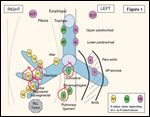
FIGURE 1

Lymph Node Stations Within the Thorax
The patient did not have a biopsy of the primary lesion prior to surgery. In an outside facility, a right lower lobectomy via open thoracotomy and selected lymph node staging biopsies were performed. Pathology revealed a 2.3-cm moderately differentiated adenocarcinoma, with negative surgical margins. Lymph node examination revealed malignancy involving one right lower lobe peribronchial lymph node, one subcarinal lymph node, one of two bronchus intermedius lymph nodes, and no disease in a sampled right middle lobe bronchial node, or in an inferior pulmonary vein lymph node (Figure 1). A magnetic resonance imaging (MRI) scan of the brain showed no evidence of intracranial metastases. The tumor was pathologically staged as pT1, N2, M0, or stage IIIA.
Overall, the patient tolerated the surgery well. It is now 5 weeks since her operation. She has already seen one oncologist, who proposed giving her four cycles of carboplatin and paclitaxel as adjuvant chemotherapy. She has come to the University of Colorado Thoracic Oncology Program seeking a second opinion on her management. Her Eastern Cooperative Oncology Group (ECOG) performance status is zero, and she has no significant past medical history.
Discussion
Pathology
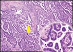
FIGURE 2

Papillary Adenocarcinoma Seen on Histology
Dr. D. Ross Camidge (Medical Oncologist): Dr. Franklin, can you review the patient’s pathology?
Dr. Wilbur Franklin (Pathologist): The right lower lobe specimen containing a 2.3-cm mass was examined. The mass proved to be a well-differentiated papillary adenocarcinoma (Figure 2) consistent with a primary lung cancer. It is increasingly well established that lung adenocarcinomas are heterogeneous in their histologic appearances. One pattern that is the object of ongoing evaluation is referred to as “true papillary.”[1] In this histologic pattern, the usual concave contours of the alveolar surfaces are replaced with irregular, complex vascularized projections (papillary structures) lined by multilayered epithelium. This pattern must be present in > 75% of the cross-sectional area of the tumor to qualify as true papillary.
A second, more common “micropapillary” pattern has been recently described, consisting of avascular cellular tufts projecting from alveolar surfaces.[2] Together these two subtypes occur in up to 37% of lung adenocarcinomas and are more likely to harbor activating mutations in the epidermal growth factor receptor (EGFR) than other adenocarcinomas.[3]
Dr. Scott Kono (Medical Oncology Fellow): Can you comment on the molecular diagnostic panel run on the tumor specimen?
Dr. Franklin: A number of different molecular tests particularly associated with sensitivity or resistance of non–small-cell lung cancer (NSCLC) to EGFR tyrosine kinase inhibitors have been explored in the past few years.[4] As part of our standard molecular diagnostic panel at the University of Colorado, we now evaluate all NSCLCs for EGFR protein expression by immunohistochemistry (IHC), EGFR gene copy number increases by fluorescence in situ hybridization (FISH), and EGFR and KRAS mutational status by direct sequencing techniques. This patient strongly expressed EGFR in nearly all tumors cells, and had a high EGFR copy number (high polysomy) by FISH. Specific exon sequencing for somatic mutations revealed wild-type KRAS (exon 1) and EGFR (exons 19-21).
Although the significance of the different analytic techniques and test results continues to be debated, the most clinically significant test in an untreated patient currently relates to the presence or absence of an activating mutation in the EGFR. Such activating mutations have been associated with dramatically increased sensitivity to EGFR tyrosine kinase inhibitors such as erlotinib (Tarceva) in the advanced disease setting, as well as with an overall improved prognosis.[5] Activating EGFR mutations such as exon 19 deletions are more common in adenocarcinomas than in other NSCLC variants, especially the papillary subtype. However, the fact that an activating EGFR mutation is not exhibited in this patient reinforces the idea that histologic and demographic factors are only guides to the likelihood of such sequence abnormalities being present. They are not a replacement for molecular testing.
Surgical Management
Dr. Camidge: Dr. Weyant, can you comment on whether a standard approach exists for lung and mediastinal lymph node (MLN) sampling in relation to surgery for lung cancer?
Dr. Michael Weyant (Thoracic Surgeon): MLN sampling plays a critical role in evaluating NSCLC, as nodal status has direct therapeutic and prognostic significance. Unfortunately, no single standard exists among surgeons with regard to sampling mediastinal lymph nodes. In a recent American College of Surgeons survey, only 27% of lung cancer patients underwent preoperative mediastinoscopy, and only 58% had MLNs removed at the time of resection.[6] The rates of MLN sampling differed significantly among centers, with academic centers having the highest rate, followed by community comprehensive cancer centers, and then community cancer centers.
Dr. Kono: Can you describe the different operative approaches used in evaluating MLNs?
Dr. Weyant: The gold standard in evaluating NSCLC for MLN metastases is cervical mediastinoscopy, at which time stations 2L, 2R, 4L, 4R, and 7 are sampled (Figure 1). Video-assisted thoracoscopic surgical (VATS) procedures may be used to access additional lymph node basins and augment the cervical mediastinoscopy.[7] Ultrasound-guided transbronchial or transesophageal lymph node biopsies are also potentially useful new techniques.[8] Cervical mediastinoscopy can be performed either as a separate procedure or during the same surgical setting with the use of frozen sections, to be reviewed by an experienced pathologist.
Dr. Kono: This patient’s preoperative PET/CT scan showed no evidence of mediastinal or hilar lymphadenopathy. Although the technology is continually evolving, preoperative PET/CT scans have recently been reported to have a sensitivity and specificity of 85% and 80%, with a negative predictive value of 95% in evaluating mediastinal disease.[9] Dr. Weyant, given the increasing use of preoperative PET/CT scanning and its high negative predictive value, how would you use this information to guide the timing and type of procedures to choose for assessing the mediastinum?
Dr. Weyant: One of the functions of MLN assessment is to determine in which patients a thoracotomy would be futile, due to the presence of multistation N2/N3 disease. In these situations, either radical radiotherapy or chemoradiotherapy would usually be considered the more appropriate approach. Although no definite rules exist, in the presence of a negative preoperative PET/CT scan, given that the chances of involved lymph nodes are low, it would be reasonable to avoid a second anesthetic procedure and to perform MLN assessments at the time of the primary lung surgery.
Lymph Node Sampling vs Dissection
Dr. Kono: Our patient did not undergo preoperative mediastinoscopy. Can you comment on MLN sampling at the time of surgical resection?
Dr. Weyant: Preresection mediastinoscopy with MLN assessment is critical in the operative setting, even with a negative preoperative PET scan. As a standard approach, we examine the lymph nodes obtained at mediastinoscopy by frozen section early on during the operative procedure. If the mediastinal nodes are positive we generally abort the surgery and await final pathologic review and discussion at our multidisciplinary tumor board.
A patient may have a VATS resection of a small previously undiagnosed lung nodule that turns out to be a primary lung cancer. In this setting, I would perform an ipsilateral VATS mediastinal lymph node sampling at the time of the VATS lobectomy. If there is concern or a need to obtain contralateral mediastinal nodes, a subsequent mediastinoscopy can then be performed.
Considerable evidence suggests that at the time of tumor resection, the more lymph nodes assessed, the better the outcome for patients with early-stage disease.[10,11] In part, this simply reflects accurate staging of N0 disease, but there is ongoing debate about the relative merits of purely prognostic mediastinal lymph node sampling (MLNS) vs the potentially additional therapeutic benefit offered by mediastinal lymph node dissection (MLND).[12]
MLNS has been defined as sampling nodes from stations 2R, 4R, 7, and 10R (Figure 1) in right-sided tumors, and stations 2L, 4L, 5, 6, 7, 10 L in left-sided tumors. Conversely, MLND has been defined in right-sided tumors as removal of all lymphatic tissue bound by the right upper lobe bronchus, innominate artery, the superior vena cava, and trachea. In left-sided tumors, the area is bound by the phrenic nerve, vagus nerve, aortic arch, and left mainstem bronchus. For tumors on either side, all lymphatic tissue at stations 7, 8, 9, 11, and 12 are removed.[13]
The major fear relating to inadequate MLN assessment is for stage migration, or basing treatment on a presumed clinical stage, when the pathologic stage is actually higher. MLNS or MLND alleviates this fear and allows for stage-appropriate treatment. A theoretic concern about MLND is a higher operative mortality compared to MLNS, due to the extensive nature of the procedure. However, multiple observational studies and randomized controlled trials have shown no significant difference in mortality rate between MLND and MLNS.
Dr. Kono: Do you feel this patient had “adequate” lymph node sampling?
Dr. Weyant: This patient did not have adequate lymph node sampling. Despite having a negative preoperative PET scan, a more thorough attempt at lymph node sampling should have been performed. The operative note describes several lymph nodes being sampled, specifically N1 lymph nodes at levels 11 and 12 and the most easily accessible N2 lymph nodes at levels 7 and 9. However, the important right paratracheal (level 4) and the contralateral level 4 areas were not sampled. It is important to note that approximately 30% of lower lobe tumors manifest mediastinal lymph node involvement without hilar involvement.[14]
Role of Postoperative Radiotherapy
Dr. Camidge: Dr. Gaspar, what is the role of postoperative radiation therapy in the treatment of this patient with apparent stage IIIA NSCLC, given the incomplete MLN sampling data? Dr. Laurie Gaspar (Radiation Oncologist): The role of postoperative radiation therapy (PORT) in N2, stage IIIA NSCLC remains controversial, as there is no clear evidence to guide therapy. In 1986, the Lung Cancer Study Group published a randomized study of PORT in completely resected stage II and III patients with squamous cell carcinoma. When analyzed for local recurrence, the rate declined from 41% to 3% in all node-positive patients. Despite this, stage II patients did not see an increase in survival with radiation therapy. However, there was a trend toward increased survival in patients with N2 disease.[15]
The controversy about PORT arose primarily after publication of a meta-analysis in 1998, whereby survival was decreased in stage I and II patients who received radiation therapy. However, a nonsignificant trend for increased survival was seen in the T2, N2 population.[16,17] Inferior radiation technology and technique have been proposed as potential reasons for the detrimental effect of PORT in some of these older studies.
More recently, the Adjuvant Nalvebine International Trialists Association (ANITA) study randomly assigned 840 patients to receive postoperative cisplatin/vinorelbine or observation. PORT was not mandated in the study, but institutions had to declare themselves as either giving, or not giving, PORT. In a retrospective analysis, patients with N2 disease who received PORT did demonstrate a 5-year overall survival benefit-independent of chemotherapy-compared to those who did not receive PORT (47% vs 34% in the chemotherapy arm, and 21% vs 17% in the no chemotherapy arm).[18,19] Also, when the Surveillance, Epidemiology, and End Results (SEER) database was used to examine over 7,400 patients retrospectively, investigators found increased survival rates associated with N2 patients who received PORT as opposed to those who did not.[20]
PORT is not currently recommended for node-positive patients in guidelines published by the American Society of Clinical Oncology.[21] However, given advances in planning techniques, a general drift toward concentrating PORT only to the fields at highest risk (specifically ipsilateral as opposed to bilateral mediastinum), and the SEER and ANITA data, I routinely discuss the pros and cons of PORT with these patients. I eagerly await the results of a phase III randomized European trial employing modern radiotherapy techniques that is currently reexploring the value of PORT in NSCLC patients.
Role of Adjuvant Chemotherapy
Dr. Kono: Dr. Camidge, given that the initial opinion the patient received focused on adjuvant chemotherapy, can you comment on the role of adjuvant chemotherapy in this setting? Dr. Camidge: The role of three to four cycles of adjuvant cisplatin-based chemotherapy to increase the cure rate in fit patients with resected stage IIIA NSCLC is well established.[22] Current controversies relate to the benefit in earlier stages of disease (stage IA/IB), to which drugs to use in unselected populations, and to the role, if any, of additional molecular testing of the resected tumor, in helping to guide which patients should receive which drugs.
The initial oncology opinion in this case recommended a carboplatin-based doublet for adjuvant therapy. All of the positive adjuvant trials have used cisplatin-based regimens.[23] The only adjuvant trial to assess carboplatin and paclitaxel was the Cancer and Leukemia Group B (CALGB) 9633 trial in stage IB disease. This trial failed to show a survival advantage from the chemotherapy, although whether this truly reflects the effect of carboplatin (as opposed to the stage of disease assessed or the small size of the study) remains controversial.[24]
In the current Intergroup ECOG 1505 study exploring the benefit of adding bevacizumab (Avastin) to adjuvant chemotherapy, only cisplatin doublets are being used, with several different partner drugs permissible within the doublet. Chemotherapeutic agents combined with cisplatin in this trial include vinorelbine, gemcitabine (Gemzar), pemetrexed (Alimta, for use in nonsquamous histology only), and docetaxel (Taxotere). Unfortunately, as stated in the ECOG 1505 protocol, patients with right-sided tumors are required to have had mediastinal node sampling of both levels 4 and 7; our patient is not eligible for this trial, given her inadequate mediastinal lymph node sampling.
Outside the constraints of a clinical trial, I tend to treat such patients with a cisplatin/docetaxel doublet, based on the TAX 326 trial in which cisplatin/docetaxel appeared marginally more effective than cisplatin/vinorelbine for advanced disease.[25] However, recent data in the advanced-disease setting have demonstrated that not all doublets are the same when histology is considered, specifically with regard to pemetrexed vs gemcitabine for adenocarcinomas.[26] A direct head-to-head comparison between a pemetrexed- and a taxane-containing doublet in adenocarcinoma patients has not yet been reported. I would discuss with this patient the different anticipated side effects of the two regimens and, in line with the ECOG 1505 schema, would certainly be agreeable to considering a pemetrexed/cisplatin doublet for her in the adjuvant setting.
Use of Biomarkers
Dr. Kono: Dr. Camidge, how would the results of our molecular diagnostic panel, or any other molecular tests you might suggest, affect your management in this case?
Dr. Camidge: The molecular biology and precise methods used to analyze and categorize NSCLC are becoming increasingly important in the management of this disease. A series of biomarkers associated with sensitivity or resistance to cytotoxic drugs within chemotherapy doublets are being explored as methods to select patients within both adjuvant and advanced-disease trials. Most prominently, the excision repair cross-complimentation group 1 (ERCC1) enzyme is known to play a central role in the nucleotide excision repair of DNA damaged by platinum agents.
As has been noted in previous advanced disease and adjuvant studies, positive ERCC1 expression in NSCLC assessed via either IHC or real-time quantitative polymerase chain reaction (PCR) has been associated with cisplatin resistance.[27-30] In a retrospective analysis of patients with material available for testing within the International Adjuvant Lung Cancer Trial (IALT), only those with ERCC1-negative tumors by IHC appeared to derive a survival benefit from adjuvant platinum-based chemotherapy (hazard ratio [HR] =0.65; 95%confidence interval [CI] = 0.50–0.86; P = .002).[27] However, the preferred prospective methodology and the optimal cutpoint for delineating positive vs negative ERCC1 findings remain to be determined, and these tests are not currently ready for routine clinical use. In contrast, biomarkers relating to EGFR have been far more extensively examined.[31,32] The ongoing RADIANT study is looking at the value of erlotinib vs placebo given instead of or after adjuvant chemotherapy for NSCLC. It is open to patients with stage IB to IIIA tumors that have been completely removed, where the tumor expressed EGFR positivity by IHC or increased gene copy number by FISH. The RADIANT protocol requires all patients to have had either a mediastinal node dissection or at least sampling of two mediastinal nodal stations (specifically, levels 4, 7, and 9 for right-sided tumors are strongly suggested). In contrast to the more strictly worded ECOG 1505 trial, the inclusion of level 9 as a valid N2 station would make our patient potentially eligible for this study.
Of note, neither the RADIANT study nor ECOG 1505 currently permit PORT. Should this patient decide on PORT after discussion with Dr. Gaspar, we would not pursue RADIANT enrollment but would recommend only four cycles of an adjuvant cisplatin-containing doublet followed by PORT, and then ongoing surveillance for recurrence.
Additional Surgery?
Dr. Kono: Dr. Weyant, given all the advantages you have outlined about the significance of adequate MLN assessment and the possibility of MLND being therapeutic as well as diagnostic, would you consider recommending a postlobectomy mediastinoscopy in this patient?
Dr. Weyant: What you are asking is: given what we already know, would the additional information obtained from MLNS-or the potential therapeutic benefit of MLND-justify the risks of an additional surgical procedure? The first thing to point out is that MLND is a more involved procedure than simple MLNS and is best performed at the time of thoracotomy. Full MLND is not feasible using just a cervical approach. With regard to the potential therapeutic role of MLND, it is unknown to what degree this would hold true following adjuvant chemotherapy and/or radiotherapy. Therefore, for this patient, from a purely surgical perspective, the strength of evidence probably doesn’t justify administering an additional anesthetic to dissect the mediastinum more fully after the fact.
Impact of Lymph Node Status on Radiotherapy
Dr Camidge: From a chemotherapeutic perspective, this woman clearly has N2 disease, and while knowledge of the involvement of additional lymph node stations may adjust our assessment of her personal prognosis, it will not alter the core recommendation for cisplatin-based adjuvant therapy. Radiotherapy might be a different story, however. Dr. Gaspar, would additional knowledge of her N2 and N3 lymph node status affect your recommendations with regard to PORT?
Dr. Gaspar: Based on information regarding patterns of locoregional failure postresection, I tend to administer PORT to the bronchial stump, ipsilateral hilum, ipsilateral paratracheal lymph nodes, and subcarinal regions.[33] Older methods employed more extensive fields including the bronchial stump, the ipsilateral hilum, the entire mediastinum, and sometimes the supraclavicular fossa, which, in conjunction with less advanced planning techniques, may have contributed to the deleterious effect of PORT in some of the original studies.
If we knew that this patient had more extensive microscopic N2 disease involving multiple ipsilateral stations, this wouldn’t necessarily change the radiation fields but it might alter the patient’s interpretation of the pros and cons of PORT. I suspect she may be more likely to agree to PORT, in light of such information. However, if she had proven microscopic N3 disease, the discussion regarding PORT or no PORT would probably be replaced by a strong recommendation for definitive chemoradiotherapy. The chances of microscopic N3 disease being present, given a negative preoperative PET/CT scan but biopsy-proven microscopic N2 disease, are unknown. They are probably small, given that the rationale underlying the use of ipsilateral mediastinal fields for PORT is that these fields represent the dominant sites of recurrence.[33]
In a study of 61 resected patients, 6 patients with right lower lobe tumors developed locoregional recurrence, and none of this group experienced a recurrence in the contralateral level 2 or 4 region. In contrast, left-sided tumors had a higher rate of contralateral recurrence. Larger but older studies of patterns of lymph node spread have suggested that in patients with right upper and right lower lobe tumors with known mediastinal involvement, contralateral disease is present in only 5% and 7% of patients, respectively.[34] Given that these studies were performed long before the availability of additional risk reduction with a known negative PET/CT scan, the risks in our patient are probably much less. While, it would be reasonable to at least discuss this with the patient, I wouldn’t argue very strongly for a mediastinoscopy at this point.
Summary
Dr. Camidge: To summarize, this patient is a relatively young, otherwise healthy female who is status post lobectomy. She has been diagnosed with stage IIIA NSCLC that is EGFR-positive by IHC and FISH, but EGFR and KRAS wild type. Optimal MLNS or MLND is ideally performed in the pre- and/or perioperative setting, but this did not happen in this case. While more extensive examination of the mediastinum after the primary operation could be undertaken-removing some of the unknowns about this patient’s true stage and prognosis-the relative risks and benefits of such an additional procedure in the setting of PET-negative microscopic level 7 and 9 disease probably do not justify such an approach.
Assuming the absence of additional information, the recommendation of the University of Colorado Clinical Thoracic Oncology program is to commence a course of four cycles of adjuvant cisplatin-based chemotherapy. The pros and cons of postoperative radiotherapy will be discussed with the patient. If she wishes to proceed with PORT, this will commence following the adjuvant chemotherapy. Due to the inadequate lymph node sampling, she would not be eligible for adjuvant therapy as part of the ECOG 1505 trial of cisplatin chemotherapy with or without bevacizumab, but if she declines PORT, she may be eligible for the RADIANT trial of erlotinib vs placebo following her adjuvant cisplatin-based therapy.
Financial Disclosure:The participants in this conference have no significant financial interest or other relationship with the manufacturers of any products or providers of any service mentioned in this article.
References:
1. Silver S, Askin F: True papillary carcinoma of the lung: A distinct clinicopathologic entity. Am J Surg Pathol 21:43-51, 1997.2. Miyoshi T, Satoh Y, Okumura S, et al: Early-stage lung adenocarcinomas with a micropapillary pattern, a distinct pathologic marker for a significantly poor prognosis. Am J Surg Pathol 27:101-109, 2003.3. Motoi N, Szoke J, Riely G, et al: Lung adenocarcinoma: modification of the 2004 WHO mixed subtype to include the major histologic subtype suggests correlations between papillary and micropapillary adenocarcinoma subtypes, EGFR mutations and gene expression analysis. Am J Surg Path 32:810-827, 2008.4. Capuzzo F: EGFR FISH versus mutation: different tests, different end-points. Lung Cancer 60:160-165, 2008.5. Ciardiello F, Tortora G: EGFR antagonists in cancer treatment. N Engl J Med 358:1160-1174, 2008.6. Little AG, Rusch VW, Bonner JA, et al: Patterns of surgical care of lung cancer patients. Ann Thorac Surg 80:2051-2056, 2005. 7. Massone PPB, Lequaglie C, Magnani B, et al: The real impact and usefulness of video-assisted thoracoscopic surgery in the diagnosis and therapy of clinical lymphadenopathies of the mediastinum. Ann Surg Oncol 10:1197-1202, 2003.8. Toloza EM, Harpole L, Detterbeck F, et al: Invasive staging of non-small cell lung cancer: A review of the current evidence. Chest 123:157Sâ166S, 2003.9. Lee B, Von Haag D, Lown T, et al: Advances in positron emission tomography technology have increased the need for surgical staging in non-small cell lung cancer. J Thorac Cardiovasc Surg 133:746-752, 2007.10. Doddoli C, Aragon A, Barlesi F, et al: Does the extent of lymph node dissection influence outcome in patients with stage I non-small cell lung cancer? Eur J Cardiothorac Surg 27:680-685, 2005.11. Ludwig MS, Goodman M, Miller DL, et al: Postoperative survival and the number of lymph nodes sampled during resection of node-negative non-small cell lung cancer. Chest 128:1545-1550, 2005.12. Lardinois D, Suter H, Hakki H, et al: Morbidity, survival, and site of recurrence after mediastinal lymph-node dissection versus systematic sampling after complete resection for non-small cell lung cancer. Ann Thorac Surg 80:268-274, 2005.13. Allen MS, Darling GE, Pechet TT, et al: Morbidity and mortality of major pulmonary resections in patients with early-stage lung cancer: Initial results of the randomized, prospective ASCOSOG Z0030 trial. Ann Thorac Surg 81:1013-1019, 2006.14. Riquet M, Assouad J, Bagan P, et al: Skip mediastinal lymph node metastasis and lung cancer: A particular N2 subgroup with a better prognosis. Ann Thorac Surg 79:225-233, 2005.15. Lung Cancer Study Group: Effects of postoperative mediastinal radiation on completely resected stage II and stage III epidermoid cancer of the lung. N Engl J Med 315:1377-1381, 1986.16. PORT Meta-Analysis Trialists Group: Postoperative radiotherapy in non-small-cell lung cancer: Systematic review and meta-analysis of individual patient data from nine randomised controlled trials. Lancet 352:257-263, 1998.17. Stephens RJ, Girling DJ, Bleehen NM, et al: The role of post-operative radiotherapy in non-small-cell lung cancer: A multicentre randomised trial in patients with pathologically staged T1-2, N1-2, M0 disease. Medical Research Council Lung Cancer Working Party. Br J Cancer 74:632-639, 1996. 18. Douillard JY, Rosell R, De Lena M, et al: Adjuvant vinorelbine plus cisplatin versus observation in patients with completely resected stage IBâIIIA non-small-cell lung cancer (Adjuvant Navelbine International Trialist Association [ANITA]): A randomised controlled trial. Lancet 7:719-727, 2006.19. Douillard JY, Rosell R, De Lena M, et al: Impact of postoperative radiation therapy on survival in patients with complete resection and stage I, II, or IIIA non-small cell lung cancer treated with adjuvant chemotherapy: The adjuvant Navelbine International Trialist Association (ANITA) Randomized Trial. Int J Radiat Oncol Biol Phys 72:695-701, 2008.20. Lally BE, Zelterman D, Colasanto JM, et al: Postoperative radiotherapy for stage II or III non-small-cell lung cancer using the surveillance, epidemiology, and end results database. J Clin Oncol 24:2998-3006, 2006.21. Pisters K, Evans W, Azzoli C, et al: Cancer Care Ontario and American Society of Clinical Oncology adjuvant chemotherapy and adjuvant radiation therapy for stages I-IIIA resectable nonâsmall-cell lung cancer guideline. J Clin Oncol 25:5506-5518, 2007.22. Weiss G, Camidge DR, Bunn P: From radiotherapy to targeted therapy: 20 Years in the management of non-small cell lung cancer. Oncology (Williston Park) 20:1515-1535 (incl discussion), 2006.23. Pignon JP, Tribodet H, Scagliotti GV, et al: Lung adjuvant cisplatin evaluation: A pooled analysis by the LACE Collaborative Group. J Clin Oncol 26:3552-3559, 2008.24. Strauss GM, Herndon JE, Maddau MA, et al: Adjuvant paclitaxel plus carboplatin compared with observation in stage IB non-small-cell lung cancer: CALGB 9633 with the Cancer and Leukemia Group B, Radiation Therapy Oncology Group, and North Central Cancer Treatment Group Study Groups. J Clin Oncol 26:5043-5051, 2008.25. Fossella F, Pereira J, Von Pawel J, et al: Randomized, multinational, phase III study of docetaxel plus platinum combinations versus vinorelbine plus cisplatin for advanced non-small-cell lung cancer: The TAX 326 study group. J Clin Oncol 21:3016-3024, 2003.26. Scagliotti GV, Parikh P, Von Pawel J, et al: Phase III study comparing cisplatin plus gemcitabine with cisplatin plus pemetrexed in chemotherapy-naive patients with advanced-stage non-small-cell lung cancer. J Clin Oncol 26:3543-3551, 2008.27. Olaussen KA, Dunant A, Fouret P, et al: DNA repair by ERCC1 in non-small-cell lung cancer and cisplatin-based adjuvant chemotherapy. N Engl J Med 355:983-991, 2006.28. Yu D, Zhang X, Liu J, et al: Characterization of functional excision repair cross-complementation group 1 variants and their association with lung cancer risk and prognosis. Clin Cancer Res 14:2878-2886, 2008.29. Zheng Z, Chen T, Haura E, et al: DNA synthesis and repair genes RRM1 and ERCC1 in lung cancer. N Engl J Med 356:800-808, 2007.30. Cobo M, Isla D, Massuta B, et al: Customizing cisplatin based on quantitative excision repair cross-complementing 1 mRNA expression: A phase III trial in nonâsmall cell lung cancer. J Clin Oncol 25:2747-2754, 2007.31. Eberhard DA, Johnson BE, Amler LC, et al: Mutations in the epidermal growth factor receptor and in KRAS are predictive and prognostic indicators in patients with non-small-cell lung cancer treated with chemotherapy alone and in combination with erlotinib. J Clin Oncol 23:5900-5909, 2005.32. Rosell R, Cuello M, Cecere F, et al: Usefulness of predictive tests for cancer treatment. Bull Cancer 93:E101-E108, 2006.33. Kelsey C, Light K, Marks L, et al: Patterns of failure after resection of nonâsmall-cell lung cancer: Implications for postoperative radiation therapy volumes. Int J Radiat Oncol Biol Phys 65:1097-1105, 2006.34. Nohl-Osler HC: An investigation of the anatomy of the lymphatic drainage of the lungs as shown by the lymphatic spread of bronchial carcinoma. Ann R Coll Surg Engl 51:157-176, 1972.