Minimizing Morbidity in Melanoma Surgery
Here we review some of the most significant changes in the surgical management of melanoma that have reduced morbidity and thereby improved patient outcomes.
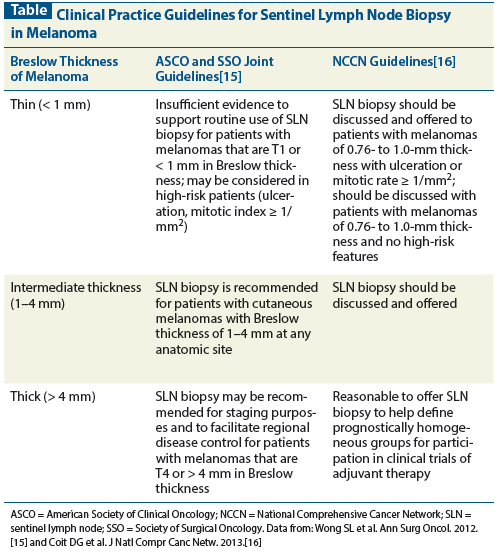
Table: Clinical Practice Guidelines for Sentinel Lymph Node Biopsy in Melanoma

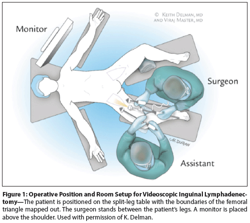
Figure 1: Operative Position and Room Setup for Videoscopic Inguinal Lymphadenectomy

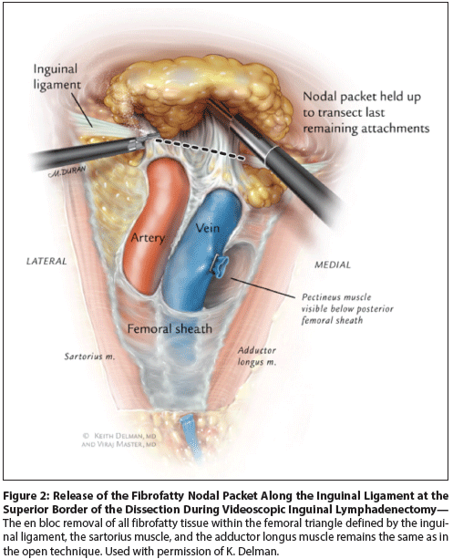
Figure 2: Release of the Fibrofatty Nodal Packet Along the Inguinal Ligament at the Superior Border of the Dissection During Videoscopic Inguinal Lymphadenectomy

The selection of a treatment modality involves a balance between risk and benefit. Surgical decision making is intrinsically dependent on potential morbidity; therefore, the desire to minimize adverse outcomes remains paramount in the effort to provide patients with the widest range of therapeutic options. The adoption of sentinel lymph node biopsy for the evaluation of regionally metastatic melanoma has reduced the number of complete lymphadenectomies and their attendant comorbidities. For patients who require completion lymphadenectomy, selective lymphadenectomy and, more recently, videoscopic inguinal lymphadenectomy have been shown to further reduce wound-related complications, while maintaining equivalent regional control and lymph node yield, respectively. Finally, in carefully selected patients laparoscopic metastasectomy can increase survival with less impact on quality of life than open extirpation. Ongoing trials, such as the Multicenter Selective Lymphadenectomy Trial II (MSLT-II), and research into gene profiling may improve the selection of patients for surgery. Obviating the need for surgery may offer the greatest reduction in morbidity of all.
Introduction
Surgical intervention is a Machiavellian endeavor: the ends justify the means. Conceptually, this mandates that we minimize the risk of the procedure as a justification for the gains offered by intervention. Progress in medical knowledge and technology, improvements in anesthesia, and our increased understanding of the biology of disease have all led to better selection of patients, better intraoperative management, and concomitant reductions in morbidity.
Despite significant advances in recent years in the treatment of melanoma, this disease still lags well behind many others in the options for systemic therapy for advanced disease. As a result, surgery remains a significant component in the armamentarium of the clinician who treats melanoma.
While surgery remains a “best choice” for many patients, and the standard of care for those with lymph node metastases, as many as 50% of patients who should receive surgical lymphadenectomy do not undergo this procedure.[1] A suggested motivation behind the failure to refer patients for appropriately indicated surgery is the concern about morbidity from the procedure. This is especially true for inguinal lymphadenectomy, which has reported rates of wound complications of more than 50% in most studies.[2-5]
During the past 2 decades, methods that have been implemented to reduce morbidity from the surgical management of melanoma have included (1) the use of sentinel node biopsy to avoid the need for completion lymphadenectomy, (2) novel applications of minimally invasive surgical techniques, and (3) improvement in the selection of patients for surgery. Here we review some of the most significant changes that have reduced morbidity and thereby improved patient outcomes.
Sentinel Lymph Node Biopsy
Cutaneous melanomas have specific patterns of lymphatic spread.[6] The status of regional lymph nodes is crucial to determining prognosis, controlling involvement of these nodes, and guiding further treatment decisions. Historically, complete dissection of the regional lymph node basin was performed via elective lymphadenectomy. However, 84% of patients with intermediate-thickness (1.2–3.5 mm) tumors do not have nodal involvement; thus, many of these patients may be subjected to unnecessary surgery and its attendant risks.[7]
Sentinel lymph node (SLN) biopsy is an approach to regional lymph node assessment that applies the principles of lymphatic spread to identify the first node or nodes involved within a lymph node basin. Introduced in 1992 by Morton et al,[8] this less invasive approach has transformed the care of patients with melanoma. In initial studies in which SLN biopsy was followed by completion lymph node dissection, the sentinel node was successfully identified in 75% to 90% of cases.[6,8] A large meta-analysis of over 25,000 patients showed the actual false-negative rate averaged 12.5%, decreasing with the proportion of patients whose nodal basin was successfully mapped.[9] Perhaps more important, the failure rate of a mapped nodal basin (in contrast to the false-negative rate) has been routinely reported to be 2% or less.[10,11]
The identification and status of the SLN is now considered the most important prognostic factor for patients with localized melanoma.[12] The Multicenter Selective Lymphadenectomy Trial (MSLT-I) is the largest study to evaluate the ability of SLN biopsy to affect prognosis and survival.[7] This study of 1,269 patients with intermediate-thickness (1.2–3.5 mm) melanomas randomly assigned patients to observation or SLN biopsy. The mean estimated 5-year disease-free survival rate for the biopsy group (78.3% ± 1.6%) was significantly higher than the rate for the observation group (73.1 ± 2.1%). In addition, among patients with nodal metastases, the 5-year survival rate was higher for those who underwent immediate lymphadenectomy (72.3 ± 4.6%) as opposed to delayed lymphadenectomy (52.4 ± 5.9%).
SLN biopsy is associated with fewer complications than regional lymphadenectomy. In the Sunbelt Melanoma Trial, at a median follow-up of 16 months the overall complication rate was significantly lower for SLN biopsy alone (5% vs 23% for SLN biopsy plus completion lymphadenectomy). The decreased rate of complications included wound infection, lymphedema, hematoma/seroma, and sensory nerve injury.[13] Recently published data from MSLT-I suggest even higher morbidity after completion lymphadenectomy following positive SLN biopsy.[14]
The American Society of Clinical Oncology (ASCO) and the Society of Surgical Oncology released joint clinical practice guidelines in 2012 on the use of SLN biopsy for patients with melanoma.[15] The Table summarizes current recommendations, including the 2013 National Comprehensive Cancer Network guidelines.[16]
Selective Lymphadenectomy
While data about selective lymphadenectomy are not widely published, the procedure has received increased attention of late. The interest in selective lymphadenectomy is mainly for cervical dissections, although more recently some authors have proposed a limited dissection in the axilla.[17] Most head and neck surgeons have largely abandoned radical neck dissection, and melanoma surgeons have followed a similar approach to cervical dissection, frequently leaving the parotid gland in patients whose tumors did not arise on the scalp or face. This selective approach is associated with lower morbidity and regional control similar to that of a more radical approach.
Videoscopic Inguinal Lymphadenectomy
Outside of clinical trials, inguinal lymphadenectomy is the standard of care for patients with melanoma who have clinically palpable lymphadenopathy or a positive biopsy of an SLN in the groin. Nevertheless, up to 50% of patients with positive SLN biopsies do not undergo completion lymphadenectomy.[1] This is probably because of the high morbidity associated with open inguinal lymphadenectomy. The complication rate after the procedure is 50% or higher; a large proportion of the complications are related to the groin incision, including infection, dehiscence, and skin flap necrosis.[2-5]
Videoscopic inguinal lymphadenectomy (VIL) has been developed as a minimally invasive approach to superficial groin dissection. The technique was first used by Bishoff in patients with penile cancer, and subsequent experience has shown reductions in wound-related morbidity [18-20].
On the basis of these results, Delman et al[21,22] extended and modified the procedure for melanoma dissection. This approach utilizes standard laparoscopic instrumentation and techniques to remove the inguinal nodal packet under videoscopic guidance. The standard three-incision VIL technique (Figure 1) has been described in detail elsewhere.[21] The en bloc removal of all fibrofatty tissue within the femoral triangle defined by the inguinal ligament, the sartorius muscle, and the adductor longus muscle remains the same as in the open technique (Figure 2). Additional tissue is removed as necessary; the tissue 5 cm onto the external oblique aponeurosis and any enlarged or pigmented lymph nodes are routinely resected.
Initial results of VIL in patients with melanoma have been reported.[22,23] Eighteen patients with a median primary Breslow depth of 2.8 mm (range, 0.6–9.9 mm) underwent VIL at Emory University Hospital.[22] In 74% of these patients, the indication was a positive SLN biopsy. Two of the procedures were converted to open dissection, owing to high end-tidal carbon dioxide levels in one patient and restricted hip mobility and anatomic uncertainty in the other. The median node count was 11 (range, 4–24), and the largest node removed was 5.6 cm.
Abbott et al[23] reported similar lymph node yields after VIL in patients with melanoma. When lymph node yield is used as an indirect measure of adequate oncologic resection, VIL appears similar to open superficial inguinal lymphadenectomy. Two-year follow-up data detailing oncologic outcomes in patients with melanoma have been submitted as an abstract, and the manuscript is in preparation.
Significantly fewer complications occur after VIL than after open superficial inguinal lymphadenectomy. In the largest reported series (N = 29), complications were noted in 42% of patients who underwent VIL; the majority were classified as minor.[24] This analysis used a comprehensive and broad definition of complication; seroma and lymphocele were included as significant complications, and a subjective definition of lymphedema was used. Minor complications included superficial wound infection (2.6%), seroma/lymphocele (12%), and mild to moderate lymphedema (12%). The most common major complication was readmission for IV antibiotic administration (10.5% of patients). Reduced complication rates translate into decreased length of hospitalization.[23] At present, the Emory investigators have completed more than 100 minimally invasive lymphadenectomies and are analyzing the data for publication.
Minimally Invasive Metastasectomy
Most cases of malignant melanoma are diagnosed at an early stage, during which resection may be curative. However, resection for disseminated disease is rarely curative because of widespread micrometastases. Carefully selected patients may still benefit from surgical intervention; 5-year survival rates after complete resection of metastatic gastrointestinal lesions range from 28% to 41%.[25] In contrast, median survival among those who undergo palliative or no resection ranges from 5.4 to 8.4 months.[26] Patient selection measures include adequate control of the primary site, ability to tolerate the planned surgery, and a metastatic pattern amenable to resection. In this setting, minimally invasive approaches to metastasectomy complement further goals of reducing morbidity and improving quality of life for patients with stage IV melanoma.
Intra-abdominal metastases are particularly amenable to laparoscopic removal. In a series comparing laparoscopic vs open adrenalectomy for adrenal metastases, including melanoma, no differences in local recurrence, disease-free interval, or overall survival were found. Patients who underwent laparoscopic resection had significantly shorter operative times, less estimated blood loss, shorter length of hospital stay (2.8 vs 8.0 days), and fewer total complications.[27] Numerous case reports detail the laparoscopic removal of melanoma metastatic to the spleen, small bowel, and gallbladder.[26,28,29] Further study is needed to confirm that oncologic outcomes are equal to those achieved with open surgery.
Respiratory failure is a frequent cause of death from metastatic melanoma and is another setting in which metastasectomy may provide benefit. In carefully selected patients (time to pulmonary metastases > 36 months and a single lesion) surgical removal of lung metastases can increase 5- and 10-year survival when complete resection can be accomplished.[30] A survey found that approximately 40% of thoracic surgeons use video-assisted thoracoscopic surgery rather than open thoracotomy for the resection of pulmonary metastases.[31] Controversy about this approach continues to exist because metastases may be missed as a result of constraints on bimanual palpation.[32]
Novel Considerations
The Multicenter Selective Lymphadenectomy Trial II (MSLT-II) potentially represents the next step in reducing surgical morbidity-and obviating the need for surgery altogether in selected populations.[33] About 70% to 80% of patients with micrometastases at the time of SLN biopsy will not have further lymph node involvement at completion lymphadenectomy.[34] MSLT-II aims to determine whether completion lymphadenectomy confers a melanoma-specific or disease-free survival advantage over observation with serial ultrasound examinations. This study stands to have a significant impact on the management of patients with regionally metastatic melanoma.
Further work, such as reverse transcription polymerase chain reaction (RT-PCR)–based gene expression profiling of SLN biopsy specimens to predict metastases, continues. Recently, results of genetic profiling of primary tumors, reported at the ASCO 2013 annual meeting, was shown to successfully classify primary lesions into high- and low-risk disease patterns.[35] This type of profiling may hold promise for other efforts in the genetic characterization of tumors and may even lead to an enhanced selection of patients for SLN biopsy. The approach with the least surgical morbidity is not to perform surgery at all.
Conclusion
Efforts at minimizing morbidity in melanoma surgery are ongoing. The desire to reduce wound complications related to open surgery has been a particularly potent catalyst for the development of less morbid and less invasive approaches, such as VIL. However, it is important to recognize that equivalent disease-specific outcome measures must be determined prior to widespread acceptance. Similarly, oncologic outcomes must be weighed against the additional benefits of decreased postoperative pain, reduced cardiopulmonary complications, and shorter hospitalizations in selected patients undergoing laparoscopic resection of distant metastatic disease.
SLN biopsy followed by completion lymphadenectomy for positive biopsy results has become the standard of care for patients with regionally metastatic melanoma. This approach was validated in a well-designed, prospective randomized trial,[7] which has undergone extensive analysis and thorough follow-up. This transformative technique is now being further analyzed to determine whether even completion lymphadenectomy can be avoided in selected patients. Genetic profiling and improved analytic techniques, used in both primary lesions and nodal metastases, may help determine which patients can avoid surgery altogether. Ultimately, this may represent the greatest step toward reducing surgical morbidity.
Disclosures:
The authors have no significant financial interest or other relationship with the manufacturers of any products or providers of any service mentioned in this article.
References:
1. Bilimoria KY, Balch CM, Bentrem DJ, et al. Complete lymph node dissection for sentinel node-positive melanoma: assessment of practice patterns in the United States. Ann Surg Oncol. 2008;15:1566-76.
2. Coit DG, Peters M, Brennan MF. A prospective randomized trial of perioperative cefazolin treatment in axillary and groin dissection. Arch Surg. 1991;126:1366-71.
3. Beitsch P, Balch C. Operative morbidity and risk factor assessment in melanoma patients undergoing inguinal lymph node dissection. Am J Surg. 1992;164:462-5.
4. Sabel MS, Griffith KA, Arora A, et al. Inguinal node dissection for melanoma in the era of sentinel lymph node biopsy. Surgery. 2007;141:728-35.
5. Chang SB, Askew RL, Xing Y, et al. Prospective assessment of postoperative complications and associated costs following inguinal lymph node dissection (ILND) in melanoma patients. Ann Surg Oncol.
2010;17:2764-72.
6. Reintgen D, Cruse CW, Wells K, et al. The orderly progression of melanoma nodal metastases. Ann Surg. 1994;220:759-67.
7. Morton DL, Thompson JF, Cochran AJ, et al. Sentinel-node biopsy or nodal observation in melanoma. N Engl J Med. 2006;355:1307-17.
8. Morton DL, Wen DR, Wong JH, et al. Technical details of intraoperative lymphatic mapping for early stage melanoma. Arch Surg. 1992;127:392-9.
9. Valsecchi ME, Silbermins D, de Rosa N, et al. Lymphatic mapping and sentinel lymph node biopsy in patients with melanoma: a meta-analysis. J Clin Oncol. 2011;29:1479-87.
10. Gershenwald JE, Tseng CH, Thompson W, et al. Improved sentinel lymph node localization in patients with primary melanoma with the use of radiolabeled colloid. Surgery. 1998;124:203-10.
11. Carlson GW, Murray DR, Greenlee R, et al. Management of malignant melanoma of the head and neck using dynamic lymphoscintigraphy and gamma probe-guided sentinel lymph node biopsy. Arch Otolaryngol Head Neck Surg. 2000;126:433-7.
12. Balch CM, Soong SJ, Gershenwald JE, et al. Prognostic factors analysis of 17,600 melanoma patients: validation of the American Joint Committee on Cancer melanoma staging system. J Clin Oncol. 2001;19:3622-34.
13. Wrightson WR, Wong SL, Edwards MJ, et al. Complications associated with sentinel lymph node biopsy for melanoma. Ann Surg Oncol. 2003;10:676-80.
14. Faries MB, Thompson JF, Cochran A, et al. The impact on morbidity and length of stay of early versus delayed complete lymphadenectomy in melanoma: results of the Multicenter Selective Lymphadenectomy Trial (I). Ann Surg Oncol. 2010;17:3324-9.
15. Wong SL, Balch CM, Hurley P, et al. Sentinel lymph node biopsy for melanoma: American Society of Clinical Oncology and Society of Surgical Oncology joint clinical practice guideline. Ann Surg Oncol. 2012;19:3313-24.
16. Coit DG, Andtbacka R, Anker CJ, et al. Melanoma, version 2.2013: featured updates to the NCCN guidelines. J Natl Compr Canc Netw. 2013;11:395-407.
17. Nessim C, Law C, McConnell Y, et al. How often do level III nodes bear melanoma metastases and does it affect patient outcomes? Ann Surg Oncol. 2013;20:2056-64.
18. Bishoff JT, Basler JW, Teichman JM, Thompson IM. Endoscopic subcutaneous modified inguinal lymph node dissection (ESMIL) for squamous cell carcinoma of the penis. J Urol. 2003;169:78.
19. Sotelo R, Sanchez-Salas R, Carmona O, et al. Endoscopic lymphadenectomy for penile carcinoma. J Endourol. 2007;21:364-7.
20. Tobias-Machado M, Tavares A, Molina WR, Jr, et al. Video endoscopic inguinal lymphadenectomy (VEIL): initial case report and comparison with open radical procedure. Arch Esp Urol. 2006;59:849-52.
21. Delman KA, Kooby DA, Ogan K, et al. Feasibility of a novel approach to inguinal lymphadenectomy: minimally invasive groin dissection for melanoma. Ann Surg Oncol. 2010;17:731-7.
22. Delman KA, Kooby DA, Rizzo M, et al. Initial experience with videoscopic inguinal lymphadenectomy. Ann Surg Oncol. 2011;18:977-82.
23. Abbott AM, Grotz TE, Rueth NM, et al. Minimally invasive inguinal lymph node dissection (MILND) for melanoma: experience from two academic centers. Ann Surg Oncol. 2013;20:340-5.
24. Master VA, Jafri SM, Moses KA, et al. Minimally invasive inguinal lymphadenectomy via endoscopic groin dissection: comprehensive assessment of immediate and long-term complications. J Urol. 2012;188:1176-80.
25. Sosman JA, Moon J, Tuthill RJ, et al. A phase 2 trial of complete resection for stage IV melanoma: results of Southwest Oncology Group Clinical Trial S9430. Cancer. 2011;117:4740-06.
26. Kasza J, Espinel F, Khambaty F, et al. Laparoscopy for stage IV melanoma in two organs. Surg Laparosc Endosc Percutan Tech. 2010;20:295-7.
27. Strong VE, D’Angelica M, Tang L, et al. Laparoscopic adrenalectomy for isolated adrenal metastasis. Ann Surg Oncol. 2007;14:3392-400.
28. Trindade MRM, Blaya R, Trindade EN. Melanoma metastasis to the spleen: laparoscopic approach. J Minim Access Surg. 2009;5:17-9.
29. Marone U, Caraco C, Losito S, et al. Laparoscopic cholecystectomy for melanoma metastatic to the gallbladder: is it an adequate surgical procedure? Report of a case and review of the literature. World J Surg Oncol. 2007;5:141.
30. Leo F, Cagini L, Rocmans P, et al. Lung metastases from melanoma: when is surgical treatment warranted? Br J Cancer. 2000;83:569-72.
31. Internullo E, Cassivi SD, Van Raemdonck D, et al. Pulmonary metastasectomy: a survey of current practice amongst members of the European Society of Thoracic Surgeons. J Thorac Oncol. 2008;3:1257-66.
32. Eckardt J, Licht PB. Thoracoscopic versus open pulmonary metastasectomy: a prospective, sequentially controlled study. Chest. 2012;142:1598-602.
33. Morton DL. Overview and update of the phase III Multicenter Selective Lymphadenectomy Trials (MSLT-I and MSLT-II) in melanoma. Clin Exp Metastasis. 2012;29:699-706.
34. van Akkooi AC, de Wilt JH, Verhoef C, et al. Clinical relevance of melanoma micrometastases (<0.1 mm) in sentinel nodes: are these nodes to be considered negative? Ann Oncol. 2006;17:1578-85.
35. Lawson DH, Russell M, Wilkinson J, et al. Gene expression profile of primary cutaneous melanomas to distinguish between low and high risk of metastasis. J Clin Oncol. 2013;31(suppl):Abstr 9022.