Leukocytoclastic Vasculitis Induced by Immune Checkpoint Inhibition in a Patient With Advanced Renal Cell Carcinoma
In this installment of Clinical Quandaries, Justine Panian, BS, and colleagues present a case of a 60-year-old Mexican woman with fevers, abdominal pain, and hypertension.
Oncology (Williston Park). 2022;36(5):316-320.
DOI: 10.46883/2022.25920957
A Mexican woman, aged 60 years, presented with fevers and abdominal pain. She had initially presented to an outside emergency department with weakness, malaise, nausea, vomiting, tachycardia to 110s, and fever to 102 °F. Her medical history was relevant for hypertension, prediabetes, and tobacco use (4-5 cigarettes/day for 12 years). There was no significant family history. Pertinent labs included hemoglobin 8.0 g/dL, white blood cells 13.1 × 10⁹/L, absolute neutrophil count 10.2 × 10⁹/L, creatinine 1.3 mg/dL, calcium 9.2 mg/dL, and lactate dehydrogenase 682 U/L. Initial imaging showed a large 14-cm right renal mass, with tumor in vein in the right renal vein and inferior vena cava, and extensive bilateral pulmonary emboli. A pulmonary thrombectomy was performed, with pathology on the right lung thrombus consistent with metastatic clear cell renal cell carcinoma (RCC), cT4N0M1, categorized as intermediate risk per the International Metastatic RCC Database Consortium.
Around 1 month following RCC diagnosis, the patient was started on first-line systemic treatment with nivolumab (Opdivo) 3 mg/kg plus ipilimumab (Yervoy) 1 mg/kg every 3 weeks (day 1). Fourteen days after her initial dose of nivolumab/ipilimumab, she presented to the oncology clinic with scattered petechiae and palpable purpura on her bilateral lower extremities, with 30% to 40% of total body surface area involved (grade 2 skin toxicity). In light of the grade 2 toxicity, immunotherapy was held and the patient was referred to dermatology for a diagnostic biopsy.
At the dermatology visit on day 19, the patient reported developing “red spots” on her lower extremities 1 to 2 weeks prior without associated itching or pain. Physical exam showed purpuric macules and thin papules spanning the lower legs and thighs, including the soles and distal toes, associated with significant symmetrical edema (Figure 1A). The hands, face, chest, abdomen, lower back, and flanks were clear of any lesions. Two punch biopsies of the right thigh were performed for hematoxylin and eosin and direct immunofluorescence (DIF) staining. The patient was also prescribed topical triamcinolone 0.1% ointment to be applied twice a day.
Figure 1A. Clinical examination before prednisone treatment.
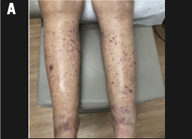
On day 29, the patient reported progressively worsening rash on the lower extremities with new areas of ulceration. Oral prednisone 60 mg was prescribed. The patient was seen by dermatology again on day 34 after starting oral prednisone and continuing the topical treatment. She reported mild pain in her lower legs and slow improvement of symptoms. Physical exam showed overall fewer purpuric macules and thin papules on the lower legs and thighs, but areas were becoming more confluent with new vesiculation (Figure 1B). Large, ulcerating plaques were present on the calves, shins, and upper thighs with areas of purpura and ecchymosis on the back of the thighs. Significant pitting edema was present from the ankles proximally. There were no pertinent lab abnormalities to suggest a clotting abnormality (platelet count persistently 200-300 × 109/L). Topical and oral steroids were continued following this visit. Ulcerating plaques improved by day 49 (Figure 1C). A timeline of the immunotherapy and vasculitis treatment is shown in Figure 2.
(B) Clinical examination after 1 week of prednisone treatment, showing persistent vesiculation and ulceration but no new lesions.
(C) Clinical examination 1 month after treatment showing improvement in ulcerations.
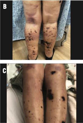
FIGURE 2. Timeline of Immunotherapy, Onset of Vasculitis, and Vasculitis Treatment

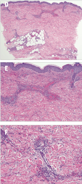
Initial morphology was consistent with a cutaneous small vessel vasculitis, and dermatopathology showed leukocytoclastic vasculitis (LCV) due to the presence of neutrophils in and around lumens of the superficial and deep perivascular plexus (Figure 1D, 1E, 1F). Immunoglobulin A (IgA) vasculitis was also on the differential, but the DIF was negative for any deposition of IgG, IgM, IgA, complement 3 (C3), and complement component 1q (C1q) with nonspecific fibrinogen positivity in the dermis. There were no signs of medium or large vessel vasculitis.
(D), (E), (F) Histology of leg biopsy showed diapedesis of neutrophils in and around the lumens of the superficial and deep perivascular plexus. Neutrophilic dust is apparent. Fibrin is evident within a few small vessels.
Further blood work was ordered to rule out other causes of LCV, including connective tissue disease and infection. Labs included antinuclear antibody (ANA), rheumatoid factor, cryoglobulins, C3, antistreptolysin O, hepatitis B serologies, hepatitis C antibody, and quantiferon-gold; all were unremarkable. A urinalysis did not show early proteinuria. Antineutrophil cytoplasmic antibodies (ANCA) were slightly positive with a titer of 1:20. Based on the pathology results and no other clear cause of LCV, the presumed diagnosis was immunotherapy-induced LCV.
What is the next step in management for a patient on immune checkpoint inhibition (ICI) who presents with severe palpable purpura?
Discussion
Immunotherapy has revolutionized the treatment of RCC and several other cancer types due to its ability to promote antitumor activity via activated T lymphocytes, resulting in significantly improved outcomes for patients with cancer. Immune system evasion by overstimulating immune pathways that downregulate T-cell activation is a mechanism of RCC pathogenesis that has recently been exploited through pioneering work by 2018 Nobel Prize awardees James Allison, PhD, and Tasuku Honjo, MD, PhD. The mechanisms of ICI available in clinical practice include blockade of cytotoxic T-lymphocyte–associated antigen 4 (CTLA-4) and programmed cell death protein 1 (PD-1)/programmed cell death protein–ligand 1 pathways using monoclonal antibodies. This leads to activation of T cells, which then generates an immune response targeting tumor-associated antigens that allows cancer cells to be identified and destroyed.1
Although immunotherapy has significantly impacted cancer therapeutics in the last decade, a well-known consequence of ICI is overactivation of T cells; this can lead to the unwanted inflammation of any organ and development of autoimmune diseases. Immunotoxicity often leads to delay or permanent discontinuation of immunotherapy and/or initiation of high-dose steroids and other immunosuppressive medications.
The combination of nivolumab (PD-1 inhibitor) and ipilimumab (CTLA-4 inhibitor), the regimen that our patient received, was studied in a landmark phase 3 clinical trial comparing the efficacy and safety profile of this combination vs that of sunitinib (Sutent) in patients with previously untreated advanced RCC (CheckMate 214; NCT02231749). This study showed significantly higher overall survival (OS) and overall response rates (ORRs) with nivolumab plus ipilimumab compared with sunitinib. The updated 5-year data on the intent-to-treat population showed a median OS of 55.7 months in the immunotherapy group vs 38.4 months in the sunitinib group, and ORRs of 39% in the immunotherapy group vs 32% in the sunitinib group. These study results ultimately led to the FDA approval of nivolumab plus ipilimumab for therapeutic use in advanced RCC. Regarding immunotoxicity, about 80% of patients developed treatment-mediated adverse events (AEs) and of these, 35% required treatment with high-dose glucocorticoids; less than 1% of immune AEs were skin toxicities.2 No new safety signals emerged in the extended 5-year follow-up.
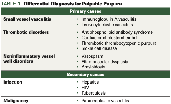
Vasculitis is defined as inflammation of a single or multiple blood vessels of various sizes leading to end-organ damage, and it can often be a clinical conundrum in its initial presentation. The classic physical exam finding of cutaneous involvement is palpable purpura, with other skin findings such as nodules, ulceration, and livedo depending on vessel size and organ involvement. The differential for palpable purpura (Table 1) is broad and can mimic several other diagnoses; these include infection, thrombotic disorders (eg, antiphospholipid antibody syndrome, cardiac or cholesterol emboli, thrombotic thrombocytopenic purpura, sickle cell disease),3-6 and noninflammatory vessel wall disorders (eg, vasospasm, fibromuscular dysplasia, amyloidosis).7,8 Secondary causes are also important to rule out during workup for suspected vasculitis; these include viral infections such as hepatitis and HIV, as well as tuberculosis.9 Identifying an underlying infection is crucial, given that the treatment for primary vasculitis is immunosuppression with systemic steroids, which could lead to worsening infection if not recognized and treated early.
TABLE 1. Differential Diagnosis for Palpable Purpura
Malignancy can cause paraneoplastic vasculitis via decreased clearance of immune complexes and deposition of IgA antibodies and tumor antigens in the vascular walls. Diagnostic criteria include pertinent clinical findings such as palpable purpura and gastrointestinal symptoms, in addition to presence of IgA immune complex deposition seen on a biopsy specimen with histopathologic evidence of vasculitis. While this is a rare complication that accounts for only 2.5% to 5% of all cases of adult vasculitis and typically occurs in the setting of solid tumors, early diagnosis is crucial to prevent severe complications such as gastrointestinal perforation or necrotizing enterocolitis.10,11
In patients receiving ICI, the incidence of immune-mediated vasculitis is rare and on the order of case reports.12 Both nivolumab and ipilimumab are associated with a less than 1% risk of vasculitis across clinical trials of both drugs administered as either monotherapy or in combination.13,14 The mechanism of immune-related vasculitis is thought to be from sustained T-cell activity; this causes B-cell activation and antibody production leading to immune complex deposition in vessel walls and to direct damage of vessel walls from highly activated T cells.15
Vasculitis of small, medium, and large vessels in the setting of ICI has been documented in the literature.12 A systematic review of 20 case reports of immune-mediated vasculitis demonstrated that the most common cancer type was melanoma, and the predominant types of vasculitis were those affecting large vessels (ie, giant cell arteritis, aortitis). No reported vasculitis-related deaths were observed, and only 1 patient died of disease progression; the remainder of patients who had available data on outcomes either went into remission or had partial tumor response.12
The timing of dermatologic manifestations from immune-mediated mechanisms is broad; it can range from 2 weeks to 38 months after treatment initiation per 1 retrospective observational study of 17 patients who presented with cutaneous AEs associated with PD-1 inhibitor therapy. Our patient fell on the early end of the spectrum and developed skin findings consistent with vasculitis 2 weeks after immunotherapy initiation. Cutaneous toxicities described in the study included eczema, sarcoidosis, lichenoid dermatitis, bullous pemphigoid, and erythema multiforme. It is difficult to map out a timeline for these AEs since they occurred at various time points (for example, bullous pemphigoid was seen at 1.5 months and 20.4 months). It is important to note that cutaneous toxicities can develop after treatment discontinuation, as PD-1 inhibitors have durable effects on tumor responses and also on AEs.16
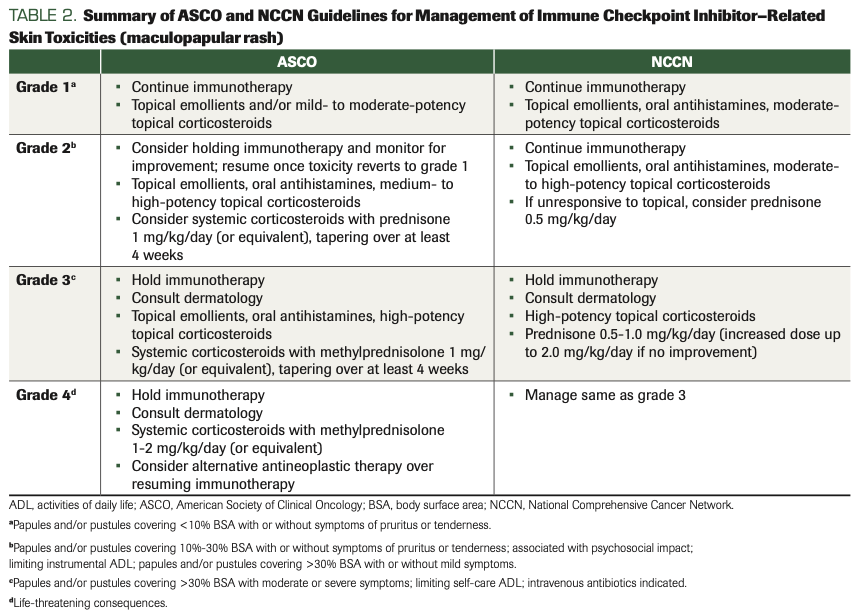
It is important to have a high index of suspicion for vasculitis when a patient presents with palpable purpura in the setting of ICI therapy, as early biopsy and management are necessary for preventing severe complications. If vasculitis is high on the differential, clinically monitoring without further diagnostic studies or starting topical steroids empirically may delay diagnosis and treatment, which is why answer choices A and B are incorrect. When histopathological confirmation of vasculitis in the setting of immunotherapy is obtained, management depends on the Common Terminology Criteria for Adverse Events grade of the rash. Grading is based on the percent total body surface area that the rash covers. Per the FDA, ipilimumab should be held for grade 2 rash and nivolumab should be held for grade 3, and both should be permanently discontinued in the setting of grade 4 disease. The American Society of Clinical Oncology (ASCO) clinical practice guidelines recommend that grade 1 disease can be managed with topical emollients or corticosteroids while continuing the ICI. Immunotherapy needs to be held for grade 2 or higher cutaneous toxicity. Failure to respond to topicals is an indication to start glucocorticoids at a prednisone equivalent of 1 to 2 mg/kg, and intravenous steroids should be considered for grade 3 or higher toxicity with a slow taper over at least 4 weeks upon symptomatic improvement.17 Additionally, the National Comprehensive Cancer Network (NCCN) management of immune checkpoint inhibitor–related toxicities guidelines include recommendations to continue immunotherapy for grade 1/2 disease and managing with topicals, and holding immunotherapy for grade 3 or higher toxicity while starting systemic steroids.18 Table 2 summarizes the NCCN and ASCO guidelines. Therefore, choice C is the correct answer.
TABLE 2. Summary of ASCO and NCCN Guidelines for Management of Immune Checkpoint Inhibitor–Related Skin Toxicities (maculopapular rash)
Methotrexate has not been shown to have clinical benefit in treatment of immune-mediated vasculitis; however, it can be used in certain forms of vasculitis such as giant cell arteritis or granulomatosis with polyangiitis. Typically, these conditions are managed with high-dose glucocorticoids for remission induction, and methotrexate is used for remission maintenance.19,20 Because methotrexate would not be used first-line for immune-mediated vasculitis and would delay proper diagnosis or potentially cause more toxicity than benefit, answer choice D is incorrect.
While the timing of vasculitis onset was somewhat early after initiation of immunotherapy, the lack of IgA complex deposition on biopsy and the negative diagnostic workup led us to suspect this was vasculitis: immune-mediated and likely drug-induced, rather than paraneoplastic. However, the exact etiology remains unclear and may be multifactorial in the setting of immunotherapy exposure and cancer progression. Vasculitic complications of ICI are a rare phenomenon that remain to be further studied, and we present our case report to contribute to the medical community’s knowledge regarding early recognition and treatment of immune-mediated vasculitis.
Outcome of This Case
More than 2 months after the initial immunotherapy dose, the patient presented to the emergency department with altered mental status and had a witnessed seizure in the setting of hypertensive emergency. Brain imaging demonstrated evidence of posterior reversible encephalopathy syndrome (PRES). She had a complicated hospital stay that involved intensive care unit (ICU) admission, intubation, a nicardipine drip for hypertension, and cardioversion for atrial fibrillation with rapid ventricular rate. Other causes of encephalopathy and multiorgan failure were investigated and included workup for infection, metabolic derangement, and drug-induced toxicities. Evaluation for rheumatologic causes included tests of ANCA and ANA that were both negative, which suggested that an immune systemic vasculitis was less likely. The etiology of PRES was likely multifactorial and related to immunotherapy and/or hypertension from steroid treatment for the vasculitis. Immunotherapy was discontinued. Her mental status improved and she was eventually extubated and discharged on oral prednisone 1 mg/kg daily with a plan to taper over several weeks.
Given the presence of significant immune-related toxicities, immunotherapy was permanently discontinued with a plan to transition therapy to a vascular endothelial growth factor tyrosine kinase inhibitor upon recovery from her acute illness. However, 2 weeks after hospital discharge, the patient was readmitted with failure to thrive, septic shock secondary to gram-negative rod bacteremia from urinary etiology, acute liver and kidney injury, thrombocytopenia, and rapidly progressive disease. She was shortly transferred to the ICU with multiorgan failure. Unfortunately, the patient died from disease progression approximately 9 weeks following the initial immunotherapy dose.
Disclosure: RRM is a consultant/advisory board member for AstraZeneca, Aveo, Bayer, Bristol Myers Squibb, Calithera, Caris, Dendreon, Exelixis, Janssen, Pfizer, Merck, Myovant, Novartis, Sanofi, and Tempus; and receives institutional research funding from Bayer and Tempus.
Author affiliations:
Justine Panian, BS1*; Elizabeth Pan, MD1*; Veronica Shi, MD2; Brian Hinds, MD2; Rana R. McKay, MD1,3
1Department of Medicine, Division of Hematology-Oncology, University of California San Diego, San Diego, CA
2Department of Dermatology, University of California San Diego, San Diego, CA
3Department of Urology, University of California San Diego, San Diego, CA
*These authors contributed equally to this work.
About the series editors:
Maria T. Bourlon, MD, is associate professor, head Urologic Oncology Clinic; national researcher, Instituto Nacional de Ciencias Médicas y Nutrición Salvador Zubirán, Mexico City, Mexico. She is also a member of ASCO’s IDEA Working Group.
E. David Crawford, MD, is chairman, Prostate Conditions Education Council; editor in chief, Grand Rounds in Urology; and professor of Urology, University of California San Diego, La Jolla.
References
- Töpfer K, Kempe S, Müller N, et al. Tumor evasion from T cell surveillance. J Biomed Biotechnol. 2011;2011:918471. doi:10.1155/2011/918471
- Motzer RJ, Tannir NM, McDermott DF, et al; CheckMate 214 Investigators. Nivolumab plus ipilimumab versus sunitinib in advanced renal-cell carcinoma. N Engl J Med. 2018;5;378(14):1277-1290. doi:10.1056/NEJMoa1712126
- Ronthal M, Gonzalez RG, Smith RN, Frosch MP. Case records of the Massachusetts General Hospital. weekly clinicopathological exercises. case 21-2003. a 72-year-old man with repetitive strokes in the posterior circulation. N Engl J Med. 2003;349(2):170-180. doi:10.1056/NEJMcpc020034
- Mayer SA, Aledort LM. Thrombotic microangiopathy: differential diagnosis, pathophysiology and therapeutic strategies. Mt Sinai J Med. 2005;72(3):166-175.
- Boussen K, Moalla M, Blondeau P, Ben Ayed H, Lie JT. Embolization of cardiac myxomas masquerading as polyarteritis nodosa. J Rheumatol. 1991;18(2):283-285.
- Cappiello RA, Espinoza LR, Adelman H, Aguilar J, Vasey FB, Germain BF. Cholesterol embolism: a pseudovasculitic syndrome. Semin Arthritis Rheum. 1989;18(4):240-246. doi:10.1016/0049-0172(89)90044-9
- Siegert CE, Macfarlane JD, Hollander AM, van Kemenade F. Systemic fibromuscular dysplasia masquerading as polyarteritis nodosa. Nephrol Dial Transplant. 1996;11(7):1356-1358.
- Rao JK, Allen NB. Primary systemic amyloidosis masquerading as giant cell arteritis. case report and review of the literature. Arthritis Rheum. 1993;36(3):422-425. doi:10.1002/art.1780360320
- Luqmani RA, Pathare S, Kwok-Fai TL. How to diagnose and treat secondary forms of vasculitis. Best Pract Res Clin Rheumatol. 2005;19(2):321-336. doi:10.1016/j.berh.2004.11.002
- Mitsui H, Shibagaki N, Kawamura T, Matsue H, Shimada S. A clinical study of Henoch-Schönlein Purpura associated with malignancy. J Eur Acad Dermatol Venereol. 2009;23(4):394-401. doi:10.1111/j.1468-3083.2008.03065.x
- Ota S, Haruyama T, Ishihara M, et al. Paraneoplastic IgA vasculitis in an adult with lung adenocarcinoma. Intern Med. 2018;57(9):1273-1276. doi:10.2169/internalmedicine.9651-17
- Daxini A, Cronin K, Sreih AG. Vasculitis associated with immune checkpoint inhibitors – a systematic review. Clin Rheumatol. 2018;37(9):2579-2584. doi:10.1007/s10067-018-4177-0
- Ipilimumab. Prescribing information. Bristol Myers Squibb; 2011. Accessed March 21, 2022. https://bit.ly/3inkJN1
- Nivolumab. Prescribing information. Bristol Myers Squibb; 2014. Accessed March 21, 2022. https://bit.ly/3qoU2Ma
- Kang A, Yuen M, Lee DJ. Nivolumab-induced systemic vasculitis. JAAD Case Rep. 2018;4(6):606-608. doi:10.1016/j.jdcr.2018.03.013
- Wang LL, Patel G, Chiesa-Fuxench ZC, et al. Timing of onset of adverse cutaneous reactions associated with programmed cell death protein 1 inhibitor therapy. JAMA Dermatol. 2018;154(9):1057-1061. doi:10.1001/jamadermatol.2018.1912
- Brahmer JR, Lacchetti C, Thompson JA. Management of immune-related adverse events in patients treated with immune checkpoint inhibitor therapy: American Society of Clinical Oncology Clinical Practice Guideline summary. J Oncol Pract. 2018;14(4):247-249. doi:10.1200/JOP.18.00005
- NCCN. Clinical Practice Guidelines in Oncology. Management of immune checkpoint inhibitor–related toxicities, version 4.2021. Accessed March 21, 2022. https://bit.ly/3qnzRhy
- Mahr AD, Jover JA, Spiera RF, et al. Adjunctive methotrexate for treatment of giant cell arteritis: an individual patient data meta-analysis. Arthritis Rheum. 2007;56(8):2789-2797. doi:10.1002/art.22754
- Guillevin L, Pagnoux C, Karras A, et al; French Vasculitis Study Group. Rituximab versus azathioprine for maintenance in ANCA-associated vasculitis. N Engl J Med. 2014;371(19):1771-1780. doi:10.1056/NEJMoa1404231
