Predicting Endocrine Therapy Responsiveness in Breast Cancer
This article reviews ongoing progress in the effort to identify predictors of endocrine therapy responsiveness for breast cancer and discusses the value of “pre-treatment” vs “on-treatment” tumor profiling for predicting outcomes.
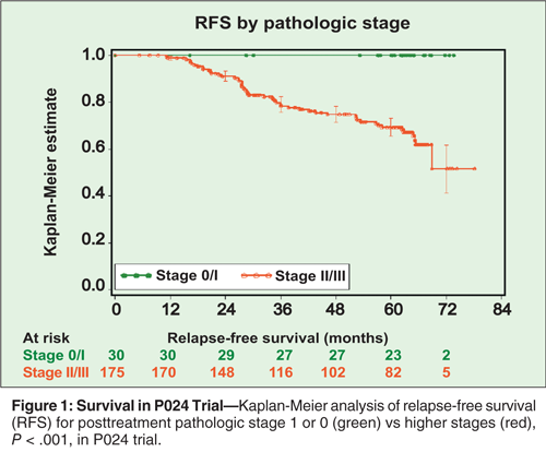
Survival in P024 Trial

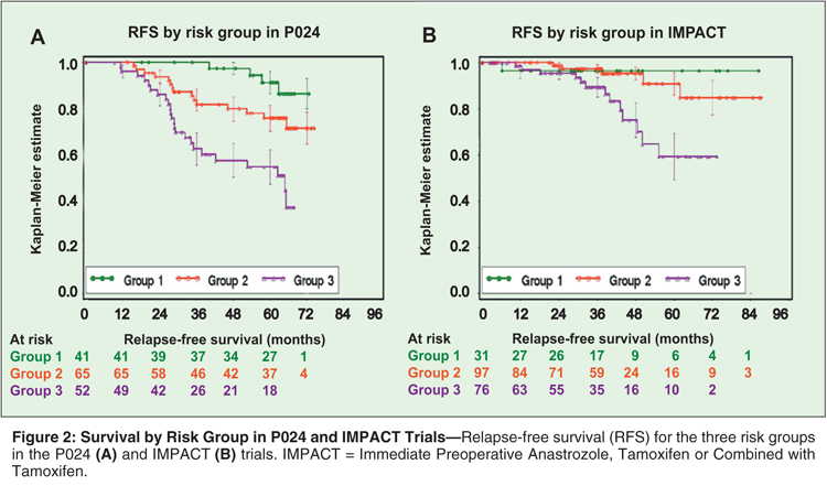
Survival by Risk Group in P024 and IMPACT Trials

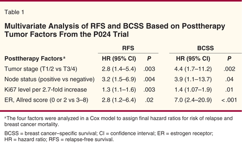
Multivariate Analysis of RFS and BCSS Based on Posttherapy Tumor Factors From the P024 Trial

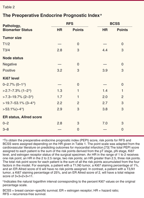
The Preoperative Endocrine Prognostic Index

Endocrine therapy is one of the most effective treatment strategies for breast cancer. However, in the adjuvant setting, up to 40% to 50% of patients with estrogen receptor (ER)-positive breast cancers relapse despite these interventions. Although ER and HER2 analysis has increased our ability to predict which patients will benefit from endocrine therapy, further improvement is needed, most specifically for patients with ER-positive, HER2-negative disease. Recent advances in genomic technology have made it possible to classify breast cancers into risk categories with significant prognostic implications. However, the predictive value of these tests remains the subject of investigation. Long-term follow-up of neoadjuvant endocrine therapy studies suggests that the in vivo assessment of therapeutic efficacy provided by this treatment approach is also valuable in predicting outcome. Indeed, the Preoperative Endocrine Prognostic Index (PEPI), based on tumor pathologic staging and expression levels of ER and Ki67 following 3 to 4 months of neoadjuvant endocrine therapy, reproducibly predicts long-term outcomes of hormone receptor–positive breast cancer. This article reviews ongoing progress in the effort to identify predictors of endocrine therapy responsiveness for breast cancer and discusses the value of “pre-treatment” vs “on-treatment” tumor profiling for predicting outcomes.
The hormone-dependent nature of breast cancer was first described in the literature by Beatson in 1896.[1] Since then, a number of pharmacologic agents have been developed to either modulate tumor cell estrogen receptor (ER) function or to reduce the levels of circulating estrogens. Among these agents are the selective estrogen receptor modulators (SERMs: tamoxifen, raloxifene [Evista], and toremifene [Fareston]), pure antiestrogens (fulvestrant [Faslodex]), luteinizing hormone-releasing hormone agonists (leuprolide, goserelin [Zoladex]), and third-generation selective aromatase inhibitors (anastrozole [Arimidex], letrozole [Femara], exemestane [Aromasin]). The widespread application of endocrine therapy with these agents has led to a significant reduction in breast cancer mortality.[2] However, up to 50% of women with breast cancers that are hormone receptor (HR)-positive do not derive benefit from these treatments, either due to intrinsic resistance or acquired resistance following prolonged use.[3,4] Furthermore, endocrine therapy is associated with vasomotor symptoms (tamoxifen), musculoskeletal discomfort (aromatase inhibitors) and occasionally more serious side effects (thrombosis and endometrial cancer from tamoxifen or osteoporotic fracture from aromatase inhibitors). These problems can affect the overall quality of life and even reduce life expectancy.[5] Identifying predictors of endocrine responsiveness is therefore important to avoid unnecessary toxicities and to promote the selection of alternative treatment strategies for patients with endocrine-resistant tumors. In this review, we will discuss recent studies in this area and debate the status of these tests in current clinical practice.
Primary Tumor Biomarker Characteristic
Most studies have investigated biomarkers on primary tumors collected before endocrine treatment, with a focus on ER, progesterone receptor (PR), HER2, Ki67, and, more recently, multigene profiles that incorporated additional genes.
Estrogen and Progesterone Receptors
ER and PR are well recognized predictors of response to endocrine therapy.[4,6] The prerequisite of a positive ER and/or PR test for endocrine responsiveness was initially observed for patients with advanced disease[6,7] and was further demonstrated in the early-stage disease setting.
• Role of ER Status-In the quinquennial overview of randomized adjuvant trials by the Early Breast Cancer Trialists’ Collaborative Group (EBCTCG), the use of 5 years of tamoxifen in patients with early-stage breast cancer was associated with a 41% reduction in the annual risk of relapse, and a 34% reduction in the annual death rate for women with ER-positive disease, but little benefit was observed for those with ER-poor disease.[2,8,9] In addition, multiple studies have revealed that the degree of ER positivity is directly related to tumor responsiveness to antiestrogen therapy. In the earlier EBCTCG Overview analysis, women with tumors that had 2+ ER staining derived a significantly larger reduction in the risk of death from 5 years of tamoxifen compared to those with 1+ staining. Similarly, patients who had tumors with an Allred score of 6 and above-calculated as the sum of an intensity score (range, 1–3) and a frequency score (range, 0–5) of ER staining-are most likely to respond to treatment.[10]
In the National Surgical Adjuvant Breast and Bowel Project (NSABP) B-14 trial, a randomized phase III study of tamoxifen vs observation in women with HR-positive breast cancer, the levels of ER expression, analyzed by quantitative reverse transcriptase polymerase chain reaction (qRT-PCR) analysis, was also predictive of tamoxifen benefit.[11] A relationship between ER expression and response to endocrine therapy was also observed in neoadjuvant endocrine studies of aromatase inhibitors including letrozole and anastrozole.[12-14] Interestingly, compared with tamoxifen in the neoadjuvant setting, aromatase inhibitors may be able to induce a response in tumors with lower levels of ER, although the sample sizes in these studies do not allow for robust conclusions in this regard.[12]
Despite the clear value of ER expression analysis, the methodologies to evaluate ER expression and the cutoffs used to determine endocrine sensitivity are not standardized in clinical trials or in clinical practice. Conventional techniques, including the ligand-binding assay, which employs a radiolabeled steroid ligand to ER, and immunohistochemistry (IHC), which involves the use of specific antibodies to ER, have many problems. Some are pre-analytical (ie, related to poor specimen processing) and others are analytic in nature (ie, lack of assay standardization, lack of robust internal controls). The accuracy and reproducibility of scoring as well as the cutoff points for ER positivity vary among different laboratories.[15] A newer method, which measures ER mRNA levels by qRT-PCR, allows a more quantitative and objective evaluation of ER expression and may be more accurate than conventional techniques. However, this method is only routinely available in the context of the Oncotype assay, rather than as a stand-alone test.
• Role of PR Status-In ER-positive breast cancer, the contribution of PR to the prediction of endocrine therapy responsiveness has been a subject of controversy. Since PR expression is regulated by ER, it was thought that the absence of PR likely reflects a nonfunctional ER pathway. It has also been observed that PR-negative tumors are generally associated with hyperactive growth factor signaling.[16,17] Compared to ER-positive/PR-positive tumors, ER-positive/PR-negative tumors have twice as many DNA copy number gains or losses and are frequently associated with upregulation of specific oncogenic pathways, including PI3K/Akt/mTOR.[18] As assessed by gene expression profiling, these tumors form a distinct subset of breast cancer that is associated with aggressive pathologic features and poor outcome.[18]
Therefore, it was hypothesized that the absence of PR in ER-positive tumor likely entails endocrine resistance. Consistent with the hypothesis, patients with tumors that are ER-positive/PR-negative have been found to be much less likely to benefit from endocrine therapy than those with ER-positive/PR-positive tumors in the metastatic setting.[7,19,20] However, studies in patients with early-stage breast cancer have failed to demonstrate a relationship between PR expression and endocrine responsiveness. In the EBCTCG Overview analysis, PR played no role in ER-positive tumors in predicting benefit to adjuvant tamoxifen therapy.[21] In the ER-positive group, PR-positive and PR-negative patients showed similar benefit from tamoxifen (relative risk [RR] = 0.81; 95% confidence interval [CI] = 0.65–1.02; and RR = 0.70; 95% CI = 0.49–0.99, respectively).[21]
Based on data collected in case record forms, it was initially reported that the relative benefit from anastrozole was substantially higher in the PR-negative subgroup in the Arimidex, Tamoxifen, Alone or in Combination (ATAC) adjuvant breast cancer trial.[22] However, upon central review of the tumor blocks collected from 2,006 of the 5,880 patients enrolled in the ATAC trial, the quantitative expression of PR or HER2 did not identify patients with differential relative benefit from anastrozole over tamoxifen.[23] Time to recurrence was longer for anastrozole than for tamoxifen in all molecular subgroups. Similarly, in the Breast International Group (BIG) 1-98 trial, letrozole was superior to tamoxifen regardless of PR status.[24]
HER2/neu
HER2 positivity has generally been accepted as a marker of endocrine resistance. HER2 (HER2/neu or cerbB2) is a proto-oncogene and HER2 overexpression/amplification is associated with higher histologic grade, low expression of ER and PR, and worse clinical outcome.[25,26] About 10% of ER-positive breast cancer involves HER2 gene amplification.[25] Preclinical studies demonstrated that HER2 overexpression reduces dependence on estrogen.[27,30] The resistance to endocrine interventions was correlated with hyperactivation of MAPK and downregulation of ER.[30]
In the metastatic setting, HER2 overexpression is associated with reduced response to tamoxifen.[31] In the neoadjuvant setting, while the amplification of HER2 does not seem to affect the clinical response to letrozole, suppression of Ki67 is significantly less in these tumors, suggesting therapeutic resistance.[32]
The relationship between endocrine resistance and HER2 positivity has further been demonstrated in adjuvant endocrine therapy studies. In the adjuvant tamoxifen studies, HER2-positive cancers did not derive significant benefit from tamoxifen.[21,33] In the randomized, four-arm, phase III adjuvant BIG 1-98 trial, patients with ER-positive/HER2-positive tumors experienced inferior disease-free survival (DFS) compared to those with ER-positive/HER2-negative tumors regardless of treatment assignment,[34] underscoring the endocrine therapy–resistant properties of ER-positive/HER2-positive tumors.[35] HER2-positive status is therefore a bona fide marker for poor outcome in ER-positive disease and warrants treatment with trastuzumab (Herceptin), which improves outcome in HER2-positive breast cancer regardless of ER status.[36]
Ki67
An increased expression of proliferation markers has generally been accepted to be a predictor of worse clinical outcomes in breast cancer. IHC staining of Ki67, a nuclear antigen that is present only in proliferating cells,[37] has been shown to be a reliable methodology to enumerate the growth fraction of normal or neoplastic cell populations,[38] and the Ki67 labeling index-the percentage of cells with positive Ki67 nuclear staining-correlates well with the S phase fraction and mitotic index.[39]
Multiple studies have shown that baseline tumor Ki67 is a prognostic factor for breast cancer.[40,41] In a meta-analysis of 46 studies of over 12,000 patients with early-stage breast cancer, Ki67 positivity, defined by individual studies (cutoff ranges from 3.5 to 34%), is associated with a higher probability of relapse (hazard ratio [HR] = 1.93; 95% CI = 1.74–2.14; P < .001) and worse survival (HR = 1.95; 95% CI = 1.70–2.24; P < .001).[42] However, routine use of Ki67 for prognostic assessment of ER-positive breast cancers has not been considered a standard practice.[43] Most of the studies are retrospective and differ in the type of antibody used, the cutoff value selected to define high vs low proliferative activity, and the number of cells counted. Moreover, investigators lack an internationally standardized method for antigen retrieval, staining procedures, and scoring methods.
The value of Ki67 in predicting responses to systemic therapy has also been evaluated. Several studies in the neoadjuvant setting suggest that tumors with high Ki67 expression are more likely to respond to neoadjuvant chemotherapy.[44,45] However, in a retrospective analysis of 1,924 patients who were enrolled in two randomized International Breast Cancer Study Group (IBCSG) trials of adjuvant chemoendocrine therapy vs endocrine therapy alone for node-negative breast cancer-the IBCSG VIII and IX trials-a high Ki67 labeling index was associated with a worse prognosis, but did not predict an added benefit of chemotherapy to endocrine therapy.[46] Interestingly, in the BIG 1-98 trial-a randomized, double blind, phase III study that showed letrozole improved DFS compared to tamoxifen for postmenopausal women with hormone receptor–positive disease[47,48]-the investigators found a greater benefit from letrozole compared to tamoxifen in tumors with a higher Ki67 labeling index.[49,50] The hazard of a DFS event for letrozole was half that for tamoxifen (HR, letrozole:tamoxifen = 0.53; 95% CI 0.39–0.72).[49,50] Therefore, high Ki67 labeling index levels may identify a patient group that particularly benefits from initial letrozole adjuvant therapy, but more studies are needed to confirm these findings.
Gene-Expression Profiling
The introduction of genome-wide profiling technology has facilitated the development of several multigene assays that have been shown to predict breast cancer outcome and treatment response.[51,52] Using microarray analysis, ER-positive tumors have been subclassified into at least two “intrinsic” subtypes (luminal subtype A and subtype B).[52,53] The luminal subtype A tumors demonstrate the highest expression of the ER and ER-associated genes. On the other hand, the luminal subtype B tumors have low-to-moderate expression of luminal-specific genes but express some of the genes that are characteristic of ER-negative tumor, with more frequent TP53 mutation compared to luminal subtype A tumors. Patients with luminal subtype B tumors manifest significantly worse relapse-free and overall survival than those with luminal subtype A ER-positive tumors.[52-54]
These data suggest that prognosis of ER-positive breast cancer is determined by the presence of not only ER but also several other genes that work in concert with ER to regulate the response to estrogen. Low expression of any of these factors could lead to estrogen-independent growth and endocrine therapy resistance.
• MammaPrint-The MammaPrint assay is the first commercialized microarray-based multigene assay for prognostic prediction in patients under age 61 with lymph node–negative breast cancer, regardless of ER status. The 70-gene signature predominantly comprises genes related to proliferation, with additional genes involved in invasion, metastasis, and angiogenesis.[55] The test gives a dichotomized result, indicating either a high or low risk of disease recurrence. In conjunction with Adjuvant! Online[56] the utility of the MammaPrint assay in predicting the benefit of chemotherapy is being studied in the ongoing Microarray in Node-Negative Disease May Avoid Chemotherapy Trial (MINDACT).[57] This test has not been fully evaluated to predict endocrine therapy responsiveness.
• Oncotype DX-The Oncotype DX assay is a multigene analysis performed to predict outcomes for patients with ER-positive, lymph node–negative breast cancer. The 21-gene list was derived from outcome analyses of an initial 250 candidate genes published in literature and genomic databases. Of the 16 cancer-related genes selected for the Oncotype DX assay, 8 were already reported to distinguish between luminal A and luminal B subtypes. The Oncotype DX recurrence score (RS) is weighted heavily on gene expression related to ER, HER2, and proliferation.
In the retrospective analysis of patients on the tamoxifen arm of the NSABP B-14 trial, the 10-year distant recurrence rates for those at low (RS < 18), intermediate (RS 18–30), and high risk (RS ≥ 31) as predicted by the RS, were 6.8%, 14.3%, and 30.5%, respectively, independent of age and tumor size but not grade.[51] Importantly, RS appears to predict clinical benefits from tamoxifen and chemotherapy.[58,59] Patients with high quantitative ER expression and low RS were found to benefit most from treatment with adjuvant tamoxifen (NSABP B-14 trial), and those with high RS were found to benefit the most from chemotherapy (NSABP B-20 trial).[58,59] In this study, approximately 50% of women with node-negative, HR-positive breast cancer had an excellent prognosis after use of tamoxifen and may not need chemotherapy.[58,59]
The prospective study of the Oncotype DX test as an approach to tailoring chemotherapy decisions, the Trial Assigning Individualized Options for Treatment (Rx), or TAILORx trial, is currently accruing. Pending the results of this study, critics of the assay point out that the utility of Oncotype DX has not been replicated in a second large retrospective study of endocrine therapy with or without chemotherapy in node-negative patients, and therefore there remains uncertainty about how reproducible the relationship between RS scores and the outcome of chemotherapy actually is. Furthermore, by general consensus generated in the design phase of the TAILORx trial, this test is not valuable in the setting of a HER2-positive breast cancer, and so ER-positive, HER2-positive breast cancer is excluded from the study. In current clinical practice, Oncotype DX should not be ordered in the setting of a HER2-positive breast cancer since a HER2-positive test indicates the need for the use of trastuzumab, usually in combination with chemotherapy, regardless of the RS category.
• PAM50-The direct translation of the original observation of intrinsic breast cancer subtypes into a clinical assay has been challenging because the hierarchical clustering approach with which these observations were made are better suited to the organization of large numbers of samples and genes than to the classification of individual samples using small gene sets. However, Parker et al recently identified a minimum set of 50 intrinsic genes using prediction analysis of microarray (PAM50) and developed a method with which to objectively assign subtypes independent of clustering.[60] The intrinsic subtypes defined by this model showed prognostic significance in multivariate analyses that incorporated standard clinical parameters (ER, histologic grade, tumor size, and nodal status). The combined model (radius of rotation, or ROR), using tumor size and intrinsic subtype information, showed significantly better prognostic performance than a straight clinical model that incorporated histologic grade, tumor size, and ER status. In addition, intrinsic subtype as defined by PAM50 classification was highly related to effectiveness of neoadjuvant chemotherapy, as measured in terms of complete pathologic response, with a predicted positive value of 97%. In other words, patients with luminal A tumors with a low ROR score had virtually a zero chance of experiencing a pathologic complete response with chemotherapy. The utility of PAM50 breast cancer subtype assignment in predicting endocrine responsiveness remains under investigation in large cohorts of patients who received tamoxifen in the adjuvant setting.
Four-Marker IHC Panel to Predict Luminal B Subtype
Recent studies indicate that a four-marker surrogate IHC panel (ER, PR, HER2, and Ki67) was able to define luminal B subtype when tested against the PAM50.[61] Luminal B tumors were defined as ER- or PR-positive, HER2-negative, and Ki67 high, whereas the luminal A subtype was defined as ER- or PR-positive, HER2-negative, and Ki67 low. The optimal cutoff point of Ki67 proliferation index was determined to be 13.25% to distinguish luminal B from luminal A, with a sensitivity of 77%, specificity of 78%, and error rate of 23%. Therefore, Ki67 of 14% or higher was used to identify luminal B tumors. A separate category of luminal/HER2-positive tumors was assigned to those that were HER2-positive and ER- or PR-positive.
The breast cancer subtypes defined by the four-marker IHC panel was found to have significant prognostic values in an independent tissue microarray series of 4,046 breast cancers, which included 2,598 cases of HR-positive cases. Compared to luminal A tumors, luminal B and luminal/HER2-positive tumors were associated with worse relapse-free and breast cancer–specific survival, both in the presence and the absence of adjuvant systemic treatment with either tamoxifen alone or chemotherapy (AC [doxorubicin (Adriamycin), cyclophosphamide], FAC [fluorouracil (5-FU), doxorubicin, cyclophosphamide], or CMF [cyclophosphamide, methotrexate, 5-FU]) followed by tamoxifen in patients with ER-positive breast cancer.
When the four-marker IHC panel was analyzed on tissues from Breast Cancer International Research Group (BCIRG) 001, a phase III study to compare FAC vs TAC (docetaxel [Taxotere], doxorubicin, cyclophosphamide) as adjuvant chemotherapy for early-stage breast cancer, TAC was found to be superior to FAC in patients with the luminal B subtype but not in those with luminal A, suggesting a potential value of breast cancer subtype in predicting benefit from treatment with more intense chemotherapy regimens.[62] These studies again demonstrated the need to incorporate several molecular markers in better predicting clinical outcomes. The poor outcome of patients with luminal B tumors in the presence of adjuvant tamoxifen suggests resistance to endocrine therapy.
Compared to multigene analysis, the IHC panel is easier to apply clinically and may save costs, depending on charges for the execution and clinical interpretation of multiple separate tests as opposed to a single format that runs these biomarkers simultaneously. Relying on multiple tests, each associated with a significant error rate, compounds the problem of misclassification and decision error. Ideally, a study should be conducted to compare an IHC approach to subtyping with a multigene assay in order to define which methodology provides the most value in prognostication and prediction of tumor responsiveness to systemic therapy.
Neoadjuvant Therapy as an Approach for In Vivo Assessment of Endocrine Responsiveness
Neoadjuvant Endocrine Therapy
Traditionally, neoadjuvant endocrine therapy has been used as an alternative to neoadjuvant chemotherapy for the management of locally advanced HR-positive breast cancer in elderly or frail individuals, to improve surgical outcomes.[63] As more recent studies in younger and healthier postmenopausal patients have shown its effectiveness in promoting superior surgical outcomes, neoadjuvant endocrine therapy has gained more acceptance.[13,64] From the research point of view, neoadjuvant endocrine therapy provides a unique opportunity to assess tumor response and molecular changes induced by treatment and to search for molecular biomarkers that might predict long-term outcomes based on an analysis of tumors that have been sampled after therapy has been started.
The P024 trial was a multicenter, randomized phase III study comparing the efficacy of 4 months of letrozole and tamoxifen in postmenopausal women with ER-positive and/or PR-positive primary breast cancer ineligible for breast-conserving surgery.[64] Efficacy analysis of data from the 324 patients enrolled in the study concluded that letrozole was superior to tamoxifen in clinical response (60% vs 41%, P = .004) and rate of breast-conserving surgery (48% vs 36%, P = .036).
The Immediate Preoperative Anastrozole, Tamoxifen or Combined with Tamoxifen (IMPACT) trial is a multicenter, double-blind randomized study in postmenopausal women with ER-positive, operable or locally advanced potentially operable breast cancer who were randomly assigned to receive anastrozole, tamoxifen, or the combination for 12 weeks before surgery. The trial was designed to be the neoadjuvant equivalent to the Arimidex, Tamoxifen, Alone or in Combination (ATAC) adjuvant therapy trial, testing the hypothesis that the clinical and/or biologic effects of neoadjuvant endocrine therapy before surgery might predict for outcome in the ATAC study. A total of 330 patients were treated. Although no significant differences in tumor response were observed among the different treatment arms, suppression of Ki67 was greater with anastrozole than tamoxifen at both 2 and 12 weeks (before surgery), which predicted for recurrence-free survival in the ATAC trial.[14]
Posttherapy Ki67 and ER
Since endocrine therapy decreases cell proliferation, changes in Ki67 scoring after short-term (2 or 12 weeks) endocrine manipulation has been tested as a pharmacodynamic marker of efficacy[65-67] and a predictor of long-term outcome.[66,68,69] As noted above, the IMPACT trial showed that 2 weeks of treatment with anastrozole induced a more dramatic suppression of Ki67 expression than with tamoxifen alone or tamoxifen in combination with anastrozole.[66] Both the percentage change in tumor Ki67 expression and the absolute level of tumor Ki67 expression at 2 weeks of treatment in the IMPACT trial predicted the superiority of anastrozole over tamoxifen in DFS in the adjuvant ATAC trial.[14,70] Long-term follow-up of patients treated in the IMPACT and P024 trial indicated that Ki67 following neoadjuvant treatment (rather than the baseline measurement) is predictive of long-term outcomes.[68,69,71] In the IMPACT trial, the 5-year recurrence-free survival rates were 85%, 75%, and 60% for the lowest, middle, and highest values of 2-week Ki67 expression, respectively. In the multivariable analysis, 2-week Ki67 expression remained statistically significantly associated with recurrence-free survival (HR = 1.95; 95% CI = 1.23–3.07; P = .004). In the P024 trial, while baseline Ki67 was not associated with relapse, posttreatment Ki67 levels had a robust association with relapse-free survival (HR = 1.4, CI = 1.2–1.6 per log unit increase; P < .001), and breast cancer–specific survival (HR = 1.4, CI = 1.1–1.7; P = .009).[69]
ER loss after neoadjuvant endocrine therapy has also been identified as an independent prognostic marker for relapse in both the IMPACT trial and the P024 trial. In the IMPACT trial, the levels of ER at 2-weeks postneoadjuvant therapy were significantly associated with recurrence-free survival (HR = 0.98; 95% CI = 0.62–0.99; P = .04).[66,68] Similarly, in the P024 trial, patients with posttreatment ER-negative tumors had worse relapse-free survival (HR of relapse = 2.4; 95% CI = 1.0–5.3; P = .03) and breast cancer–specific survival (HR of breast cancer death = 4.3; 95% CI = 1.6–11.7; P = .002) than patients with tumors that retained ER-positive status after treatment.[69]
To examine the molecular features of tumors that exhibited the switch from positive to negative ER status, Agilent 44K whole-genome array analysis was performed on tumor specimens collected fresh at baseline prior to therapy and on treatment at 1 month and/or at the time of surgery in a multicenter phase II study of neoadjuvant letrozole.[72] About 10% of ER-positive tumors underwent a transition to an ER-negative state early after the initiation of endocrine treatment. Tumors that lose ER expression with neoadjuvant endocrine treatment was found to lack both basal and luminal gene expression. These unclassified proliferative tumors exhibited a fulminant clinical course.[72]
PEPI Score
Ellis et al recently reported a prognostic model (Preoperative Endocrine Prognostic Index, or PEPI score) based on results from the P024 trial, which incorporated pathologic tumor size, lymph node status, and tumor expression of Ki67 and ER following 3 to 4 months of neoadjuvant endocrine therapy to predict the long-term outcome of ER-positive breast cancer patients.[69] Tumors from 228 postmenopausal women in the P024 trial were studied for posttreatment ER status, Ki67 proliferation index, histologic grade, pathologic tumor size, nodal stage, and treatment response. Cox proportional hazards were used to identify factors associated with relapse-free survival and breast cancer–specific survival in 158 women.
The median follow-up was 61.2 months. No relapses were recorded in the group of 29 patients with stage 0/I disease vs 53 of the 175 patients (30% relapse incidence) with stage II/III disease (Figure 1). Multivariable testing of posttreatment tumor characteristics revealed that pathologic tumor stage, nodal status, Ki67 level, and ER status remained independently associated with both relapse-free and breast cancer–specific survival (Table 1). Clinical response and histologic grade were not independently associated with relapse-free or breast cancer–specific survival. Based on this information, PEPI score was derived as an arithmetic sum of risk points weighted by the size of the hazard ratio assigned to each statistically significant factor.[73] The scoring process outlined in Table 2 produced three groups (risk score 0, 1–3, and ≥ 4) that were associated with relapse risks of 10%, 23%, and 48% (P < .001; Figure 2A) and breast cancer death risk of 2%, 11%, and 17% (P < .001).
The use of PEPI score as a prognostic model was subsequently validated in the IMPACT study.[69] Data on pathologic stage, ER status, and Ki67 intervals were available for 203 surgical specimens from the IMPACT trial. The model produced a statistically significant separation of the three PEPI risk groups (risk score 0, 1–3, and ≥ 4) and predicted relapse-free survival in the IMPACT trial (P = .002, Figure 2B). As with the P024 study, IMPACT patients with stage 0 or I tumors had a more favorable outcome than patients with stage II or III disease. There were no relapses in either study for the stage 0/I group with a PEPI risk score of 0, with a combined median follow-up of 60.3 months (range = 4.5–86.5 months). The group with intermediate (1–3) PEPI scores consisted of patient with early-stage tumor and adverse biomarkers or higher-stage tumor and favorable biomarkers. Both trials agree in that clinical response was not a good determinant of long-term outcomes, but “dynamic factors”-ie, changes in Ki67 and ER expression are strongly associated with prognosis.
Taken together, these studies indicate that PEPI score obtained after a course of neoadjuvant endocrine therapy can effectively assess the endocrine responsiveness of HR-positive breast cancer on an individual basis.
Posttherapy Gene-Expression Profiling
In addition to the analysis of ER, Ki67, and tumor staging on post-neoadjuvant specimens, several ongoing studies are investigating gene-expression profiling as a predictor of endocrine responsiveness. Ellis et al conducted a phase II study of preoperative letrozole in postmenopausal patients with ER-positive, stage II/III breast cancer, in which fresh biopsy specimens taken at baseline prior to therapy, 1 month on therapy, and at the time of surgery after 4 to 6 months of treatment were collected for quality RNA extraction. Agilent 44K whole-genome array analysis was performed for 65 tumor samples. PAM50 ROR calculated at 1 month on therapy, rather than at baseline, strongly predicted the risk of future disease relapse.[72] This study further demonstrates the powerful predictive value of markers performed on specimens after neoadjuvant endocrine therapy.
Future Directions
REFERENCE GUIDE
Therapeutic Agents
Mentioned in This Article
Anastrozole (Arimidex)
Cyclophosphamide
Docetaxel (Taxotere)
Doxorubicin
Exemestane (Aromasin)
Fluorouracil (5-FU)
Fulvestrant (Faslodex)
Goserelin (Zoladex)
Letrozole (Femara)
Leuprolide
Methotrexate
Raloxifene (Evista)
Tamoxifen
Toremifene (Fareston)
Trastuzumab (Herceptin)
Brand names are listed in parentheses only if a drug is not available generically and is marketed as no more than two trademarked or registered products. More familiar alternative generic designations may also be included parenthetically.
The highly predictive value of posttreatment molecular markers on long-term outcomes of patients with HR-positive breast cancer raises the hope that therapy could be tailored after a course of neoadjuvant endocrine therapy. Peri-Operative Endocrine Therapy for Individualizing Care (POETIC) has been proposed to prospectively evaluate this further. Patients will be treated with an aromatase inhibitor for 2 weeks prior to surgery, with Ki67 values and molecular profiling assessed at baseline and 2-week samples to correlate with long-term outcome.[3]
The ongoing American College of Surgeons Oncology Group (ACOSOG) Z1031 is a three-arm phase III study randomizing postmenopausal women with HR-positive (and with ER or PR Allred score of 6–8), stage II/III breast cancer to receive either anastrozole, letrozole, or exemestane for 4 months before surgery. Fresh tumor biopsies are collected at baseline and prior to surgery for predictive marker analysis. The study is expected to generate baseline and posttreatment molecular markers as predictors of endocrine responsiveness.
Conclusions
Despite the recognition of ER as the best predictive factor for endocrine response, a significant number of patients with ER-positive breast cancer do not benefit from endocrine therapy, and the optimum strategy to predict endocrine responsiveness is yet to be defined. Single markers are unlikely to provide precise outcome predictions. Multigene analysis reflecting tumor proliferation and ER pathway genetic activities analyzed by either IHC (Ki67, ER, PR, HER2) or more sophisticated gene-expression profiling (Oncotype DX, MammaPrint, PAM50) on baseline tumors have added prognostic value and are being tested for their utility in predicting the efficacy of systemic therapy. Promising results have also been obtained with the PEPI score, which incorporates tumor size, lymph node status, and Ki67 and ER expression into a prognostic index that is applied to the surgical sample after a patient has received neoadjuvant endocrine treatment.
One can reasonably conclude from this review that even a minimal 2 to 4 weeks of endocrine therapy before surgery will provide additional information over that obtained from a baseline specimen alone, and a large trial has been activated in the UK to address this hypothesis. Ongoing neoadjuvant studies with correlative endpoints and long-term follow-up are critical in addressing these issues as well as getting us to the next step-a complete mechanistic understanding of the reasons for the success or failure of endocrine treatment, so that new mechanism-based therapeutic approaches can emerge.
Financial Disclosure:Dr. Ellis has received speaking honoraria, consulting fees, and clinical trial support from AstraZeneca, Novartis, and GlaxoSmithKline. He has also been a speaker and consultant for Pfizer and Genentech, a consultant for Merck, Monogram Biosciences, and Qiagen GmbH, and has received clinical trial support from Taiho. He is a founder of University Genomics, which has licensing rights to the PAM50 intrinsic subtyping test for breast cancer (for which he is also on the patent as an inventor).
References:
1. Beatson G: On the treatment of inoperable cases of carcinoma of the mamma: Suggestions for a new method of treatment with illustrative cases. Lancet 2:104, 1896.
2. Effects of chemotherapy and hormonal therapy for early breast cancer on recurrence and 15-year survival: An overview of the randomised trials. Lancet 365:1687-1717, 2005.
3. Anderson H, Bulun S, Smith I, et al: Predictors of response to aromatase inhibitors. J Steroid Biochem Mol Biol 106:49-54, 2007.
4. Goldhirsch A, Glick JH, Gelber RD, et al: Meeting highlights: International expert consensus on the primary therapy of early breast cancer 2005. Ann Oncol 16:1569-1583, 2005.
5. Perez EA: Appraising adjuvant aromatase inhibitor therapy. Oncologist 11:1058-1069, 2006.
6. Osborne CK, Yochmowitz MG, Knight WA, 3rd, et al: The value of estrogen and progesterone receptors in the treatment of breast cancer. Cancer 46:2884-2888, 1980.
7. Bezwoda WR, Esser JD, Dansey R, et al: The value of estrogen and progesterone receptor determinations in advanced breast cancer. Estrogen receptor level but not progesterone receptor level correlates with response to tamoxifen. Cancer 68:867-872, 1991.
8. Early Breast Cancer Trialists’ Collaborative Group: Tamoxifen for early breast cancer: An overview of the randomised trials. Lancet 351:1451-1467, 1998.
9. Early Breast Cancer Trialists’ Collaborative Group: Effects of chemotherapy and hormonal therapy for early breast cancer on recurrence and 15-year survival: An overview of the randomised trials. Lancet 365:1687-1717, 2005.
10. Harvey JM, Clark GM, Osborne CK, et al: Estrogen receptor status by immunohistochemistry is superior to the ligand-binding assay for predicting response to adjuvant endocrine therapy in breast cancer. J Clin Oncol 17:1474-1481, 1999.
11. Baehner FL, Habel LA, Quesenberry CP, et al: Quantitative RT-PCR analysis of ER and PR by Oncotype DX indicates distinct and different associations with prognosis and prediction of tamoxifen benefit (abstract 45). Breast Cancer Res Treat 100(suppl 1):S20, 2006.
12. Ellis MJ, Coop A, Singh B, et al: Letrozole is more effective neoadjuvant endocrine therapy than tamoxifen for ErbB-1- and/or ErbB-2-positive, estrogen receptor-positive primary breast cancer: Evidence from a phase III randomized trial. J Clin Oncol 19:3808-3816, 2001.
13. Ellis MJ, Ma C: Letrozole in the neoadjuvant setting: The P024 trial. Breast Cancer Res Treat 105(suppl 1):33-43, 2007. 14. Smith IE, Dowsett M, Ebbs SR, et al: Neoadjuvant treatment of postmenopausal breast cancer with anastrozole, tamoxifen, or both in combination: The Immediate Preoperative Anastrozole, Tamoxifen, or Combined with Tamoxifen (IMPACT) multicenter double-blind randomized trial. J Clin Oncol 23:5108-5116, 2005.
15. Allred DC: Problems and solutions in the evaluation of hormone receptors in breast cancer. J Clin Oncol 26:2433-2435, 2008. 16. Bamberger AM, Milde-Langosch K, Schulte HM, et al: Progesterone receptor isoforms, PR-B and PR-A, in breast cancer: Correlations with clinicopathologic tumor parameters and expression of AP-1 factors. Horm Res 54:32-37, 2000.
17. Arpino G, Weiss H, Lee AV, et al: Estrogen receptor-positive, progesterone receptor-negative breast cancer: Association with growth factor receptor expression and tamoxifen resistance. J Natl Cancer Inst 97:1254-1261, 2005.
18. Creighton CJ, Kent Osborne C, van de Vijver MJ, et al: Molecular profiles of progesterone receptor loss in human breast tumors. Breast Cancer Res Treat Apr 19, 2008 (epub ahead of print).
19. Elledge RM, Green S, Pugh R, et al: Estrogen receptor (ER) and progesterone receptor (PgR), by ligand-binding assay compared with ER, PgR and pS2, by immuno-histochemistry in predicting response to tamoxifen in metastatic breast cancer: A Southwest Oncology Group study. Int J Cancer; 89:111-117, 2000.
20. Ravdin PM, Green S, Dorr TM, et al: Prognostic significance of progesterone receptor levels in estrogen receptor-positive patients with metastatic breast cancer treated with tamoxifen: Results of a prospective Southwest Oncology Group study. J Clin Oncol 10:1284-1291, 1992.
21. Dowsett M, Houghton J, Iden C, et al: Benefit from adjuvant tamoxifen therapy in primary breast cancer patients according oestrogen receptor, progesterone receptor, EGF receptor and HER2 status. Ann Oncol 17:818-826, 2006.
22. Dowsett M, Cuzick J, Wale C, et al: Retrospective analysis of time to recurrence in the atac trial according to hormone receptor status: An hypothesis-generating study. J Clin Oncol 23:7512-7517, 2005.
23. Dowsett M, Allred C, Knox J, et al: Relationship between quantitative estrogen and progesterone receptor expression and human epidermal growth factor receptor 2 (HER-2) status with recurrence in the Arimidex, Tamoxifen, Alone or in Combination trial. J Clin Oncol 26:1059-1065, 2008.
24. Mauriac L, Keshaviah A, Debled M, et al: Predictors of early relapse in postmenopausal women with hormone receptor-positive breast cancer in the BIG 1-98 trial. Ann Oncol 18:859-867, 2007.
25. Lal P, Tan LK, Chen B: Correlation of HER-2 status with estrogen and progesterone receptors and histologic features in 3,655 invasive breast carcinomas. Am J Clin Pathol 123:541-546, 2005.
26. Kaptain S, Tan LK, Chen B: Her-2/neu and breast cancer. Diagn Mol Pathol 10:139-152, 2001.
27. Pietras RJ, Arboleda J, Reese DM, et al: Her-2 tyrosine kinase pathway targets estrogen receptor and promotes hormone-independent growth in human breast cancer cells. Oncogene 10:2435-2446, 1995.
28. Liu Y, el-Ashry D, Chen D, et al: MCF-7 breast cancer cells overexpressing transfected c-erbB-2 have an in vitro growth advantage in estrogen-depleted conditions and reduced estrogen-dependence and tamoxifen-sensitivity in vivo. Breast Cancer Res Treat 34:97-117, 1995.
29. Benz CC, Scott GK, Sarup JC, et al: Estrogen-dependent, tamoxifen-resistant tumorigenic growth of MCF-7 cells transfected with HER2/neu. Breast Cancer Res Treat 24:85-95, 1992.
30. Oh AS, Lorant LA, Holloway JN, et al: Hyperactivation of MAPK induces loss of ERalpha expression in breast cancer cells. Mol Endocrinol 15:1344-1359, 2001.
31. Wright C, Nicholson S, Angus B, et al: Relationship between c-erbB-2 protein product expression and response to endocrine therapy in advanced breast cancer. Br J Cancer 65:118-121, 1992.
32. Ellis MJ, Tao Y, Young O, et al: Estrogen-independent cell proliferation occurs in the majority of estrogen receptor positive (ER+)/HER2 gene-amplified primary breast cancers: Evidence from a combined analysis of two independent neoadjuvant letrozole studies (abstract 9538). J Clin Oncol 23(16S):846s, 2005.
33. De Placido S, De Laurentiis M, Carlomagno C, et al: Twenty-year results of the naples gun randomized trial: Predictive factors of adjuvant tamoxifen efficacy in early breast cancer. Clin Cancer Res 9:1039-1046, 2003.
34. Rasmussen BB, Regan MM, Lykkesfeldt AE, et al: Adjuvant letrozole versus tamoxifen according to centrally-assessed erbB2 status for postmenopausal women with endocrine-responsive early breast cancer: Supplementary results from the BIG 1-98 randomised trial. Lancet Oncol 9:23-28, 2008.
35. Viale G, Regan MM, Maiorano E, et al: Prognostic and predictive value of centrally reviewed expression of estrogen and progesterone receptors in a randomized trial comparing letrozole and tamoxifen adjuvant therapy for postmenopausal early breast cancer: BIG 1-98. J Clin Oncol 25:3846-3852, 2007.
36. Piccart-Gebhart MJ, Procter M, Leyland-Jones B, et al: Trastuzumab after adjuvant chemotherapy in HER2-positive breast cancer. N Engl J Med 353:1659-1672, 2005.
37. Gerdes J, Schwab U, Lemke H, et al: Production of a mouse monoclonal antibody reactive with a human nuclear antigen associated with cell proliferation. Int J Cancer 31:13-20, 1983.
38. Gerdes J, Lemke H, Baisch H, et al: Cell cycle analysis of a cell proliferation-associated human nuclear antigen defined by the monoclonal antibody Ki-67. J Immunol 133:1710-1715, 1984.
39. Spyratos F, Ferrero-Pous M, Trassard M, et al: Correlation between MIB-1 and other proliferation markers: Clinical implications of the MIB-1 cutoff value. Cancer 94:2151-2159, 2002.
40. van Diest PJ, van der Wall E, Baak JPA: Prognostic value of proliferation in invasive breast cancer: A review. J Clin Pathol 57:675-681, 2004.
41. Urruticoechea A, Smith IE, Dowsett M: Proliferation marker Ki-67 in early breast cancer. J Clin Oncol 23:7212-7220, 2005.
42. de Azambuja E, Cardoso F, de Castro G Jr, et al: Ki-67 as prognostic marker in early breast cancer: A meta-analysis of published studies involving 12,155 patients. Br J Cancer 96:1504-1513, 2007.
43. Goldhirsch A, Wood WC, Gelber RD, et al: Meeting highlights: Updated international expert consensus on the primary therapy of early breast cancer. J Clin Oncol 21:3357-3365, 2003.
44. Chang J, Ormerod M, Powles TJ, et al: Apoptosis and proliferation as predictors of chemotherapy response in patients with breast carcinoma. Cancer 89:2145-2152, 2000.
45. Archer CD, Parton M, Smith IE, et al: Early changes in apoptosis and proliferation following primary chemotherapy for breast cancer. Br J Cancer 89:1035-1041, 2003.
46. Viale G, Regan MM, Mastropasqua MG, et al: Predictive value of tumor Ki-67 expression in two randomized trials of adjuvant chemoendocrine therapy for node-negative breast cancer. J Natl Cancer Inst 100:207-212, 2008.
47. Thurlimann B, Keshaviah A, Coates AS, et al: A comparison of letrozole and tamoxifen in postmenopausal women with early breast cancer. N Engl J Med 353:2747-2757, 2005.
48. Coates AS, Keshaviah A, Thurlimann B, et al: Five years of letrozole compared with tamoxifen as initial adjuvant therapy for postmenopausal women with endocrine-responsive early breast cancer: Update of study BIG 1-98. J Clin Oncol 25:486-492, 2007.
49. Viale G, Giobbie-Hurder A, Regan MM, et al: Prognostic and predictive value of centrally reviewed Ki-67 labeling index in postmenopausal women with endocrine-responsive breast cancer: Results from Breast International Group trial 1-98 comparing adjuvant tamoxifen with letrozole. J Clin Oncol 26:5569-5575, 2008.
50. Viale G, Giobbie-Hurder A: Value of centrally-assessed Ki-67 labeling index as a marker of prognosis and predictor of response to adjuvant endocrine therapy in the BIG 1-98 trial of postmenopausal women with estrogen receptor-positive breast cancer (abstract 64). Presented at the 30th Annual San Antonio Breast Cancer Symposium. San Antonio, Texas; Dec 13-16, 2007.
51. Paik S, Shak S, Tang G, et al: A multigene assay to predict recurrence of tamoxifen-treated, node-negative breast cancer. N Engl J Med 351:2817-2826, 2004.
52. Perou CM, Sorlie T, Eisen MB, et al: Molecular portraits of human breast tumours. Nature 406:747-752, 2000.
53. Sorlie T, Perou CM, Tibshirani R, et al: Gene expression patterns of breast carcinomas distinguish tumor subclasses with clinical implications. PNAS 98:10869-10874, 2001.
54. Sorlie T, Tibshirani R, Parker J, et al: Repeated observation of breast tumor subtypes in independent gene expression data sets. PNAS 100:8418-8423, 2003.
55. van de Vijver MJ, He YD, van’t Veer LJ, et al: A gene-expression signature as a predictor of survival in breast cancer. N Engl J Med 347:1999-2009, 2002.
56. Ravdin PM, Siminoff LA, Davis GJ, et al: Computer program to assist in making decisions about adjuvant therapy for women with early breast cancer. J Clin Oncol 19:980-991, 2001.
57. Bogaerts J, Cardoso F, Buyse M, et al: Gene signature evaluation as a prognostic tool: Challenges in the design of the mindact trial. Nat Clin Pract Oncol 3:540-551, 2006.
58. Paik S, Tang G, Shak S, et al: Gene expression and benefit of chemotherapy in women with node-negative, estrogen receptor-positive breast cancer. J Clin Oncol 24:3726-3734, 2006.
59. Paik S, Shak S, Tang G, et al: Expression of the 21 genes in the recurrence score assay and prediction of clinical benefit from tamoxifen in NSABP study B-14 and chemotherapy in NSABP study B-20 (abstract 24). Presented at the 27th Annual San Antonio Breast Cancer Symposium, Dec 8-11, 2004.
60. Parker JS, Mullins M, Cheang MC, et al: A supervised risk predictor of breast cancer based on intrinsic subtypes. J Clin Oncol. In press.
61. Cheang M, Chia SK, Voduc D: Ki67 and her2 define luminal B breast cancers with poor prognosis. J Natl Cancer Inst. In press.
62. Hugh J, Hanson J, Cheang M, et al: Breast cancer subtypes and response to docetaxel in node-positive breast cancer: Use of an immunohistochemical definition in the BCIRG 001 trial. J Clin Oncol. In press.
63. Macaskill EJ, Renshaw L, Dixon JM: Neoadjuvant use of hormonal therapy in elderly patients with early or locally advanced hormone receptor-positive breast cancer. Oncologist 11:1081-1088, 2006.
64. Eiermann W, Paepke S, Appfelstaedt J, et al: Preoperative treatment of postmenopausal breast cancer patients with letrozole: A randomized double-blind multicenter study. Ann Oncol 12:1527-1532, 2001.
65. Clarke RB, Laidlaw IJ, Jones LJ, et al: Effect of tamoxifen on Ki67 labelling index in human breast tumours and its relationship to oestrogen and progesterone receptor status. Br J Cancer 67:606-611, 1993.
66. Dowsett M, Smith IE, Ebbs SR, et al: Short-term changes in Ki-67 during neoadjuvant treatment of primary breast cancer with anastrozole or tamoxifen alone or combined correlate with recurrence-free survival. Clin Cancer Res 11:951s-958s, 2005.
67. Ellis MJ, Coop A, Singh B, et al: Letrozole inhibits tumor proliferation more effectively than tamoxifen independent of HER1/2 expression status. Cancer Res 63:6523-6531, 2003.
68. Dowsett M, Smith IE, Ebbs SR, et al: Prognostic value of Ki67 expression after short-term presurgical endocrine therapy for primary breast cancer. J Natl Cancer Inst 99:167-170, 2007.
69. Ellis MJ, Tao Y, Luo J, et al: Outcome prediction for estrogen receptor-positive breast cancer based on postneoadjuvant endocrine therapy tumor characteristics. J Natl Cancer Inst 100:1380-1388, 2008.
70. Baum M, Budzar AU, Cuzick J, et al: Anastrozole alone or in combination with tamoxifen versus tamoxifen alone for adjuvant treatment of postmenopausal women with early breast cancer: First results of the ATAC randomised trial. Lancet 359:2131-2139, 2002.
71. Dowsett M, Smith IE, Ebbs SR, et al: Short-term changes in Ki-67 during neoadjuvant treatment of primary breast cancer with anastrozole or tamoxifen alone or combined correlate with recurrence-free survival. Clin Cancer Res 11:951s-958s, 2005.
72. Ellis MJ, Tao Y, Luo J, et al: A poor prognosis ER and HER2-negative, nonbasal, breast cancer subtype identified through postneoadjuvant endocrine therapy tumor profiling (abstract 502). J Clin Oncol 26(15S):7s, 2008.
73. Morrow DA, Antman EM, Charlesworth A, et al: TIMI risk score for ST-elevation myocardial infarction: A convenient, bedside, clinical score for risk assessment at presentation: An intravenous nPA for treatment of infarcting myocardium early II trial substudy. Circulation 102:2031-2037, 2000.