Understanding Novel Molecular Therapies
The science supporting molecularly targeted therapies for the treatment of patients with solid tumors continues to evolve. Nurses are challenged to understand cell signaling, molecular targeting, and the mechanism of action of targeted agents. Two cell signal transduction pathways regulate the development, proliferation, and metastasis of solid tumors: the human epidermal growth factor (HER) receptor pathway and the vascular endothelial growth factor (VEGF) receptor pathway. Several novel pharmacologic agents with distinct indications and methods of administration target the HER and VEGF molecular pathways.
The science supporting molecularly targeted therapies for the treatment of patients with solid tumors continues to evolve. Nurses are challenged to understand cell signaling, molecular targeting, and the mechanism of action of targeted agents. Two cell signal transduction pathways regulate the development, proliferation, and metastasis of solid tumors: the human epidermal growth factor (HER) receptor pathway and the vascular endothelial growth factor (VEGF) receptor pathway. Several novel pharmacologic agents with distinct indications and methods of administration target the HER and VEGF molecular pathways.
Human cells process and respond to diverse stimulatory and inhibitory signals through multifaceted signaling pathways. A tightly regulateed system of signal transduction pathways controls cell metabolism, division, death, differentiation, and movement.[1] Malignant transformation stems from altered regulation of six essential cell behaviors: self-sufficiency in growth signals, insensitivity to antigrowth signals, evasion of apoptosis (programmed cell death), limitless replication potential, sustained angiogenesis, and tissue invasion and metastasis.[2]
Researchers have identified numerous abnormal signal transduction elements that contribute to the development of cancer. These discoveries have led to the creation of pharmacologic agents that target signal transduction elements and cell behavior.[3] Two signal transduction pathways regulate the development, proliferation, and metastasis of solid tumors: the human epidermal growth factor (HER) receptor pathway and the vascular endothelial growth factor (VEGF) receptor pathway. Each of the novel pharmacologic agents that target these pathways has unique indications and means of administration.
Human Epidermal Growth Factor Receptors
The HER family of receptors consists of four structurally related transmembrane receptors: HER1 (epidermal growth factor receptor [EGFR] or cerbB1), HER2 (cerbB2 or HER2/neu), HER3 (cerbB3), and HER4 (cerbB4). HER receptor tyrosine kinases (TKs) have an extracellular ligand-binding domain, a transmembrane domain, and an intracellular tyrosine kinase (TK) domain.[4] Research has demonstrated that HER family dysregulation is associated with atypical cell behavior,[5] and current investigations are focused specifically on the role that EGFR signaling pathways play in carcinogenesis.
EGFR is expressed in healthy cells of germ cell derivation, especially those of epithelial origin. EGFR overexpression is associated with cancers of the colon, head and neck, pancreas, lung (non-small cell), breast, kidney, bladder, and gliomas. Alterations in EGFR activity correlate with disease progression, poor prognosis, and the development of resistance to cytotoxic agents.[5]
EGFR activation begins when an extracellular ligand binds to an EGFR monomer (inactive protein). Several stimulatory ligands bind with EGFR, including the epidermal growth factor (EGF) and transforming growth factor-alpha (TGF-α). The ligand-bound receptor dimerizes or pairs with other monomers on the cell surface. EGFR can pair with another EGFR (homodimerization) or another member of the HER family (heterodimerization). Dimerization promotes transmembrane signal transduction, resulting in intracellular TK activity and phosphorylation.[5] A phosphate group from adenosine triphosphate (ATP) is transferred to the tyrosine residues on the signal transduction molecules. The phosphorylated TK residue becomes a binding site for key signal transducers that activate multiple downstream signaling pathways.[6] Significant downstream pathways include Ras-Raf-Mek-MAPK, which regulates gene transcription and proliferation, and the P13K/Akt signaling pathway, which governs cell survival.[5] The specific binding ligand and the coreceptor to which EGFR is dimerized determine the signaling pathways that EGFR activates.[7] Multiple factors contribute to upregulation of EGFR signaling, including overproduction of ligands by the tumor cell, overexpression of EGFRs on the cell surface, and mutations that initiate EGFR activity independently of ligand binding.[8]
HER2, like EGFR, is a TK receptor that is expressed on a variety of normal cells. The HER2 receptor has no known ligand and participates in signal transduction by forming heterodimers with other HER family receptors. HER2-containing heterodimers exhibit strong ligand binding, which enhances downstream signaling and delivery of proliferative signals to the nucleus.[9] Overexpression of HER2 results in the formation of HER2 homodimers that are also extremely active.[10] Gene amplification (generation of more than the normal two gene copies) and overexpression of HER2 occur in approximately 25% of breast cancers and are associated with aggressive tumor behavior and decreased overall survival.[9]
Activation of HER2 and EGFR receptors triggers multiple signaling pathways that play a critical role in cellular growth and proliferation. Tumor cells express VEGF, a protein responsible for the development of new blood vessels (angiogenesis), as a result of EGFR signaling.[6]
Vascular Endothelial Growth Factor
Like normal cells, cancer cells depend on an adequate blood supply to provide oxygen, nutrients, and other elements essential for survival and growth. Solid tumors can absorb sufficient nutrients and oxygen by diffusion until they measure 2 to 3 mm; further growth requires the formation of new blood vessels or angiogenesis.[11]
Angiogenesis is a normal physiologic response during wound healing, menstruation, and embryonic development. It is a dynamic, complex process regulated by a number of factors. VEGF, a member of the platelet-derived growth factor family, has a well-documented role in tumor angiogenesis. A number of solid tumors express VEGF; among them are glioblastomas and colon, gastric, breast, lung, brain, hepatocellular, and bladder cancers.[12]
Numerous stimuli increase VEGF expression: genetic events, hypoxia, nitric oxide, and growth factors such as platelet-derived growth factor (PDGF), epidermal growth factor (EGF), and insulin-like growth factor (IGF-1).[13] The primary source of VEGF is the tumor itself, but associated stromal and vascular endothelium cells also express VEGF, especially in the presence of hypoxia.[12]
Angiogenesis is a multistep process that begins with VEGF binding to VEGFR1 (FLT1) and VEGFR2 (KDR or Flk-1), which are located on endothelial cells found in blood vessels.[13] Receptor activation leads to TK phosphorylation, inducing multiple downstream pathways and production of proteins that promote angiogenesis. VEGF signaling increases the permeability of surrounding vasculature, proliferation of endothelial cells, and degradation of the extracellular matrix, which promotes endothelial cell migration.
Finally, VEGF inhibits endothelial cell apoptosis by stimulating the expression of antiapoptotic factors Bcl-2 and Bcl-A1. The resulting unstable vasculature is tortuous, dilated, and leaky. Despite the development of new vasculature, the tumor remains hypoxic, and angiogenesis is further stimulated. The unstable characteristic of the tumor vasculature may contribute to ineffective delivery of cytotoxic agents, resulting in poor response.[12,13]
Novel Strategies for Molecular Targeting
Signaling pathways present multiple opportunities for intervention. In the extracellular domain, altering the ligand or the receptor would prevent dimerization and associated signaling. Disruption of TK activity or the activity of secondary cytoplasmic messengers would inhibit intracellular signaling.[14] Monoclonal antibodies, TK inhibitors, and multitargeted agents are potent therapeutic weapons to counteract aberrant cellular behavior resulting from abnormal signaling.
Mechanisms of Action
Monoclonal Antibodies
The monoclonal antibody (MoAb) has a Y shape with two active sites: the Fab portion (arms of the Y), which recognizes and binds to a specific antigen, and the Fc portion (leg of the Y), which signals the immune system to eliminate the antigen or the associated cell. Each of the four types of MoAbs has a slightly different composition. Murine MoAbs, derived from mice, are limited by a short half-life and the potential to create human antimouse antibodies. In an attempt to improve efficacy and the side-effect profile, scientists have engineered MoAbs that contain fewer murine and more human components. Chimeric MoAbs are approximately 75% human; humanized MoAbs contain a small murine Fab portion and are 95% to 98% human; and the fully human MoAbs contain only the human antibody gene sequence.[15]
The monoclonal antibody primary mechanism of action lies in the extracellular domain and is directed at disrupting ligand-receptor activity. By binding to specific targets, MoAbs disrupt extracellular signaling. A MoAb can bind with a ligand and prevent ligand-receptor pairing, or it can bind with a receptor, inhibiting ligand-dependent receptor activation. A MoAb can also interfere with the activation of ligand-independent receptors. By disrupting ligand-receptor binding, MoAbs prevent phosphorylation and thereby inhibit TK signal transduction pathways.[6] MoAbs have the ability to destroy the cell associated with the antigen by eliciting an effector response from the antibody-dependent cell-mediated cytotoxicity and the complement-dependent cytotoxicity systems.[15]
Tyrosine Kinase Inhibitors
Tyrosine kinase inhibitors (TKIs) are small molecules that can cross the cell membrane and block intracellular signaling. The TKI occupies the ATP binding site on the receptor's intracellular TK domain. Blocking ATP binding prevents phosphorylation and activation of the intracellular signaling cascade. Tyrosine kinase inhibitors are oral agents that demonstrate a common mechanism of action but differ in their specificity, potency, and reversibility.[16]
Multitargeted Agents: New Treatment Options
Several characteristics of cancer, particularly the signal transduction pathways, support the development of multitargeted therapeutic interventions. Because most cancers develop as a result of multiple mutations in numerous signaling pathways, therapies aimed at simultaneous inhibition of multiple pathways may be more effective than those that inhibit a single pathway. Tumors and their supporting vasculature usually express multiple receptor TKs that regulate key cellular activities such as angiogenesis and proliferation.[17] Signaling cross-talk occurs throughout the signal transduction pathway, enabling one signal to affect the output of another. Targeting multiple receptor TKs may, therefore, elicit a vigorous and rapid biological response.
Multitargeted therapeutic agents may not only be more effective than single-target agents but may possibly decrease the occurrence of drug resistance as well. Combining single-target agents has produced an enhanced effect in clinical trials.[18] The US Food and Drug Administration has approved several multitargeted agents in the last year, and many more are in development. The question remains whether treatment is more effective with a combination of single-target agents or with one multitargeted agent. Combining multiple single-target agents would permit flexible dosing, whereas a single multitargeted agent may be more cost-effective and convenient, thereby improving patient compliance.[19]
Indications and Uses of Molecularly Targeted Agents
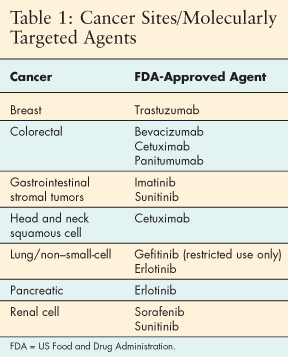
The FDA has approved nine molecularly targeted agents during the past decade for treatment of cancer patients with solid tumors. Standard clinical practice now includes use of targeted agents with at least seven types of solid tumors (Table 1). Clinical trials continue to investigate these drugs, and additional agents will be added to the cancer treatment armamentarium in the near future.
Clinicians are not only using targeted agents for management of metastatic and recurrent disease, but are also adding them to adjuvant regimens in certain tumors. Some targeted agents are efficacious alone, while others have better response rates when combined with cytotoxic chemotherapy or radiation therapy. The majority of the targeted agents that are currently approved by the FDA for solid tumors are directed toward the molecular pathways HER and VEGF (see Tables 2 and 3).
Pharmacologic Agents Targeting Human EGFR
Commercially Available Agents
Trastuzumab (Herceptin; anti-HER2) is a recombinant DNA-derived humanized MoAb that targets HER2 in the extracellular domain. The FDA approved trastuzumab for metastatic breast cancer (MBC) in 1998, making it the first molecularly targeted agent in commercial use for a solid tumor. Trastuzumab was initially approved for patients with HER2-overexpressing MBC, in combination with paclitaxel for chemo-naive patients and as a single agent for second-line MBC treatment (Table 2).[20]
Clinical trials in patients with HER2-overexpressing breast cancer have continued since the initial FDA approval. Emerging data indicate that trastuzumab shows promise in MBC when combined with other cytotoxic agents (vinorelbine, gemcitabine, capecitabine) and as adjuvant treatment in early-stage breast cancer.[21,22] The National Comprehensive Cancer Network (NCCN) guidelines recommend trastuzumab for both the metastatic and adjuvant applications.[23]
Gefitinib (Iressa), which the FDA approved in 2003 for the treatment of metastatic non-small-cell lung cancer (NSCLC), targets HER1 (Table 2). Gefitinib, the first commercially available EGFR-inhibiting agent, is an oral small-molecule TKI with activity in the intracellular domain.[24]
The results of two large trials using gefitinib combined with chemotherapy in advanced NSCLC did not demonstrate an increase in tumor response rates, time to progression, or overall survival.[25,26] Based on phase II trials conducted in patients with advanced NSCLC who had demonstrated progression after two prior regimens of a platinum drug and docetaxel, the FDA granted accelerated approval for single-agent gefitinib as third-line therapy.[27,28,29]
In June 2005, after review of the data in a failed phase III clinical trial comparing gefitinib with best supportive care (BSC) and placebo with BSC, the FDA issued new recommendations restricting the use of gefitinib (Table 2).[24,30,31] Investigation of gefitinib continues as new trials are being developed and established trials completed. Review of the resulting data will determine the drug's future.
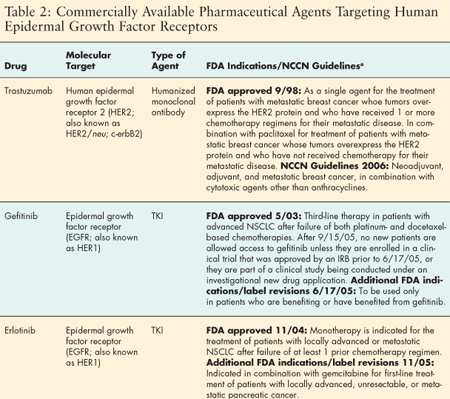
The FDA approved erlotinib hydrochloride (Tarceva) in November 2004. Erlotinib hydrochloride, like gefitinib, is an oral EGFR TKI with action in the intracellular domain.[32] Erlotinib was designed to target the same patient population as gefitinib (advanced NSCLC patients who had failed previous treatment), but erlotinib has demonstrated efficacy, while gefitinib failed to do so. A clinical trial conducted in 17 countries, involving 731 patients with locally advanced or metastatic NSCLC after failure of at least one chemotherapy regimen, showed an overall increase in survival and disease response rates for patients who received erlotinib.[33]
Although erlotinib was initially approved as monotherapy for patients with locally advanced or metastatic NSCLC after failure of at least one prior chemotherapy regimen, clinical trials continue to evaluate the most efficacious use of the drug (Table 2). Analyses of these trials indicate that certain patients-nonsmokers, women, Asians, and patients with adenocarcinoma histology-may show improved responses.[33,34]
Ongoing correlative and clinical trials are exploring the predictive value of factors such as EGFR expression, EGFR mutations, and patient characteristics in the use of EGFR TKIs in NSCLC.[35]
Erlotinib gained FDA approval for an additional indication in November 2005, in combination with gemcitabine as first-line treatment of patients with locally advanced, unresectable, or metastatic pancreatic cancer (Table 2).[36]
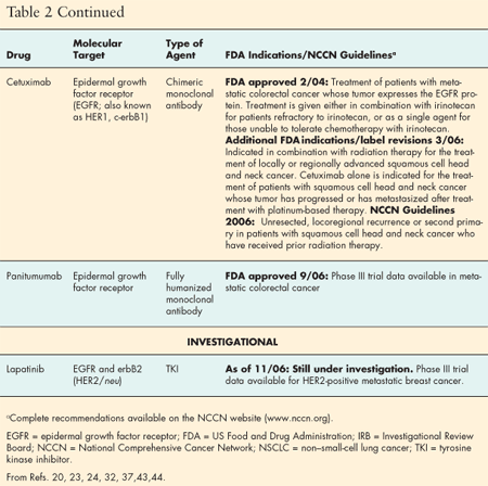
The FDA approved the EGFR-inhibiting agent cetuximab (Erbitux) in 2004 for use in metastatic colorectal cancer (Table 2). Cetuximab is a recombinant chimeric MoAb that binds to EGFR in the extracellular domain.[37] Its approval was based on early clinical data response rates in an open-label, randomized study that compared monotherapy cetuximab with cetuximab and irinotecan in patients who were diagnosed with EGFR-expressing colorectal cancer and were refractory to irinotecan. The tumor response rate of 22.9% to cetuximab in combination with irinotecan in patients with irinotecan-refractory disease was clinically important. The median time to progression and survival time were similar to that of patients who had received standard treatment.[38]
No data are available as yet to demonstrate an increased survival rate for patients receiving cetuximab, but clinical trails with colorectal cancer patients are under way to determine the most efficacious use of the drug in metastatic colorectal cancer and other solid tumors. Although the FDA approved cetuximab for EGFR-expressing metastatic colorectal cancer, NCCN guidelines state that because EGFR testing has yet to show predictive value, routine EGFR testing is not routinely recommended, and cetuximab should not be given or withheld based on EGFR test results.[23]
Based on positive data from clinical trials in patients with squamous cell carcinoma of the head and neck (SCCHN), the FDA approved additional indications for cetuximab in 2006 (Table 2). Compared with radiation therapy alone, cetuximab combined with radiation nearly doubled the survival time in a phase III international trial of patients with locally advanced head and neck cancer.[39]
A phase II trial that achieved an overall response rate of 12.6% and a disease stability rate of 33% supported the use of cetuximab monotherapy in patients with platinum-resistant recurrent/metastatic SCCHN.[40] Recent phase II trials using cetuximab and chemotherapy in patients who had progressed on the chemotherapy regimen alone show promise for the future of cetuximab in SCCHN.[41,42]
Evolving science has prompted research into the development of MoAbs with less murine and more human components. Panitumumab is the first fully human MoAb to be approved to target EGFR. It is an lgG2 MoAb that binds with high affinity to EGFR in the extracellular domain. The FDA approved it in September 2006 for patients with EGFR-expressing metastatic colorectal cancer who have failed standard chemotherapy treatment (Table 2).[43]
Researchers who conducted a phase III randomized trial using best supportive care with or without panitumumab in patients with metastatic colorectal cancer reported their findings at the annual meeting of the American Association for Cancer Research in April 2006. Panitumumab with best supportive care significantly improved progression-free survival and disease control compared with the group who received best supportive care alone. The data showed a 46% reduction in the risk of tumor progression and 8% partial response rate.[44] Ongoing trials are investigating panitumumab for metastatic colorectal cancer and other solid tumors.
Investigational Agents
Targeting the HER family of TKs remains an intriguing prospect for the treatment of solid tumors. Preclinical and clinical investigations of one such drug, lapatinib, are under way. Lapatinib (Tykerb) is a novel HER-family targeted therapy that appears promising for treatment of solid tumors.
Lapatinib is an oral agent that acts as a dual TKI in the intracellular domain. While all EGFR-inhibiting agents have been designed for single targets, lapatinib has the novel mechanism of targeting both HER1 and HER2 (Table 2). Currently under development by GlaxoSmithKline, lapatinib is available for use only in clinical trials involving patients with a variety of solid tumors. Early results for the treatment of advanced breast cancer are encouraging.[45]
In June 2006, GlaxoSmithKline shared the results from a large, randomized phase III study of lapatinib combined with capecitabine (Xeloda) vs capecitabine alone in the treatment of HER2-positive breast cancer patients who had progressed on trastuzumab. Lapatinib plus capecitabine demonstrated a longer time to disease progression than capecitabine alone (8.5 vs 4.5 months). Fewer patients receiving lapatinib developed disease recurrence in the brain. The trial data are not sufficiently mature, however, to demonstrate an improvement in overall survival.[45]
Commercially Available Pharmaceutical Agents Targeting VEGF/Receptors
Bevacizumab (Avastin; anti-VEGF), a recombinant humanized monoclonal IgG1 antibody, binds to the ligand VEGF in the extracellular domain. It prevents VEGF from interacting with the VEGF receptor on the surface of endothelial cells, thus interfering with angiogenesis. The FDA has approved bevacizumab in combination with intravenous fluorouracil for first- and second-line treatment of patients with MCRC (Table 3).[46] The new second-line approval was based on data showing significant improvement in overall survival among patients who received bevacizumab plus FOLFOX4 (fluorouracil, leucovorin, and oxaliplatin), compared with those who received FOLFOX4 alone.[47]
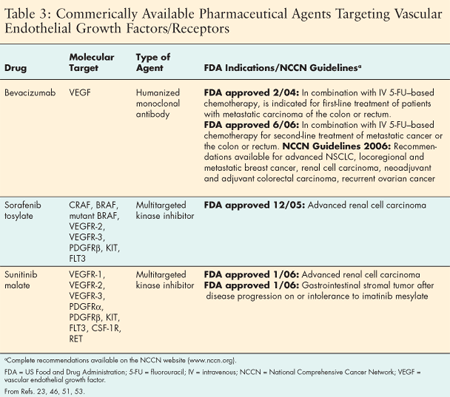
Promising data are emerging from phase II/III trials using bevacizumab in combination with cytotoxic chemotherapy in MBC and advanced NSCLC. The Eastern Cooperative Oncology Group (ECOG) trial, E2100, compared MBC patients treated with bevacizumab plus paclitaxel and patients treated with paclitaxel alone. The difference in progression-free survival was significant in the group receiving bevacizumab plus paclitaxel. Overall survival is still being evaluated, but this study is the first to demonstrate the potential benefit of antiangiogenic therapy in patients with breast cancer.[48] The NCCN's 2006 guidelines recommend bevacizumab for MBC in certain groups of patients.[23]
Recently, bevacizumab, in combination with carboplatin and paclitaxel, was approved by the FDA for first-line treatment of patients with unresectable, locally advanced, recurrent or metastatic non-squamous NSCLC. This approval was based on results from ECOG trial E4599, which compared the use of bevacizumab, paclitaxel, and carboplatin with paclitaxel and carboplatin alone in previously untreated, non-squamous cell advanced NSCLC. The results demonstrated significant improvement in response rates, progression-free survival, and overall survival.[49] The NCCN guidelines recommend the use of bevacizumab, paclitaxel, and carboplatin or cisplatin as front-line treatment for non-squamous cell NSCLC in bevacizumab-eligible patients.[23] Bevacizumab is also being used in ongoing trials investigating the potential of an antiangiogenic approach in renal cell and pancreatic cancer, as well as many other solid tumors.[50]
Sorafenib tosylate (Nexavar), an oral multikinase inhibitor, targets several serine/threonine and receptor TKs. Because sorafenib interacts with multiple cell surface kinases (Table 3), it has both antiangiogenic and antiproliferative properties. It was the first multikinase inhibitor approved and indicated for treatment of advanced renal cell cancer.[51] A phase III multicenter randomized double-blind placebo-controlled trial conducted in advanced renal cell cancer patients demonstrated sorafenib's efficacy and safety. The progression-free rate after 12 weeks was 79% for those who received sorafenib, compared with a 50% progression-free rate for those given the placebo drug. The data demonstrated that sorafenib doubled progression-free survival.[52]
Sunitinib malate (Sutent), another small-molecule oral multikinase inhibitor with antiangiogenic and antiproliferative properties, targets several receptor TKs.[53] In January 2006, sunitinib became the second multikinase inhibitor approved for the treatment of advanced renal cell cancer, based on a study that used the drug as a second-line treatment for patients with metastatic disease. Forty percent of the patients achieved partial responses, and 27% achieved stable disease.[54] A second trial comparing sunitinib with interferon-alfa in first-line metastatic RCC showed statistically significant improvement in response rates for patients on sunitinib.[55]
The FDA also approved sunitinib in January 2006 for the treatment of gastrointestinal stromal tumor in patients whose disease progressed during treatment with imatinib mesylate or who cannot tolerate imatinib mesylate (Table 3).[53] Approval was based on phase III trial data indicating that sunitinib treatment resulted in an increased time to tumor progression and a statistically significant longer overall survival time in gastrointestinal stromal tumor patients who were imatinib-resistant.[56]
Conclusion
Molecularly targeted therapies are becoming a mainstay of treatment for cancer patients. By learning the science and applications of these therapies, oncology nurses improve their own knowledge base and enhance the education of patients and their families.
Disclosures:
Teresa Knoop has acted as a consultant for Amgen and Bristol-Myers Squibb, served on advisory boards for Bristol-Myers Squibb, and served on speakers bureaus for Amgen, Bristol-Myers Squibb, and Genentech.
References:
1. Carpenter C, Cantley L: Essentials of signal transduction, in DeVita VT Jr, Hellman S, Rosenberg SA (eds): Cancer: Principles & Practice of Oncology, 6th ed, pp 31-40. Philadelphia, Lippincott Williams & Wilkins, 2001.
2. Hanahan D, Weinberg R: The hallmarks of cancer. Cell 100:50-70, 2000.
3. Rowinsky EK: Signal events: Cell signal transduction and its inhibition in cancer. Oncologist 8(3):5-17, 2003.
4. Perez-Soler R: HER1/EGFR targeting: Refining the strategy. Oncologist 9:58-67, 2004.
5. Grunwald V, Hidalgo M: Developing inhibitors of the epidermal growth factor receptor for cancer treatment. JNCI 95:851-867, 2003.
6. Herbst R, Shin D: Monoclonal antibodies to target epidermal growth factor receptor-positive tumor: A new paradigm for cancer therapy. Cancer 94:1593-1611, 2002.
7. Schlessinger J: Cell signaling by receptor tyrosine kinases. Cell 103:211-225, 2000.
8. Lenz HJ: Anti-EGFR mechanism of action: Antitumor effect and underlying cause of adverse events. ONCOLOGY 20:5-12, 2006.
9. Yarden Y: Biology of HER2 and its importance in breast cancer. ONCOLOGY 61(suppl 2):1-13, 2001.
10. Yarden Y, Baselga J, Miles D: Molecular approach to breast cancer treatment. Semin Oncol 31:6-13, 2004.
11. Kerbel R: Tumor angiogenesis: Past, present and the near future. Carcinogenesis 21:505-515, 2000.
12. Dvorak H: Vascular permeability factor/vascular endothelial growth factor: A critical cytokine in tumor angiogenesis and a potential target for diagnosis and therapy. J Clin Oncol 20:4368-4380, 2002.
13. Farrara N: Vascular endothelial growth factor as a target for anticancer therapy. Oncologist 9(suppl 1):2-10, 2004.
14. Adjei A, Hidalgo M: Intracellular signal transduction pathway proteins as targets for cancer therapy. J Clin Oncol 23:5326-5403, 2005.
15. Cheifetz A, Mayer L: Monoclonal antibodies, immunogenicity, and associated infusion reactions. Mt Sinai J Med 72:250-259, 2005.
16. Vlahovic G, Crawford J: Activation of tyrosine kinases in cancer. Oncologist 8: 531-538, 2003.
17. Patyna S, Laird AD, Mendel DB, et al: SU 14813: A novel multiple receptor tyrosine kinase inhibitor with potent antiangiogenic and antitumor activity. Mol Cancer Ther 5:1774-1782, 2006.
18. Sandler A, Herbst R: Combining targeted agents: Blocking the epidermal growth factor and vascular endothelial growth factor pathways. Clin Cancer Res 12:4421s-4425s, 2006.
19. McNeil C: Two targets, one drug for new EGFR inhibitors. NJCI 98:1103-1104, 2006.
20. Herceptin (trastuzumab) package insert. Genentech, Inc, San Francisco, CA: February 2005.
21. Jackisch C: HER2-positive metastatic breast cancer optimizing trastuzumab-based therapy. Oncologist 11(suppl 1):34-41, 2006.
22. Baselga J, Perez E, Pienkowski T, et al: Adjuvant trastuzumab: A milestone in the treatment of HER2-positive early breast cancer. Oncologist 11(suppl 1):4-12, 2006.
23. NCCN Clinical Practice Guidelines in Oncology: Available at www.nccn.org. Accessed September 19, 2006.
24. Iressa (gefitinib) tablets, package insert. AstraZeneca Pharmaceuticals, Wilmington, DE: June 2005.
25. Giaccone G, Herbst RS, Manegold C, et al: Gefitinib in combination with gemcitabine and cisplatin in advanced non-small-cell lung cancer: A phase III trial-INTACT 1. J Clin Oncol 22:777-784, 2004.
26. Herbst RS, Giaccone G, Schiller JH, et al: Gefitinib in combination with paclitaxel and carboplatin in advanced non-small-cell lung cancer: A phase III trial-INTACT 2. J Clin Oncol 22:785-794, 2004.
27. US Food and Drug Administration: Catalog of approved cancer drugs. Available at http://www.accessdata.fda.gov/scripts/cder/drugsatfda/index.cfm?fuseaction=Search.Label
ApprovalHistory#apphist Iressa. Accessed August 15, 2006.
28. Fukuoka M, Yano S, Giaccone G, et al: Multi-institutional randomized phase II trial of gefitinib for previously treated patients with advanced non-small-cell ling cancer. J Clin Oncol 21:2237-2246, 2003.
29. Kris MG, Natale RB, Herbst RS, et al: Efficacy of gefitinib, and inhibitor of the epidermal growth factor receptor tyrosine kinase, in symptomatic patients with non-small-cell lung cancer. JAMA 290:2149-2158, 2003.
30. US Food and Drug Administration: Safety alerts for drugs, biologics, medical devices, and dietary supplements. Available at http://www.fda.gov/cder/drug/infopage/gefitinib/default.htm 2005 Iressa. Accessed September 9, 2006.
31. Thatcher N, Chang A, Parikh P, et al: Gefitinib plus best supportive care in previously treated patients with refractory advanced non-small-cell lung cancer: Results from a randomized, placebo-controlled, multicenter study (Iressa survival evaluation in lung cancer). Lancet 366:1527-1537, 2005.
32. Tarceva (erlotinib) tablets, package insert. Genentech, San Francisco, CA, and OSI Oncology, Melville, NY: November 2005.
33. Shepard FA, Pereira JR, Ciuleanu T, et al, for the National Cancer Institute of Canada Trials Group: Erlotinib in previously treated non-small-cell lung cancer. N Engl J Med 353:123-132, 2005.
34. Tsao MS, Sakurada A, Cutz JC, et al: Erlotinib in lung cancer-Molecular and clinical predictors of outcome. N Engl J Med 353:133-144, 2005.
35. Vokes EE, Chu E: Anti-EGFR therapies: Clinical experience in colorectal, lung and head and neck cancer. ONCOLOGY 20(suppl 2):17-25, 2006.
36. Moore MJ, Goldstein D, Hamm J, et al: Erlotinib plus gemcitabine compared to gemcitabine alone in patients with advanced pancreatic cancer. A phase III trial of the National Cancer Institute of Canada Clinical Trials Group [NCIC-CTG] (abstract 1). J Clin Oncol 23(suppl 16S):1s, 2005.
37. Erbitux (cetuximab) package insert. ImClone Systems Inc, Branchberg, NJ, and Bristol Myers Squibb Co, Princeton, NJ: March 2006.
38. Cunningham D, Humblet Y, Siena S, et al: Cetuximab monotherapy and cextuximab plus irinotecan in irinotecan-refractory metastatic colorectal cancer. N Engl J Med 351:337-345, 2004.
39. Bonner JA, Harari PM, Giralt J, et al: Radiotherapy plus cetuximab for squamous-cell carcinoma of the head and neck. N Engl J Med 354:567-578, 2006.
40. Trigo J, Hitt R, Koralewski P, et al: Cetuximab monotherapy is active in patients (pts) with platinum refractory recurrent/metastatic squamous cell carcinoma of the head and neck (SCCHN): Results of a phase II study (abstract 5502) [and slide presentation]. Proc Am Soc Clin Oncol 23:487, 2004.
41. Baselga J, Trigo JM, Bourhis J, et al: Phase II multicenter study of the anti-epidermal growth factor receptor monoclonal antibody cetuximab in combination with platinum-based chemotherapy in patients with platinum-refractory metastatic and/or recurrent squamous cell carcinoma of the head and neck. J Clin Oncol 23:5578-5587, 2005.
42. Herbst RS, Shin DM, Dicke K, et al: Phase II multicenter study of the epidermal growth factor receptor antibody cetuximab and cisplatin for recurrent and refractory head and neck. J Clin Oncol 23:5568-5577, 2005.
43. Panitumumab: Available at http://www.amgen.com/science/pipe_panitumumab.html Accessed September 2006.
44. Peeters M, et al: A phase III, multicenter, randomized controlled trial (RTC) of panitumumab plus best supportive care (BSC) vs. BSC alone in patients (pts) with metastatic colorectal cancer (mCRC) (abstract CP-1). Presented at the Annual Meeting of the American Association for Cancer Research, 2006.
45. Geyer CE, Forster JK, Cameron DS: Phase III randomized, open-label, international study comparing lapatinib and capecitabine versus capecitabine in women with refractory advanced or metastatic breast cancer. American Society of Clinical Oncology 42nd annual meeting, Atlanta, 2006.
46. Avastin (bevacizumab) package insert. Genentech, San Francisco, CA, June 2006.
47. Benson AB, Catalano PJ, Meropol J, et al: Bevacizumab (anti-VEGF) plus FOLFOX 4 in previously treated advanced colorectal cancer (adv CRC): An interim toxicity analysis of the Eastern Cooperative Group (ECOG) study E3200 (abstract 975). Proc Am Soc Clin Oncol 22:243, 2003.
48. Miller K, Wang M, Gralow J, et al: E2100, a randomized phase III trial of paclitaxel vs. paclitaxel plus bevacizumab as first-line therapy for locally recurrent or metastatic breast cancer [presentation]. American Society of Clinical Oncology 41st annual meeting, Orlando, Florida, 2005.
49. Sandler AB, Gray R, Brahmer J, et al: Randomized phase II/III trial of paclitaxel (P) plus carboplatin with or without bevacizumab (NSC #704865) in patients with advanced non-squamous non-small cell lung cancer (NSCLC): An Eastern Cooperative Oncology Group (ECOG) Trial-E4599 (abstract LBA4). J Clin Oncol 23(suppl 16S):2s, 2005.
50. de Gramont A, Van Cutsem E: Investigating the potential of bevacizumab in other indications: Metastatic renal cell, non-small cell lung, pancreatic and breast cancer. ONCOLOGY 69(suppl 3):46-56, 2005.
51. Nexavar (sorafenib) package insert. Bayer Pharmaceuticals Corp, West Haven, CT: August 2006.
52. Escudier B, Szczylik C, Eisen T, et al: Randomized phase III trial of the Raf kinase and VEGFR inhibitor sorafenib (BAY 43-9006) in patients with advanced renal cell carcinoma (RCC). J Clin Oncol 23(16S):4510, 2005.
53. Sutent (sunitinib malate) package insert. Pfizer Labs, New York, February 2006.
54. Motzer RJ, Michaelson MD, Redman BG, et al: Activity of SU11248, a multitargeted inhibitor of vascular endothelial growth factor receptor and platelet derived growth factor receptor, in patients with metastatic renal cell carcinoma J Clin Oncol 24:16-24, 2006.
55. Motzer RJ, Hutson TE, Tomczak P, et al: Phase III randomized trial of sunitinib malate (SU 11248) versus interferon-alfa (IFN-α) as first line systemic therapy for patients with metastatic renal cell carcinoma (mRCC) (abstract LBA3). J Clin Oncol 24(suppl 18S):2s, 2006.
56. Demetri GD, van Oosteron AT, Blackstein M, et al: Phase 3, multicenter, randomized, double blind, placebo-controlled trial of SU11248 in patients (pts) following failure of imatinib for metastatic GIST (abstract 4000). J Clin Oncol 23(suppl 16S):308s, 2005.
Gedatolisib Combo With/Without Palbociclib May Be New SOC in PIK3CA Wild-Type Breast Cancer
December 21st 2025“VIKTORIA-1 is the first study to demonstrate a statistically significant and clinically meaningful improvement in PFS with PAM inhibition in patients with PIK3CA wild-type disease, all of whom received prior CDK4/6 inhibition,” said Barbara Pistilli, MD.